- Physical Examination
- Surgical Examination
- Ophthalmology
- Clinical Skills
- Orthopedics
- Surgery Videos
- Laparoscopy
- Pediatrics
- Funny Videos
- Cardiothoracic Surgery
- Nursing Videos
- Plastic Surgery
- Otorhinolaryngology
- Histology and Histopathology
- Neurosurgery
- Dermatology
- Pediatric Surgery
- Urology
- Dentistry
- Oncology and Cancers
- Anatomy Videos
- Health and Fitness
- Radiology
- Anaesthesia
- Physical Therapy
- Pharmacology
- Interventional Radiology
- Cardiology
- Endocrinology
- Gynecology
- Emergency Medicine
- Psychiatry and Psychology
- Childbirth Videos
- General Medical Videos
- Nephrology
- Physiology
- Diet and Food Health
- Diabetes Mellitus
- Neurology
- Women Health
- Osteoporosis
- Gastroenterology
- Pulmonology
- Hematology
- Rheumatology
- Toxicology
- Nuclear Medicine
- Infectious Diseases
- Vascular Disease
- Reproductive Health
- Burns and Wound Healing
- Other
Top videos
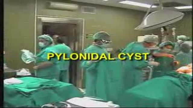
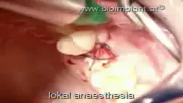
LASER SURGERY Pilonidal Cyst removal
The video will describe what is miliary tuberculosis. Please see my website for disclaimer.
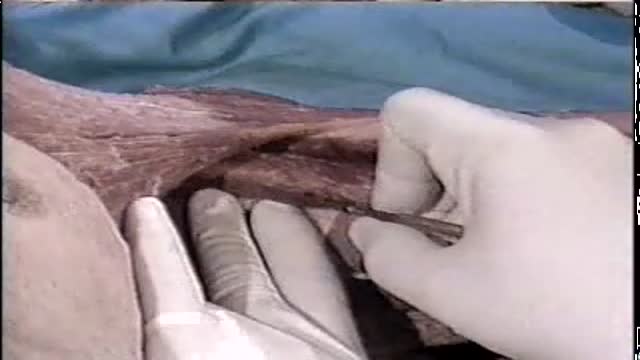
Axillary Block with a Nerve Stimulator
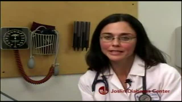
A video discussing the importance of following up the blood pressure for diabetic patients and the serious complications that they can avoid by this very simple measure.

Ultrasound Guided Sclerotherapy for Varicose Veins
WORLD'S FIRST TRUE ANATOMIC ZIRCONIA DENTAL IMPLANT SOLUTION dentistry

Open Distal Pancreatectomy Surgery Video
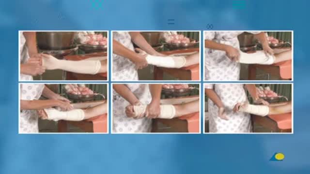
Volar Slab Splint for Forearm and Wrist Fractures and Sprains
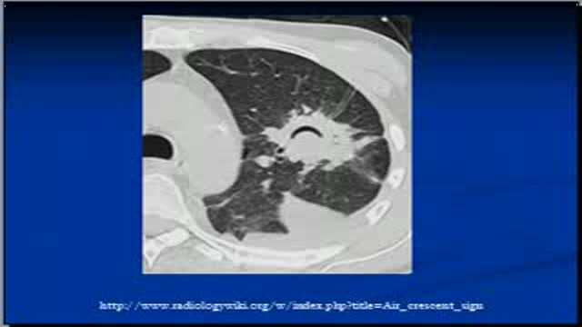
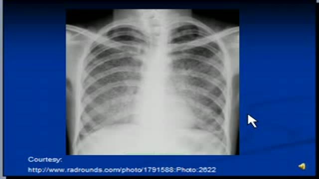
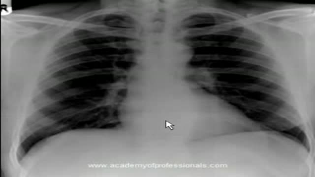
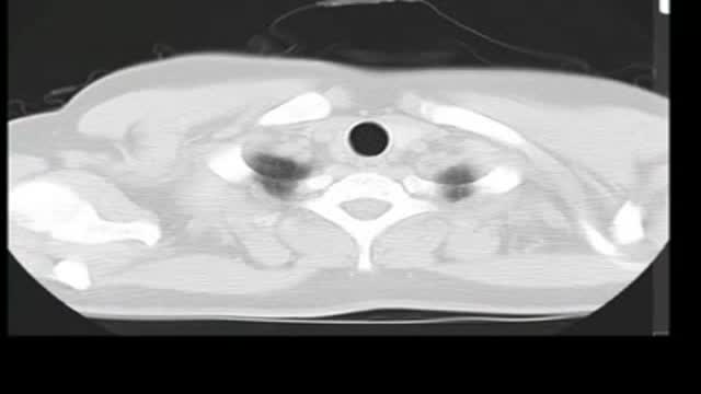
The video will describe air crescent sign on Chest x-rays and CT scans. Please see disclaimer on my website. www.academyofprofessionals.com
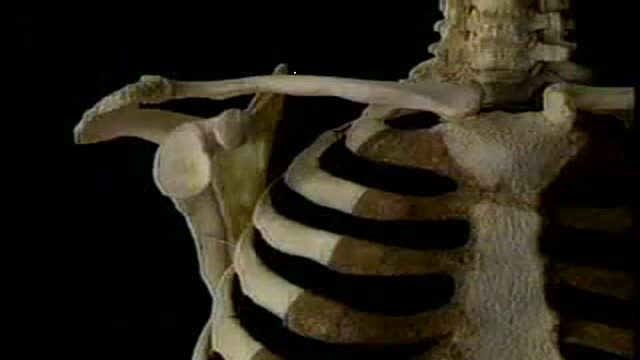
Anatomy of the shoulder joint
دكتور مصطفى ياقوت محاضرة القدم السكرى A lecture presented by Dr. Mostafa Yakoot to the Annual Congress of the Vascular Surgery, Alexandria 10/2009. Based on the original article published in JWC by: Yakoot M, Abdelatif M, Etman M.
Lecture presented by Dr. Mostafa Yakoot, to the European Multicongress of parasitology Valencia, Spain
Robot-Assisted Laparoscopic Rectal Resection for Endometriosis
artificial aortic valves. Please see disclaimer on my website. www.academyofprofessionals.com
As above. Please see disclaimer on my website. www.academyofprofessionals.com
Pulmonary alveolar proteinosis. Please see disclaimer on my website. www.academyofprofessionals.com. MCQs are also available.
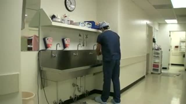
Chapel Hill Tubal Reversal Center - www.tubal-reversal.net - illustrates proper hand hygiene technique for surgical scrub to disinfect the hands prior to entering the operating room for tubal ligation reversal surgery.
Embolization is a medical advance that shrinks uterine fibroids. One tiny incision allows us to solve the problem quickly, safely and without surgery
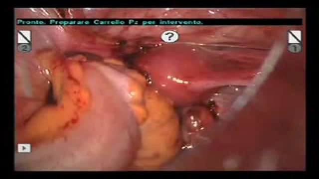
A video showing Laparoscopic Resection of Splenic Artery Aneurysm
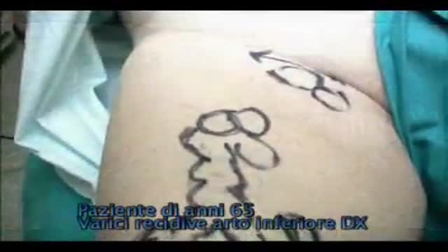
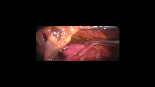
Recurrent varicose veins are a common problem. The patient in this video was operated for great saphenous vein insufficiency and a “neocrosse” occurred after few years. Surgical exploration revealed a “cavernoma” just over the nodes of the crural area, feeding varicose veins of thigh and leg.
