- Physical Examination
- Surgical Examination
- Ophthalmology
- Clinical Skills
- Orthopedics
- Surgery Videos
- Laparoscopy
- Pediatrics
- Funny Videos
- Cardiothoracic Surgery
- Nursing Videos
- Plastic Surgery
- Otorhinolaryngology
- Histology and Histopathology
- Neurosurgery
- Dermatology
- Pediatric Surgery
- Urology
- Dentistry
- Oncology and Cancers
- Anatomy Videos
- Health and Fitness
- Radiology
- Anaesthesia
- Physical Therapy
- Pharmacology
- Interventional Radiology
- Cardiology
- Endocrinology
- Gynecology
- Emergency Medicine
- Psychiatry and Psychology
- Childbirth Videos
- General Medical Videos
- Nephrology
- Physiology
- Diet and Food Health
- Diabetes Mellitus
- Neurology
- Women Health
- Osteoporosis
- Gastroenterology
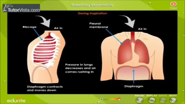
- Pulmonology
- Hematology
- Rheumatology
- Toxicology
- Nuclear Medicine
- Infectious Diseases
- Vascular Disease
- Reproductive Health
- Burns and Wound Healing
- Other
Top videos
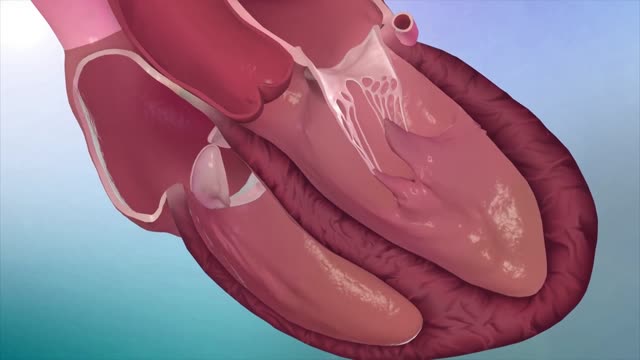
Hypertrophic cardiomyopathy (HCM) is very common and can affect people of any age. It affects men and women equally. It is a common cause of sudden cardiac arrest in young people, including young athletes. Hypertrophic cardiomyopathy occurs if heart muscle cells enlarge and cause the walls of the ventricles (usually the left ventricle) to thicken. The ventricle size often remains normal, but the thickening may block blood flow out of the ventricle. If this happens, the condition is called obstructive hypertrophic cardiomyopathy. Sometimes the septum, the wall that divides the left and right sides of the heart, thickens and bulges into the left ventricle. This can block blood flow out of the left ventricle. Then the ventricle must work hard to pump blood. Symptoms can include chest pain, dizziness, shortness of breath, or fainting. Hypertrophic cardiomyopathy also can affect the heart's mitral valve, causing blood to leak backward through the valve. Sometimes, the thickened heart muscle doesn't block blood flow out of the left ventricle. This is referred to as non-obstructive hypertrophic cardiomyopathy. The entire ventricle may thicken, or the thickening may happen only at the bottom of the heart. The right ventricle also may be affected. In both obstructive and non-obstructive HCM, the thickened muscle makes the inside of the left ventricle smaller, so it holds less blood. The walls of the ventricle may stiffen, and as a result, the ventricle is less able to relax and fill with blood.
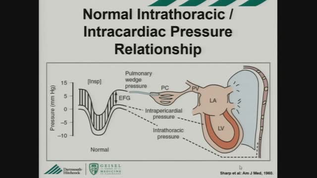
Constrictive pericarditis is the result of scarring and consequent loss of the normal elasticity of the pericardial sac. This leads to impairment of ventricular filling in mid and late diastole. As a result, the majority of ventricular filling occurs rapidly in early diastole and the ventricular volume does not increase after the end of the early filling period. Restrictive cardiomyopathy is characterized by a nondilated rigid ventricle, resulting in severe diastolic dysfunction and restrictive filling that produces hemodynamic changes similar to those in constrictive pericarditis. Constrictive pericarditis and restrictive cardiomyopathy both lead to diastolic heart failure with normal (or near normal) systolic function, and characteristically abnormal ventricular filling that results in similar clinical and hemodynamic features. However, because of their markedly different treatments, differentiating between the two conditions is critical. In some patients, the correct diagnosis may be readily suggested from the history or routine diagnostic testing. In others, however, this differentiation cannot be diagnosed before biopsy or even surgical exploration.
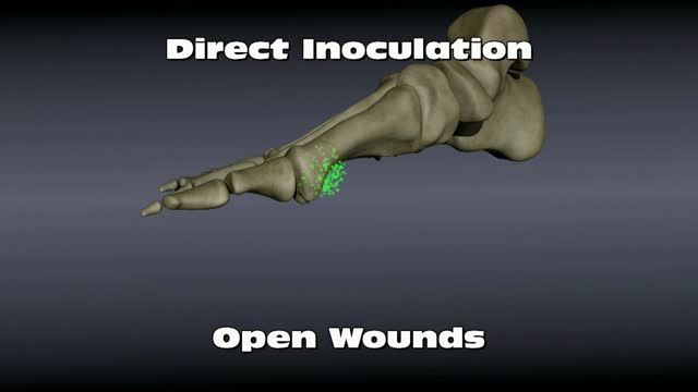
Osteomyelitis is an infection in a bone. Infections can reach a bone by traveling through the bloodstream or spreading from nearby tissue. Infections can also begin in the bone itself if an injury exposes the bone to germs. In children, osteomyelitis most commonly affects the long bones of the legs and upper arms. Adults are more likely to develop osteomyelitis in the bones that make up the spine (vertebrae). People who have diabetes may develop osteomyelitis in their feet if they have foot ulcers. Once considered an incurable condition, osteomyelitis can be successfully treated today. Most people require surgery to remove parts of the bone that have died — followed by strong antibiotics, often delivered intravenously, typically for at least four to six weeks.
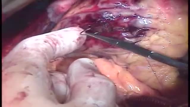
Repair of post-infarction ventricular septal defect (VSD) remains a challenging procedure with a high risk of VSD recurrence. In order to reduce this risk, a double patch and glue technique was introduced in the department in 1986. This surgical technique is hereunder presented. Since 1971, ninety-three patients have been operated on early (≪15 days) after the occurrence of a post-infarction VSD. This retrospective study allows to compare the results of this double patch and glue technique to those obtained with the conventional one, in terms of hospital death and VSD recurrence. The double patch and glue technique avoids recurrence of VSD and plays a part in reducing hospital mortality.
A stroke occurs when the blood supply to your brain is interrupted or reduced. This deprives your brain of oxygen and nutrients, which can cause your brain cells to die. A stroke may be caused by a blocked artery (ischemic stroke) or the leaking or bursting of a blood vessel (hemorrhagic stroke)
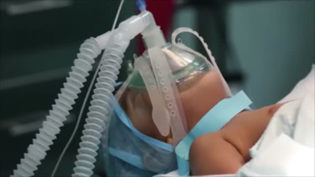
There's only one group of people who really know what happens when you die: the dead. And since the dead won't be revealing their secrets anytime soon, it's up to scientists to explain what happens when a person dies. Death, just like life, is a process, scientists say. The first stage of this process is known as clinical death. It lasts from four to six minutes, beginning when a person stops breathing and the heart stops pumping blood. During this time, there may be enough oxygen in the brain that no permanent brain damage occurs. Other organs, such as the kidneys and eyes, also remain alive throughout clinical death.

Life Before Birth
Myasthenia gravis is a chronic autoimmune neuromuscular disease characterized by varying degrees of weakness of the skeletal (voluntary) muscles of the body. The name myasthenia gravis, which is Latin and Greek in origin, literally means "grave muscle weakness."
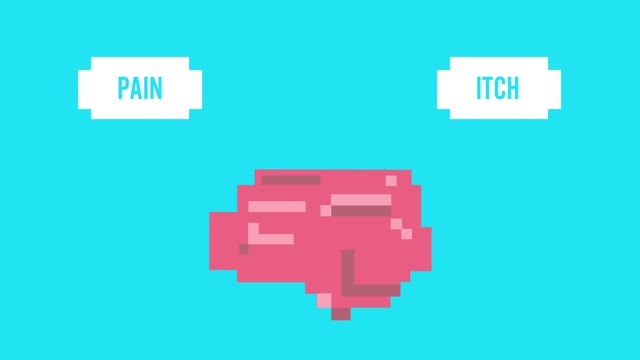
Pruritis is itchy skin that makes you want to scratch. It can be caused by many things. Normally, itchy skin isn't serious, but it can make you uncomfortable. Sometimes, itchy skin is caused by a serious medical condition. It can occur in association with a primary rash (e.g. dermatitis) or may occur because of hypersensitive nerves in the skin (neuropathic pruritus). ... Scratching a localised itch may lead to lichen simplex, prurigo or prurigo nodularis. Systemic causes of pruritus. Sytemic diseases may cause generalised pruritus.
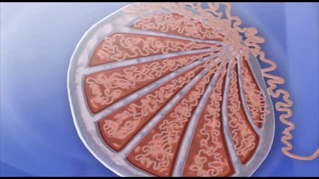
The protein inside red blood cells (a) that carries oxygen to cells and carbon dioxide to the lungs is hemoglobin (b). Hemoglobin is made up of four symmetrical subunits and four heme groups. Iron associated with the heme binds oxygen.
Spermatogenesis is the process in which spermatozoa are produced from spermatogonial stem cells by way of mitosis and meiosis. The initial cells in this pathway are called spermatogonia, which yield primary spermatocytes by mitosis.
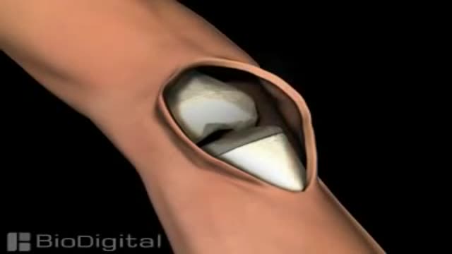
Knee replacement, also called arthroplasty, is a surgical procedure to resurface a knee damaged by arthritis. Metal and plastic parts are used to cap the ends of the bones that form the knee joint, along with the kneecap. This surgery may be considered for someone who has severe arthritis or a severe knee injury.
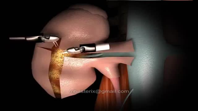
The robotic approach to renal surgery, particularly partial nephrectomy, has some inherent challenges, and some familiarity with the da Vinci robotic system is necessary. The surgeon must gain an understanding of the robotic arm movements and range of motion, especially in relation to the clutch and camera. The advent of robotically assisted prostatectomy in 2001 [23] paved the way for widespread accessibility to the da Vinci robotic unit and its application to renal surgery. Since that time, at least one multi-institutional survey has demonstrated superiority of the robotic approach when compared to laparoscopic for outcomes of blood loss, hospital stay and a substantially shorter warm ischemia time, while maintaining equivalence in positive margin rate, operative time and complications. [11] A transperitoneal approach is most commonly used. Prior abdominal operation is not necessarily a contraindication to this procedure, but access should be approached with regard for previous operation(s) by an experienced team.

Fast Lower Back Pain & Sciatica Pain Relief – Beginners Yoga Stretches and Poses
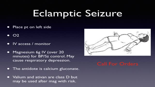
Obstetrical emergencies of pregnancy ECTOPIC PREGNANCY. ... PLACENTAL ABRUPTION. ... PLACENTA PREVIA. ... ECTOPIC PREGNANCY. ... PLACENTAL ABRUPTION. ... PLACENTA PREVIA. ... Amniotic fluid — The liquid in the placental sac that cushions the fetus and regulates temperature in the placental environment.
Trisomy 18, also called Edwards syndrome, is a chromosomal condition associated with abnormalities in many parts of the body. Individuals with trisomy 18 often have slow growth before birth (intrauterine growth retardation) and a low birth weight. Affected individuals may have heart defects and abnormalities of other organs that develop before birth. Other features of trisomy 18 include a small, abnormally shaped head; a small jaw and mouth; and clenched fists with overlapping fingers. Due to the presence of several life-threatening medical problems, many individuals with trisomy 18 die before birth or within their first month. Five to 10 percent of children with this condition live past their first year, and these children often have severe intellectual disability.
What Your Handwriting Says About You
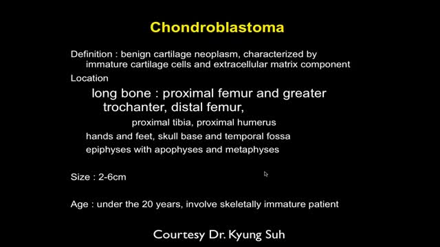
MRI of Bone Tumor
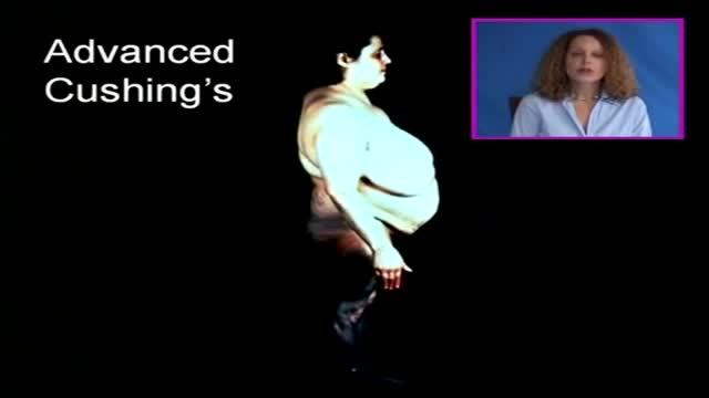
Cushing's disease is a serious condition of an excess of the steroid hormone cortisol in the blood level caused by a pituitary tumor secreting adrenocorticotropic hormone (ACTH). ACTH is a hormone produced by the normal pituitary gland. ACTH stimulates the adrenal glands (located on top of the kidneys) to produce cortisol, commonly referred to as the stress hormone.

Gastroparesis -- literally “paralyzed stomach” -- is a serious condition manifested by delayed emptying of stomach contents into the small intestine after a meal. There is no cure for gastroparesis, but treatment can speed gastric emptying and relieve gastrointestinal symptoms such as nausea and vomiting.
