Top Videos
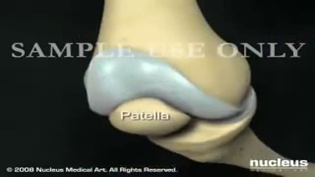
Total Knee Replacement Surgery Video
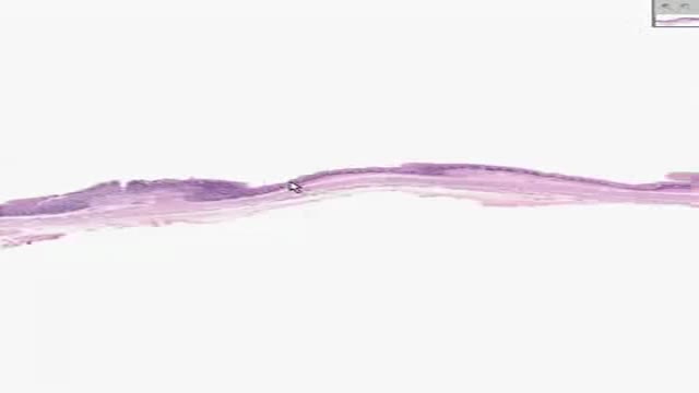
Histology of GastroEsophageal Junction
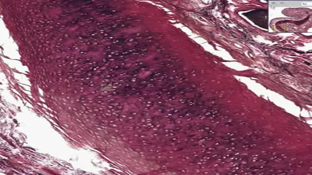
Histology of Elastic Cartilage
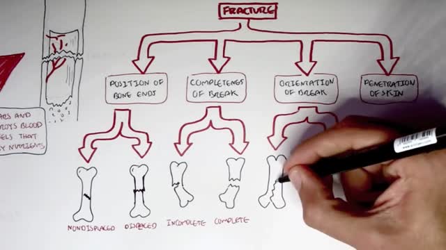
Common types of fractures include: Stable fracture. The broken ends of the bone line up and are barely out of place. Open, compound fracture. The skin may be pierced by the bone or by a blow that breaks the skin at the time of the fracture. ... Transverse fracture. ... Oblique fracture. ... Comminuted fracture.
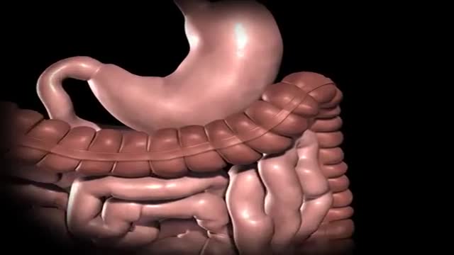
Gastric bypass is surgery that helps you lose weight by changing how your stomach and small intestine handle the food you eat. After the surgery, your stomach will be smaller. You will feel full with less food. The food you eat will no longer go into some parts of your stomach and small intestine that absorb food. Because of this, your body will not get all of the calories from the food you eat.

Amnesia refers to the loss of memories, such as facts, information and experiences. Though having no sense of who you are is a common plot device in movies and television, real-life amnesia generally doesn't cause a loss of self-identity. Instead, people with amnesia — also called amnestic syndrome — are usually lucid and know who they are, but may have trouble learning new information and forming new memories. Amnesia can be caused by damage to areas of the brain that are vital for memory processing. Unlike a temporary episode of memory loss (transient global amnesia), amnesia can be permanent. There's no specific treatment for amnesia, but techniques for enhancing memory and psychological support can help people with amnesia and their families cope.

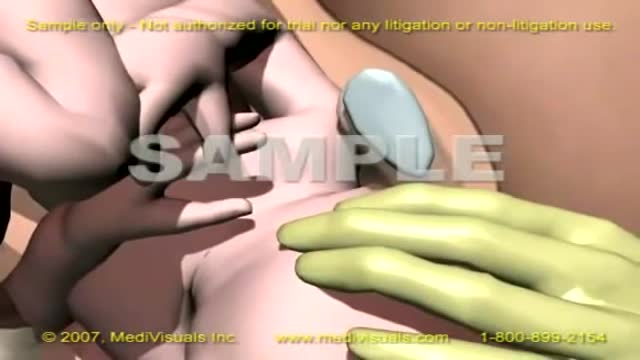
If you’ve suffered a sporting knee injury, how do you know when it’s serious? In this short video, Yorkshire Knee Clinic’s Dave Duffy reveals the two key tests that tell you whether your knee needs urgent, specialist attention.
𝗡𝗼𝘁𝗲𝘀 𝗳𝗼𝗿 𝘁𝗵𝗲 𝘀𝗾𝘂𝗲𝗮𝗺𝗶𝘀𝗵: This video features only features a model of the knee. There is no live footage from operations.
Discover more about sports knee injuries: https://yorkshirekneeclinic.com/sports-injuries/
Discover more about Dave Duffy: https://yorkshirekneeclinic.com/about/dave-duffy/
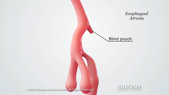
A tracheoesophageal fistula (TEF, or TOF; see spelling differences) is an abnormal connection (fistula) between the esophagus and the trachea. TEF is a common congenital abnormality, but when occurring late in life is usually the sequela of surgical procedures such as a laryngectomy.
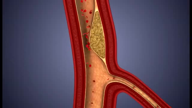
What damage does atherosclerosis cause? Plaque may partially or totally block the blood's flow through an artery in the heart, brain, pelvis, legs, arms or kidneys. Some of the diseases that may develop as a result of atherosclerosis include coronary heart disease, angina (chest pain), carotid artery disease, peripheral artery disease (PAD) and chronic kidney disease.

His father, Dr. Joseph Dello Russo, helped turn Lasik eye surgery into the widespread procedure it is today. Now he explains a new technique and how it differs.
The protein inside red blood cells (a) that carries oxygen to cells and carbon dioxide to the lungs is hemoglobin (b). Hemoglobin is made up of four symmetrical subunits and four heme groups. Iron associated with the heme binds oxygen.
Shoulder dystocia is a rare emergency that can happen during the end of the second stage of labour. It's all to do with how your baby moves down through your vagina and out into the world. Shoulder dystocia happens when your baby's head has been born, but one of her shoulders becomes stuck.

Runners Knee Overview:
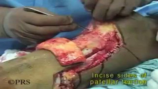
Welcome to our Patello-Femoral Rehab video. The goal of this video is to minimize pain around the kneecap and maximize recovery. This video should not be used as a substitute for regular physical therapy visits and guidance from your physician
Visit http://www.matthewboesmd.com/p....atello-femoral-rehab for more information

Let SightMD walk you through an entire LASIK procedure.
Find out more about LASIK at SightMD - https://www.sightmd.com/eye-do....ctor/lasik-eye-surge
Pathology: Previous spinal cord injury, diabetes, renal failure, dynamic knee contracture, open left ankle disarticulation for sepsis and severe foot infection
The annual incidence of primary intraspinal neoplasm is approximately five per million for females and three per million for males.[9] Spinal intradural extramedullary tumors account for two thirds of all intraspinal neoplasms and include neuromas and meningiomas.[1] Overall, meningiomas account for 25 to 46% of primary spinal neoplasms and are the second most common intradural spine tumor after neuromas.[9] Spinal meningiomas occur less frequently than intracranial ones and account for approximately 7.5 to 12.7% of all meningiomas.[25]


The common obsession among men and women of having a perfect body has lead them to many serious neurotic disorders. They are constantly exposed to the ideas of having the perfect body. We are bombarded with the images on social media which create a hype among men and women, to achieve the exact same ratio of fats in them. Body image is merely an image of your thoughts and perceptions. The way you think how people notices you can greatly impact yourself and your way of thinking about yourself. It becomes quite a big deal when you start to feel low about yourself. It leads you towards having a low self esteem and it becomes hard for you to feel worthy and confident. On contrary when you have good self esteem you feel empowered and confident. It is not the consequence of just liking your own body but its about accepting who you are and making people accept you as you are.

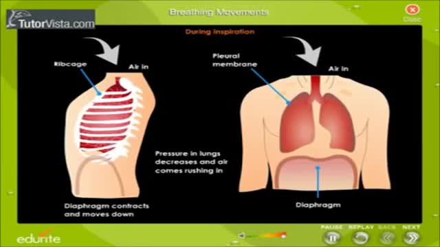
There are 3 major parts of the respiratory system: the airway, the lungs, and the muscles of respiration. The airway, which includes the nose, mouth, pharynx, larynx, trachea, bronchi, and bronchioles, carries air between the lungs and the body's exterior.

This Unorthodox Procedure Makes Short People A Foot Taller
