- Physical Examination
- Surgical Examination
- Ophthalmology
- Clinical Skills
- Orthopedics
- Surgery Videos
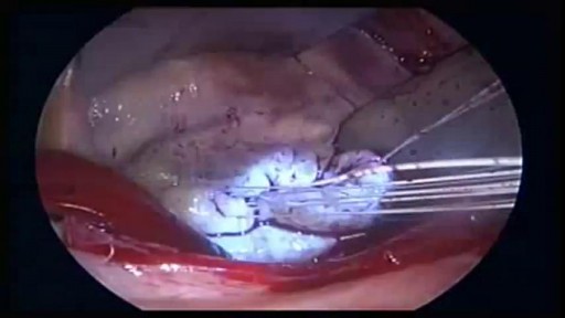
- Laparoscopy
- Pediatrics
- Funny Videos
- Cardiothoracic Surgery
- Nursing Videos
- Plastic Surgery
- Otorhinolaryngology
- Histology and Histopathology
- Neurosurgery
- Dermatology
- Pediatric Surgery
- Urology
- Dentistry
- Oncology and Cancers
- Anatomy Videos
- Health and Fitness
- Radiology
- Anaesthesia
- Physical Therapy
- Pharmacology
- Interventional Radiology
- Cardiology
- Endocrinology
- Gynecology
- Emergency Medicine
- Psychiatry and Psychology
- Childbirth Videos
- General Medical Videos
- Nephrology
- Physiology
- Diet and Food Health
- Diabetes Mellitus
- Neurology
- Women Health
- Osteoporosis
- Gastroenterology
- Pulmonology
- Hematology
- Rheumatology
- Toxicology
- Nuclear Medicine
- Infectious Diseases
- Vascular Disease
- Reproductive Health
- Burns and Wound Healing
- Other
Top videos
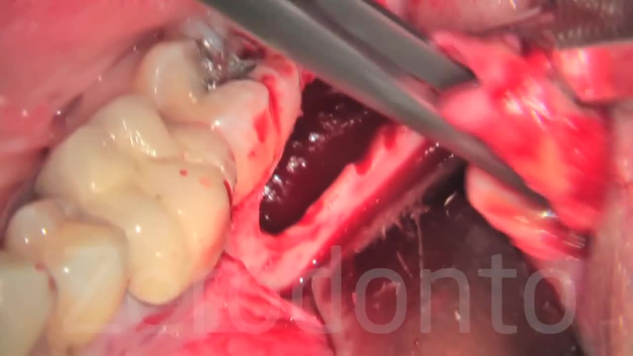
This pressure can also cause problems with crowding of the other teeth or require orthodontic treatment to straighten other teeth. Cysts. The wisdom tooth develops in a sac within the jawbone. The sac can fill with fluid, forming a cyst that can damage the jawbone, teeth and nerves.
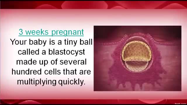
Pregnancy first Trimester
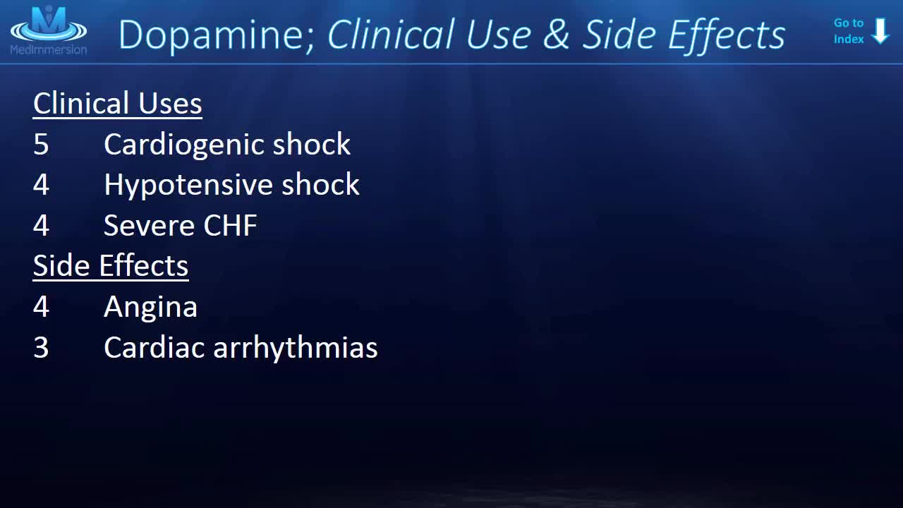
Dopamine is the one neurotransmitter that everyone seems to know about. Vaughn Bell once called it the Kim Kardashian of molecules, but I don’t think that’s fair to dopamine. Suffice it to say, dopamine’s big. And every week or so, you’ll see a new article come out all about dopamine.
What combines research opportunities, intellectual challenge, and international collaboration in the study of a disease which affects many organs of the body and all sectors of society? And demands that specialists from many different backgrounds work together to crack sometimes intractable problems? It is, of course, oncology. As a career choice, it's demanding; it takes passion coupled with a willingness to put in the hours and to learn how to discuss death honestly and sensitively. But for the right person, it can be immensely rewarding.
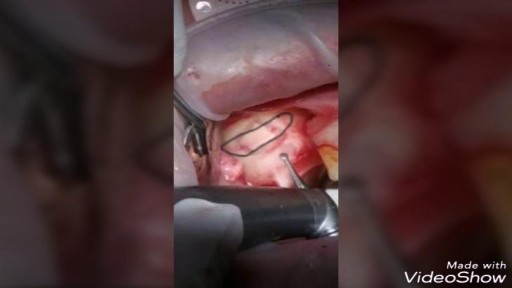
An arthroscopic meniscectomy is a procedure to remove some or all of a meniscus from the tibio-femoral joint of the knee using arthroscopic (aka 'keyhole') surgery. In a complete meniscectomy the meniscus including the meniscal rim is removed. A partial meniscectomy involves partial removal of the meniscus. This may vary from minor trimming of a frayed edge to anything short of removing the rim. This is a minimally invasive procedure often done as day suas an outpatient in a one-day clinic [1] This procedure is performed when a meniscal tear is too large to be corrected by a surgical meniscal repair.[1] When non-operative therapy provides some degree of symptom relief over the long-term, these benefits may wane with continued meniscal degeneration. In such patients, arthroscopic partial meniscectomy can be effective in improving patient quality of life.
Our results in this study of MIPO treated with conventional plates are comparable to the results of the femoral shaft fractures treated with intramedullary nailing. The technique can be used for all femoral shaft fractures. Although the biomechanics of the plate fixation are less stable compared to the intamedullary nail, the mechanical stability is stable enough for bone healing. Healing was rapid, and postoperative care was simplified. The two major complications were malalignment and screw breakage. We recommend using at least three separated screws in each fragment to prevent stress on the screw and screw breakage. Intraoperative limb length, axial alignment, and rotation must be carefully assessed to prevent malalignment. The limitations of our study include lack of a comparison group, retrospective data collection, and no randomisation in outcome evaluation
This is the future of medicine
Infection leg gets cleaning inside
There are several ways to do minimally invasive aortic valve surgery. Techniques include min-thoracotomy, min-sternotomy, robot-assisted surgery, and percutaneous surgery. To perform the different procedures: Your surgeon may make a 2-inch to 3-inch (5 to 7.5 centimeters) cut in the right part of your chest near the sternum (breastbone). The muscles in the area will be divided. This lets the surgeon reach the heart and aortic valve. Your surgeon may split only the upper portion of your breast bone allowing exposure to the aortic valve. For robotically-assisted valve surgery, the surgeon makes 2 to 4 tiny cuts in your chest. The surgeon uses a special computer to control robotic arms during the surgery. A 3D view of the heart and aortic valve are displayed on a computer in the operating room.
Anemia is a condition in which the body does not have enough healthy red blood cells. Red blood cells provide oxygen to body tissues. There are many types of anemia. Pernicious anemia is a decrease in red blood cells that occurs when the intestines cannot properly absorb vitamin B12.
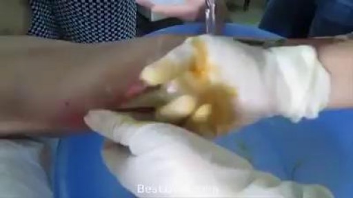
watch that video of Pulling out 1 foot long foot of gauze out from face
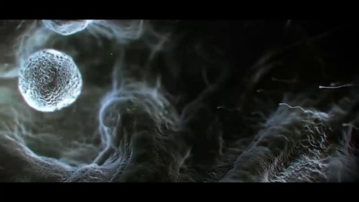
The journey of egg and sperm. There are a lot of casualties (deaths) among the sperm as they swim toward the egg. First, many get lost in the maze of a woman's uterus where they also have to contend with acidic vaginal secretions.

Functional endoscopic sinus surgery is a minimally invasive surgical treatment which uses nasal endoscopes to enlarge the nasal drainage pathways of the paranasal sinuses to improve sinus ventilation.
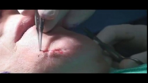
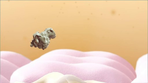
This is an introduction into mole removal through excisional means (cutting it out) or using a laser to remove the mole

http://eliminar-celulite.plus101.com --- Eliminar Celulite, O Que Fazer Para Acabar Com A Celulite, Como Tirar Celulite Das Pernas. Mas as razões que vou compartilhar são diferentes das que a maioria das outras fontes está tentando fazê-la acreditar. Há um mito fazendo com que algumas mulheres acreditem que certos alimentos e nutrientes irão “eliminar as toxinas que estão causando a celulite”. ISSO É TOTALMENTE FALSO, porque não há toxinas em ou sob sua pele. Se houvesse toxinas se acumulando e ficando presas sob sua pele, você estaria morta. Simples assim. Nosso corpo foi feito para remover toxinas com muita eficácia. Este processo fisiológico acontece 24 horas por dia, 7 dias por semana, sem parar, o tempo todo. Então, a ideia não comprovada de que “toxinas” são a causa de sua celulite significa que a celulite não pode ser revertida ao “eliminá-las” com alguns alimentos, porque elas não estão lá, para começar. Mas não se preocupe, porque eis o que o planejamento alimentar apropriado pode fazer para reverter, ou prevenir, a raiz da causa da celulite em suas pernas, bumbum, quadris e coxas. Uma verdadeira dieta contra a celulite fornece nutrientes em quantidades que impactam positivamente a regulagem e equilíbrio dos hormônios femininos. Esta é a razão principal de o Planejamento Alimentar/Dieta Contra Celulite do "Adeus Celulite" só estar disponível para mulheres que começam com o Método de Exercícios SYMULAST do programa Adeus Celulite. Então se você estiver interessada, vá para: http://eliminar-celulite.plus101.com
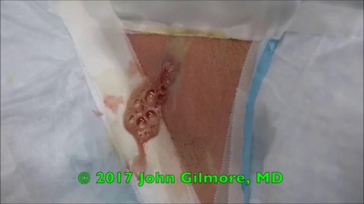
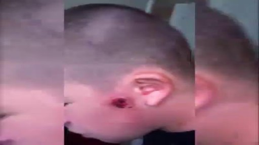
Watch that video of a Huge Cyst Infection Popping

http://cure-papiloma-humano.info-pro.co --- Sintomas Del Papiloma Humano, Sintomas De Papiloma Humano, Virus Papiloma Humano Cura. El Papiloma Humano Se Cura ¿El Papiloma Humano se Cura? Si te has encontrado recientemente con un diagnóstico positivo de VPH probablemente estas en busca de una solución para tratar este Virus. Seguramente tienes miedo de no encontrar una cura para las verrugas genitales, y que son muy difíciles de eliminar, amigo o amiga no te sientas avergonzado/a o preocupado el saber que estas infectado con este virus no es fácil, más aun ver cómo crecen verrugas en tu cuerpo, pero arriba los ánimos existen muchas cosas que puedes hacer para tratar este virus. Aparte de las verrugas genitales no hay otro síntoma que presente el Virus en tu cuerpo, puedes tratar las verrugas genitales con tratamientos naturales o los métodos actuales. Trata de no rascarse si sientes comezón en la zona afecta ya que puedes lastimarte o irritar más la piel, las verrugas genitales son altamente contagiosas, No debes tener relaciones sexuales con nadie hasta que hayas tenido tratamiento para el VPH. Hoy en Día existen varios tratamientos médicos diseñados para ayudarte a curar las verrugas genitales producidas por el papiloma humano, aunque debo aclararte que estos métodos son dolorosos y dejan cicatrices en la piel donde se encontraba la verruga. Crioterapia: Básicamente las verrugas genitales se congelan con nitrógeno líquido. Tratamiento a base de láser: se utilizan laser de CO2 para quemar las verrugas genitales, se aplica anestesia al área afectada para no sentir mucho dolor, aunque siempre existen molestias durante el procedimiento. Bisturí eléctrico: En esta técnica se utiliza una corriente eléctrica para destruir las verrugas, Se puede hacer en el consultorio con anestesia local, con este método se debe tener cierto cuidado ya que existe peligro de infección. La breve lista antes mencionada son los métodos médicos más comunes para eliminar las verrugas genitales, cuando se diagnostican verrugas genitales estos métodos son los primeros en que se piensan para curar las verrugas genitales. Aunque hay que decir la verdad, estos tratamientos no podrán eliminar el verdadero problema detrás de las verrugas genitales, el cual es el Virus del papiloma humano, aunque las verrugas se eliminan de la zona afectada el virus seguirá permaneciendo en el cuerpo de forma latente, ninguno de estos método puede garantizar que no volverá a haber otro brote de verrugas genitales. Descubre como mantener DESACTIVADO el VPH DE POR VIDA para permitirte una vida sin verrugas, sin frustraciones y sin molestias, ingresa ahora a: http://cure-papiloma-humano.info-pro.co
SINUS LIFT SURGERY surgical procedure which aims to increase the amount of bone in the posterior maxilla (upper jaw bone), in the area of the premolar and molar teeth, by lifting the lower Schneiderian membrane (sinus membrane) and placing a bone graft.

Vitiligine Cause, Vitiligine Bambini, Micropigmentazione Vitiligine, Vitiligine Trucco, Vitiligine --- http://vitiligine-cura.good-info.co --- Un Ricercatore Medico, Nutrizionista, Consulente Di Salute Ed Ex Malato Cronico Di Vitiligine Ti Spiega Come: Curare La Vitiligine E Ripristinare Il Colore Naturale Della Tua Pelle In 7 Giorni! Curare La Causa Alla Base Della Vitiligine Affrontando Le Cause Interne Di Questo Disturbo In 45 - 60 Giorni. Prevenire La Comparsa Di Cicatrici E Segni Gettare Via . Lozioni O Creme "Miracolo" E Sentirti Subito Più Fiducioso! Risparmiare Migliaia Di Euro In Farmaci, Laser E Trattamenti UV, Visite Dal Dottore O Operazioni Chirurgiche! Ripristinare Il Tuo Equilibrio Interno E Fermare I Problemi Di Salute Legati Alla Vitiligine Mantenendoli Alla Larga Per Sempre! Perdere Chili In Eccesso, Sembrare Più Giovane E Riguadagnare L'autostima Ripristinare I Livelli Di Energia E Migliorare La Qualità Della Vita Significativamente... Garantito! Se Vuoi Imparare Come Curare La Vitiligine In Modo Definitivo E Riaquistare La Tua Salute E Benessere, Senza Farmaci, Senza I Tradizionali Trattamenti Per La Vitiligine E Senza Alcun Effetto Collaterale, Allora Questa Sarà La Lettura Più Importante Che Abbia Mai Fatto. Te Lo Garantisco E Ho I Risultati Per Provartelo! http://vitiligine-cura.good-info.co
