- Physical Examination
- Surgical Examination
- Ophthalmology
- Clinical Skills
- Orthopedics
- Surgery Videos
- Laparoscopy
- Pediatrics
- Funny Videos
- Cardiothoracic Surgery
- Nursing Videos
- Plastic Surgery
- Otorhinolaryngology
- Histology and Histopathology
- Neurosurgery
- Dermatology
- Pediatric Surgery
- Urology
- Dentistry
- Oncology and Cancers
- Anatomy Videos
- Health and Fitness
- Radiology
- Anaesthesia
- Physical Therapy
- Pharmacology
- Interventional Radiology
- Cardiology
- Endocrinology
- Gynecology
- Emergency Medicine
- Psychiatry and Psychology
- Childbirth Videos
- General Medical Videos
- Nephrology
- Physiology
- Diet and Food Health
- Diabetes Mellitus
- Neurology
- Women Health
- Osteoporosis
- Gastroenterology
- Pulmonology
- Hematology
- Rheumatology
- Toxicology
- Nuclear Medicine
- Infectious Diseases
- Vascular Disease
- Reproductive Health
- Burns and Wound Healing
- Other
Top videos
The future of Medicine - Il futuro della medicina - Die Zukunft der Medizin: High Tech, Robots, VR ⚡️Anatomia Biomeccanica Fisiologia by Ticinosthetics: tutto gira attorno alla palestra ©️2017 - www.ticinostheticsgs.com
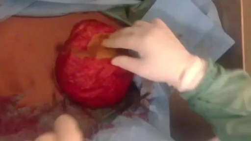
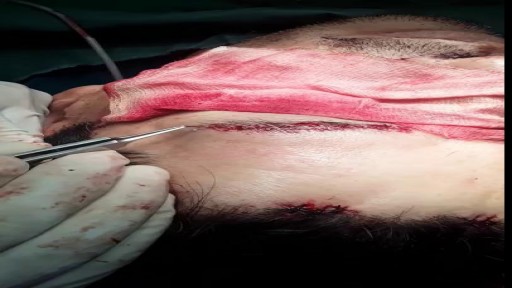
seroma 3 years after surgery
The treatment of cardiac arrest
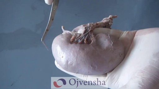
Each kidney contains around 1 million individual nephrons, the kidneys' microscopic functional units that filter blood to produce urine. The nephron is made of 2 main parts: the renal corpuscle and the renal tubule.
Watch that Massive Big Skin Wart Removal
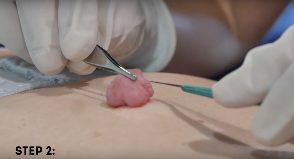
Watch that Huge Skin Tag Removal Procedure
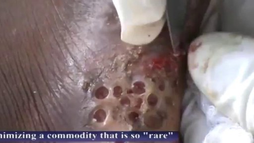
Watch that Massive Skin Jiggers Removals
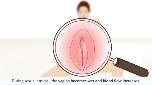
All you need to know about the female orgasm
Transcript: Body Restoration (http://stalbertphysiotherapy.com/) has treated over 12,400 patients since it opened its doors in 1992. While embracing new technology and techniques they have not left behind the basic tenets of hands-on healing. If you are injured or have chronic pain, the mission is to help you live pain-free. Relief is a click or a phone call away. Come in for your no obligation exam and find out what will work for you.

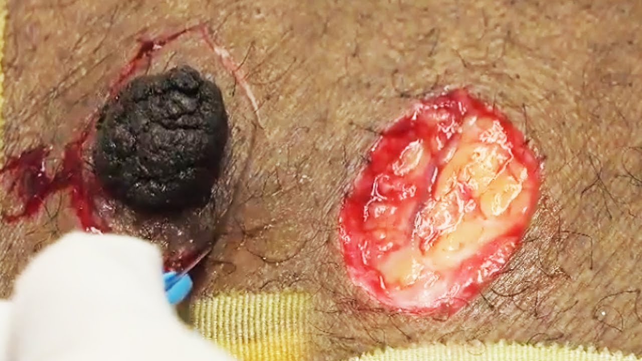
Watch that video of an Ingrown Hair Causes Huge Tumor in a Man's Stomach

Como Aumentar La Libido, Aumentar Niveles De Testosterona, Como Aumentar El Deseo Masculino ---- http://aumentar-testosterona.good-info.co/ --- ¿Se puede tener una erección con bajos niveles de testosterona? Mi libido está quedando atrás y estoy teniendo dificultades para conseguir una erección, así que estoy tratando de averiguar qué está pasando aquí. La disfunción eréctil rara vez es causada sólo por la deficiencia de testosterona. Por lo general es un grupo de cosas que funcionan en concierto juntos, que se alimentan entre sí, que conducen a la incapacidad del hombre para lograr una erección. La aterosclerosis (estrechamiento y endurecimiento de las arterias) es uno de los mayores impulsores de la disfunción eréctil, pero estas arterias dañadas no aparecen de la nada. Otras cosas tienen que estar sucediendo en el cuerpo para que ésta aterosclerosis pase, y como estamos a punto de ver, estas otras cosas contribuyen al problema también. Así que vamos a repasar esta lista… Nivel de azúcar alto – baja testosterona y disfunción eréctil La azúcar elevada en la sangre es un arma de doble filo, porque los hombres que sufren de esta condición son mucho más propensos a ser afectados por la disfunción eréctil y la testosterona baja. Una Investigación de John Hopkins encontró que las ratas diabéticas presentaron una respuesta eréctil 30% inferior, sus erecciones fueron como máximo 40% más pequeñas y las erecciones tomaron 70% más tiempo para lograrse en comparación con los controles que no eran diabéticos. Otros estudios han confirmado que los hombres con diabetes tipo 2 son dos veces más propensos a sufrir de disfunción eréctil, y la condición les golpeará una década antes, en comparación con los hombres sin tipo 2. Este vínculo es tan fuerte porque el azúcar en la sangre hace un daño directo a las arterias cuando se tiene demasiado de él, y las arterias en el pene suelen ser afectados en primer lugar, porque son muy pequeñas y estrechas. Por lo tanto, tiene todo el sentido que éstas pueden dañarse primero. El ejercicio que baja la testosterona haga click aqui http://aumentar-testosterona.good-info.co/
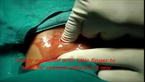
She is a twenty years young female presented with large cystic swelling in anterior aspect of neck. The swelling was of size 6cmx 6cm x5 cm ,tense tender, cystic just above sternal nutch.This was diagnosed as large neck abscess ./nRepeated aspiration done but the swelling reappeared. So Incision & Drainage planned under local anaesthesia./nPatient in supine position. Surgery part painted and draped. Local anaesthesia 2% xylocaine with adrenaline used for field block.After giving local anaesthesia, I used a no 11 blade for stab incision at the most prominent part of the swelling, where skin was thin and fluctuation present./nPus drained form that opening. Little dilatation of opening to be done with artery forceps or sinus forceps. Complete pus drainage to be ensured.Little finger can be introduced inside the pus cavity to ensure proper drainage of pus. The cavity I use to clean with a gauge piece. If necessary curette biopsy can be taken from the wall of the cavity.These wounds usually need daily proper dressing for faster healing.
Eye Brow Transplant Procedure
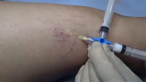
Several options are available to remove spider veins — thin red lines or weblike networks of blood vessels that appear on your legs and feet. Spider veins are usually harmless, though they can sometimes cause aching, burning or pain, especially when you've been standing for long periods. If you have symptoms or are concerned about the appearance of spider veins, treatment options include: Sclerotherapy. In this procedure, your doctor injects the veins with a solution that scars and closes those veins, causing the blood to reroute through healthier veins. In a few weeks, treated spider veins fade. Although the same vein may need to be injected more than once, sclerotherapy is usually effective if done correctly. Sclerotherapy doesn't require anesthesia and can be done in your doctor's office. Side effects include swelling, itching and skin color changes in the treated area. Laser surgery. Laser surgery works by sending strong bursts of light into the vein that make the vein slowly fade and disappear. No incisions or needles are used. The treatment is often less effective than sclerotherapy, particularly for larger veins. Side effects may include redness, bruising, itching, swelling and permanent skin tone changes. After treatment, blood vessels fade over several months, but they may not disappear completely. Also, new spider veins can develop in the same area.
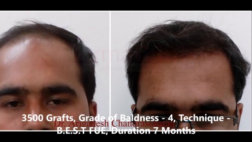
Hair Transplant Results Before and After Photos who undergone Hair Transplant. View our patient's successful results with the FUE, Bio - FUE and B.E.S.T FUE hair transplant technique. Comparable before & after photos! For More Visit Here:- https://www.hairtransplantchennai.org/hair-transplant-results-chennai.php or call us:- +91-8939636222

Case of ITP with persistent very low platelet count despite best medical management
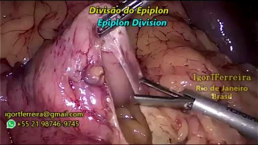
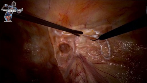
a sleeve gastrectomy with very few edditing. During the start 3 smal spleen perforations caused by Veres Needle were identified, caused by a giant spleen undentified on pre operatory ultrasound. They were controled with gauze compression and at the end of the surgery surgicel was placed and no complications were observed. Patient discharged 3 days after the surgery.
top 10 most incredible surgeries ever done
Antibiotic therapy is the mainstay of medical treatment for pediatric rhinosinusitis.] Because of increasing prevalence of beta-lactam–resistant bacteria in the community, administer antibiotics only for suspected infection as based on a careful history and physical examination. Direct the therapeutic regimen against the prevalent pathogens in the community and carefully consider suspicion for highly resistant bacteria. Typically, uncomplicated cases of acute sinusitis are responsive to amoxicillin. Most patients respond to this initial regimen. For children allergic to penicillin, a second- or third-generation cephalosporin can be used (only if the allergic reaction is not a type 1 hypersensitivity reaction). In cases of serious allergic reaction, a macrolide or clindamycin can be used.
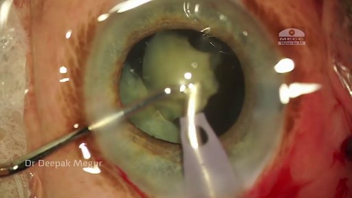
Phacolytic glaucoma usually is associated with a mature or hypermature cataract and typically occurs in elderly patients. Today, phacolytic glaucoma is rare in the United States, found primarily in areas where access to care is poor. Will the increase in the number of under- and uninsured patients lead to an increase in this condition? Evaluation and Diagnosis Signs and symptoms. Patients typically report acute-onset pain, decreased vision, tearing and photophobia. Examination will reveal injection, corneal edema, elevated IOP, anterior chamber reaction with or without pseudohypopyon, particles on the lens capsule and anterior capsule wrinkling. Patient history. The duration of symptoms should be elicited; a delayed presentation of more than five days since onset can result in glaucomatous disc damage and poorer prognosis.¹ The ocular history may reveal that the patient decided against removal of an advanced cataract. Prior intraocular surgery or trauma may have left residual lens material that could cause phacoanaphylactic glaucoma or exacerbate infectious endophthalmitis. Visual acuity and visual potential should be assessed. Exam essentials. A complete ophthalmologic examination should be done. The eye should be inflamed, and the cornea may be edematous due to the high IOP. The anterior chamber will demonstrate massive inflammation and/ or pseudohypopyon. Gonioscopy is essential; it will help rule out angle closure due to phacomorphic glaucoma or neovascularization of the angle. Assess ment of the posterior pole should be performed to rule out vitreous hemorrhage (which can result in ghost-cell glaucoma) or vitritis (which may be associated with infectious endophthalmitis or panuveitis). If the view to the fundus is obstructed, B-scan ultrasonography also should be performed. Differential diagnosis. The differential diagnosis includes infectious endophthalmitis, phacoanaphylactic glaucoma, inflammatory glaucoma, glaucoma secondary to intraocular tumor, phacomorphic glaucoma, acute-angle closure glaucoma and neovascular glaucoma. Management Medication. Medical management is used to temporarily control the glaucoma and inflammation. Initial treatment consists of hyperosmotic agents, aqueous suppressants, anti-inflammatory drugs and cycloplegics. Surgery. Definitive treatment is removal of the lens via extracapsular cataract extraction with or without an IOL. Some ophthalmologists defer placement of an IOL until after the inflammation subsides; however, there is no significant difference in final visual acuity between those patients who did receive an IOL and those who did not.¹ If the phacolytic glaucoma is of long duration (more than seven days), a combined trabeculectomy may be needed to prevent postoperative IOP spikes.² In eyes with hypermature Morgagnian cataracts, one must be especially careful, as the capsule is fragile, the zonules are weak and the view is difficult due to the white, milky cortex. Vision limited to light perception on presentation is not a contraindication to performing cataract extraction. Surgical Tips For a planned extracapsular cataract extraction with a posterior chamber IOL, fashion a superior fornix-based conjunctival flap.³ Make a partial-thickness incision along the sclerolimbal junction superiorly for 120 degrees with a No. 69 blade. Forty-five degrees away, a paracentesis should be done to decompress the eye. The anterior chamber fluid can be withdrawn for analysis, to look for macrophages and high molecular-weight proteins. Inject balanced salt solution in a cannula to wash out any residual particulate matter, then inject Healon or viscoelastic into the anterior chamber. Make an incision entering the anterior chamber at the 12 o’clock position with a keratome. A 26-gauge cystotome mounted on a syringe is then introduced through the 12 o’clock incision and used to puncture the capsular bag. The milky cortex should be aspirated as much as possible, until the nucleus is visible. Withdraw the needle through the keratome incision, then inject Healon through the 12 o’clock incision into the capsular bag. Next, enlarge the corneoscleral keratome incision with curved Westcott scissors to 120 degrees. Perform a partial V-shaped capsulotomy; this can be done either with the cystotome or with an angled Vannas scissors. Place viscoelastic under the nucleus to float the nucleus and sever any adhesions between the nucleus and the capsule. The nuclear portion of the lens can then be removed with an irrigating vectis (lens loop) with or without gentle pressure at the inferior limbus (6 o’clock). Irrigate and aspirate the residual cortex with the Simcoe cannula. Inspect the capsular bag; if it is intact, place a posterior chamber IOL into the bag. Close the incision with several interrupted 10-0 monofilament nylon sutures and reattach the conjunctival flap. Potential Sequelae and Prognosis Postoperatively, the patient should be managed with topical steroids and/or aqueous suppressants and hyperosmotics if necessary. Vitreous opacification behind the posterior capsule occurs in a small percentage of eyes. These vitreous opacities are typically absorbed by one to two weeks postoperatively. IOP usually is controlled without antiglaucoma medications after the cataract removal. A detailed glaucoma evaluation (including repeat gonioscopy to assess for peripheral anterior synechiae, visual field and optic nerve status) should be done to assess the extent of glaucomatous damage. The prognosis is dependent on the duration of elevated IOP, PAS and optic nerve damage. In one study, patients who were older than 60 and whose glaucoma was present for more than five days did significantly worse than a comparison group of younger individuals with shorter disease duration.
