En iyi videolar
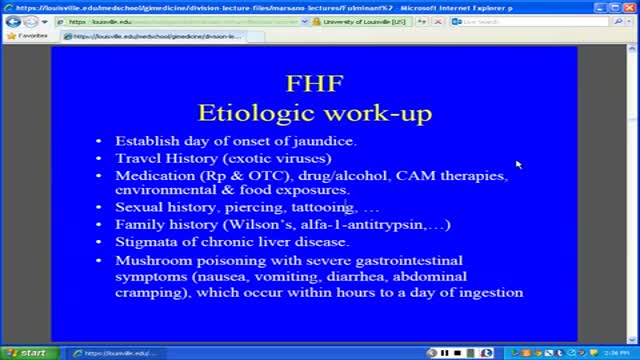
Fulminant hepatic failure (FHF) or acute liver failure (ALF) is defined as the rapid development of acute liver injury with severe impairment of the synthetic function and hepatic encephalopathy in a patient without obvious, previous liver disease.
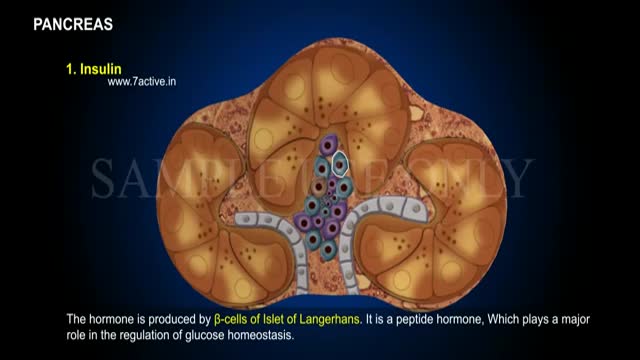
Enzymes, or digestive juices, produced by the pancreas are secreted into the small intestine to further break down food after it has left the stomach. The gland also produces the hormone insulin and secretes it into the bloodstream in order to regulate the body's glucose or sugar level.

Function. Vitamin A helps form and maintain healthy skin, teeth, skeletal and soft tissue, mucus membranes, and skin. It is also known as retinol because it produces the pigments in the retina of the eye. Vitamin A promotes good vision, especially in low light. Vitamin deficiency anemia occurs when your body doesn't have enough of the vitamins needed to produce adequate numbers of healthy red blood cells. Red blood cells carry oxygen from your lungs throughout your body. If your diet is lacking in certain vitamins, vitamin deficiency anemia can develop.

The foods for your child are easily digestible foods, such as rice cereal, pasta, breads, cooked beans, mashed potatoes, cooked carrots, applesauce, and bananas. Pretzels or salty crackers can help your child replace the salt lost from diarrhea. Foods containing large amounts of sugar or fat should be avoided.
Premature ventricular contractions (PVCs) are extra, abnormal heartbeats that begin in one of your heart's two lower pumping chambers (ventricles). These extra beats disrupt your regular heart rhythm, sometimes causing you to feel a flip-flop or skipped beat in your chest.

Knee Examination
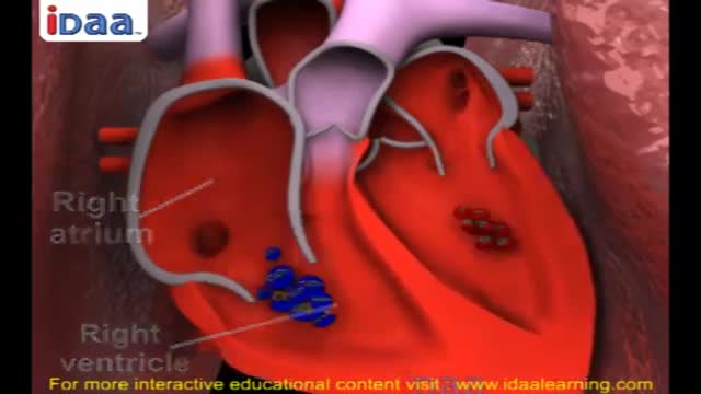
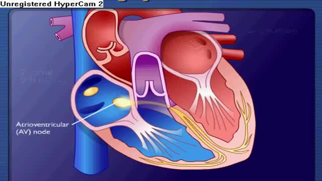
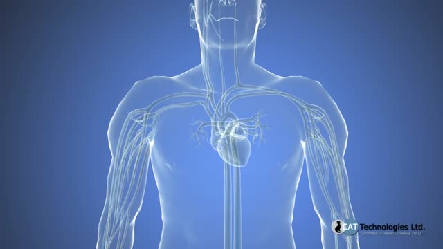
Systemic circulation carries oxygenated blood from the left ventricle, through the arteries, to the capillaries in the tissues of the body. From the tissue capillaries, the deoxygenated blood returns through a system of veins to the right atrium of the heart.
Cardiac cath is performed to find out if you have disease of the heart muscle, valves or coronary (heart) arteries. During the procedure, the pressure and blood flow in your heart can be measured. Coronary angiography is done during cardiac catheterization.

Mohs Surgeon Dr. Leslie Christenson shows entire Mohs Surgery from start to finish. This is the full procedure and includes the entire surgery. Dr. Christenson talks about the procedure as she removes the skin cancer.
Learn more about Dr. Christenson: https://www.mcfarlandclinic.co....m/doctors/leslie-chr
Learn more about Mohs Surgery: https://www.mcfarlandclinic.co....m/doctors/specialtie

A new report analyzing FDA-approved monoclonal antibodies (mAbs) produced by a select group of leading biotechnology companies shows that clinical development times – specifically the duration of Phase II and Phase III trials – are lengthening, while FDA review times have remained constant. The average time from investigational new drug (IND) filing to market was 6.7 years for 11 mABs approved between 1994 and 2003 but shot up to 8.3 years for 12 mAbs approved between 2004 and March 9, 2011, according to Deloitte Recap LLC’s analysis, Therapeutic Monoclonal Antibodies – Insights, Strategies and Data.
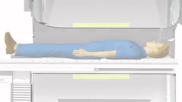
Magnetic resonance imaging (MRI) can be an important tool in the diagnosis of multiple sclerosis (MS). MRI can also be used to monitor the progression of the disease in people living with MS. How does it work? MRI uses very strong magnets, radio signals, and computer software to take 3-dimensional pictures of the inside of the body. Will I need contrast material? Maybe. Contrast material is a substance that temporarily changes the way imaging tools interact with the body. They are often used to visualize certain types of MS disease activity on the MRI. If your doctor thinks your scan requires this contrast material, you may get an injection before you get in the MRI machine. How long will it take? The time may vary based on the type of MRI. Be sure to discuss with your doctor in advance so he or she can provide you with exact timing. But don’t worry, you won’t have to stay still the whole time. The technician will let you know when they’re starting a new image.

Intelligent People Have Fewer Friends, Here's Why...

Obstetrics is the field of medicine which encompasses the care of a woman during pregnancy and childbirth. In that way it is very unique, as when assessing these patients, your actually also assessing another the child.
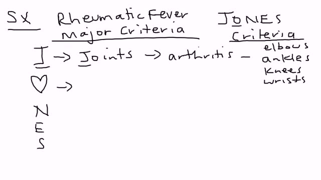
Rheumatic fever is an inflammatory disease that can develop as a complication of inadequately treated strep throat or scarlet fever. Strep throat and scarlet fever are caused by an infection with streptococcus bacteria. Rheumatic fever is most common in 5- to 15-year-old children, though it can develop in younger children and adults. Although strep throat is common, rheumatic fever is rare in the United States and other developed countries. However, rheumatic fever remains common in many developing nations. Rheumatic fever can cause permanent damage to the heart, including damaged heart valves and heart failure. Treatments can reduce damage from inflammation, lessen pain and other symptoms, and prevent the recurrence of rheumatic fever.

A narrowing of the major artery (the aorta) that carries blood to the body. This narrowing affects blood flow where the arteries branch out to carry blood along separate vessels to the upper and lower parts of the body. CoA can cause high blood pressure or heart damage.

What is your mental age?

Ehlers-Danlos syndrome is a group of disorders that affect the connective tissues that support the skin, bones, blood vessels, and many other organs and tissues. Defects in connective tissues cause the signs and symptoms of Ehlers-Danlos syndrome, which vary from mildly loose joints to life-threatening complications. Previously, there were more than 10 recognized types of Ehlers-Danlos syndrome, differentiated by Roman numerals. In 1997, researchers proposed a simpler classification that reduced the number of major types to six and gave them descriptive names: the classical type (formerly types I and II), the hypermobility type (formerly type III), the vascular type (formerly type IV), the kyphoscoliosis type (formerly type VIA), the arthrochalasia type (formerly types VIIA and VIIB), and the dermatosparaxis type (formerly type VIIC). This six-type classification, known as the Villefranche nomenclature, is still commonly used. The types are distinguished by their signs and symptoms, their underlying genetic causes, and their patterns of inheritance. Since 1997, several additional forms of the condition have been described. These additional forms appear to be rare, affecting a small number of families, and most have not been well characterized.

The DASH diet is a lifelong approach to healthy eating that's designed to help treat or prevent high blood pressure (hypertension). The DASH diet encourages you to reduce the sodium in your diet and eat a variety of foods rich in nutrients that help lower blood pressure, such as potassium, calcium and magnesium.

The menstrual cycle is the regular natural change that occurs in the female reproductive system like the uterus and ovaries that make pregnancy possible. The cycle is required for the production of ovocytes, and for the preparation of the uterus for pregnancy.

Adrenoleukodystrophy, or ALD, is a deadly genetic disease that affects 1 in 18 000 people. It most severely affects boys and men. This brain disorder destroys myelin, the protective sheath that surrounds the brain's neurons -- the nerve cells that allow us to think and to control our muscles.
