- Physical Examination
- Surgical Examination
- Ophthalmology
- Clinical Skills
- Orthopedics
- Surgery Videos
- Laparoscopy
- Pediatrics
- Funny Videos
- Cardiothoracic Surgery
- Nursing Videos
- Plastic Surgery
- Otorhinolaryngology
- Histology and Histopathology
- Neurosurgery
- Dermatology
- Pediatric Surgery
- Urology
- Dentistry
- Oncology and Cancers
- Anatomy Videos
- Health and Fitness
- Radiology
- Anaesthesia
- Physical Therapy
- Pharmacology
- Interventional Radiology
- Cardiology
- Endocrinology
- Gynecology
- Emergency Medicine
- Psychiatry and Psychology
- Childbirth Videos
- General Medical Videos
- Nephrology
- Physiology
- Diet and Food Health
- Diabetes Mellitus
- Neurology
- Women Health
- Osteoporosis
- Gastroenterology
- Pulmonology
- Hematology
- Rheumatology
- Toxicology
- Nuclear Medicine
- Infectious Diseases
- Vascular Disease
- Reproductive Health
- Burns and Wound Healing
- Other
Top videos

Multiple strains put your children and teens at risk of meningococcal meningitis. How-to ensure they are fully protected.
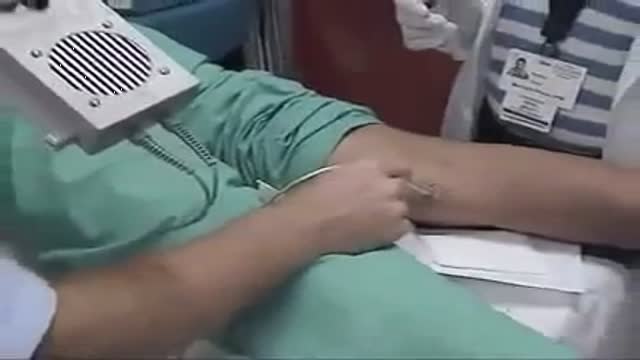
Politeal and Peroneal Nerves Block
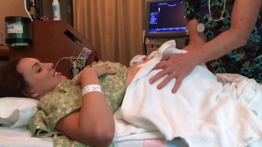
Successful External Cephalic Version (ECV) - Turning a breech baby in less than 30 seconds!
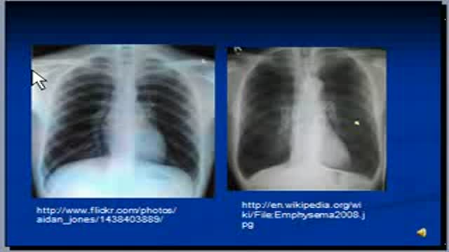
The video will describe radiologic features of Emphysema on a chest x-ray. Please see my website for disclaimer.

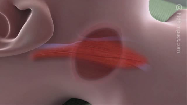
Ptosis is when the upper eyelid droops over the eye. The eyelid may droop just a little, or so much that it covers the pupil (the black dot at the center of your eye that lets light in). Ptosis can limit or even completely block normal vision. Children and adults can have ptosis. Fortunately, this condition can be treated to improve vision as well as appearance.

Extremely funny, and very in-depth look at all the parts and pieces of your blood.
Central catheters provide dependable intravenous access and enable hemodynamic monitoring and blood sampling [1-3]. The jugular veins are one of the most popular sites for central venous access due to accessibility and overall low complication rates, and are the preferred site for temporary hemodialysis.

A video showing the surgery of vaginal hysterectomy Operation

Watch that video of a Snake bite causes girl’s leg to rot away

A video shows how to deal with thermal burns

The purpose of the organs of the male reproductive system is to perform the following functions: To produce, maintain, and transport sperm (the male reproductive cells) and protective fluid (semen) To discharge sperm within the female reproductive tract during sex To produce and secrete male sex hormones responsible for maintaining the male reproductive system

Rectal bleeding can refer to any blood that passes from your anus, although rectal bleeding is usually assumed to refer to bleeding from your lower colon or rectum. Your rectum makes up the last few inches of your large intestine. Rectal bleeding may show up as blood in your stool, on the toilet paper or in the toilet bowl. Blood that results from rectal bleeding can range in color from bright red to dark maroon to a dark, tarry color.

Sexual Desire & our Eating

Emergency contraception is a method of birth control you can use if you had sex without using birth control or if your birth control method did not work correctly. You must use emergency contraception as soon as possible after unprotected sex. Emergency contraception pills are different from the abortion pill. If you are already pregnant, emergency contraception pills do not stop or harm your pregnancy. Emergency contraception has also been called the "morning-after pill," but you do not need to wait until the morning after unprotected sex to take it. Emergency contraception is not meant to be used for regular birth control. Talk to your doctor or nurse about regular birth control to help prevent pregnancy. Nearly half of all pregnancies in the United States are unplanned.1

Gallstone ileus is an important, though infrequent, cause of mechanical bowel obstruction, affecting older adult patients who often have other significant medical conditions. It is caused by impaction of a gallstone in the ileum after being passed through a biliary-enteric fistula. The diagnosis is often delayed since symptoms may be intermittent and investigations fail to identify the cause of the obstruction. The mainstay of treatment is removal of the obstructing stone after resuscitating the patient. Gallstone ileus continues to be associated with relatively high rates of morbidity and mortality.


Generic minoxidil is known to treat hair-fall issues in men and women, it is best for hair growth, hair re-development, etc. it is available in the strength of 5mg and easily available at online pharmacy store. For more information visit to http://www.medstorerx.com/generic-minoxidil.aspx

Watch that video of a Man With Pipe Penetrated His Head Inside Emergency Room

The importance of uninterrupted contact between mother and newborn SHOW MORE
