Top videos
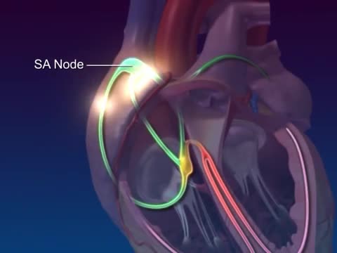
The cardiac conduction system is a group of specialized cardiac muscle cells in the walls of the heart that send signals to the heart muscle causing it to contract. The main components of the cardiac conduction system are the SA node, AV node, bundle of His, bundle branches, and Purkinje fibers.
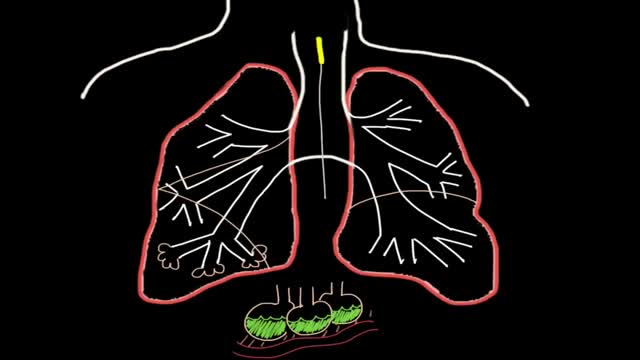
A pneumothorax can be caused by a blunt or penetrating chest injury, certain medical procedures, or damage from underlying lung disease. Or it may occur for no obvious reason. Symptoms usually include sudden chest pain and shortness of breath. On some occasions, a collapsed lung can be a life-threatening event.

A recap of Mater Hospital patient Helen's story as she progressed from experiencing chronic knee pain due to osteoarthritis, through to knee replacement treatment and ultimately a new lease on life.
Dedicated to surgical excellence and patient-centred care, the Mater Hospital North Sydney is regarded as a leading orthopaedic hospital and the only Australian hospital to be accepted into the International Society of Orthopaedic Centres.
For more information, click here: https://bit.ly/3bvhY8G
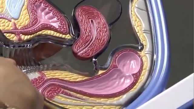
Postmenopausal bleeding (PMB) is defined for practical purposes as vaginal bleeding occurring after twelve months of amenorrhoea, in a woman of the age where the menopause can be expected.[1] Hence it does not apply to a young woman, who has had amenorrhoea from anorexia nervosa, or a pregnancy followed by lactation. However, it can apply to younger women following premature ovarian failure or premature menopause. Unscheduled bleeding in women of menopausal age taking hormone replacement therapy (HRT) should be managed in the same way from a practical perspective.[2] 'Unscheduled bleeding' is defined as non-cyclical bleeding still continuing six months after commencing HRT or after six months of amenorrhoea.
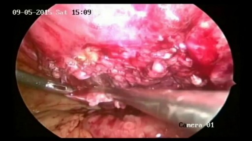
Removal of Infected Hernia Mesh
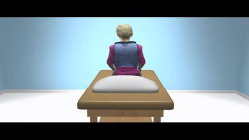
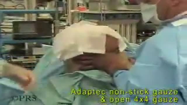
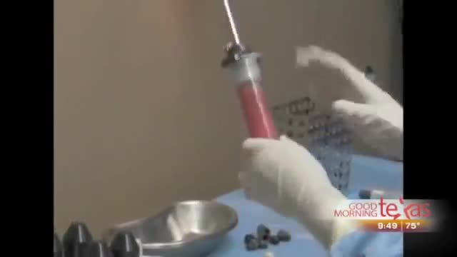
Bone marrow examination refers to the pathologic analysis of samples of bone marrow obtained by bone marrow biopsy (often called a trephine biopsy) and bone marrow aspiration. Bone marrow examination is used in the diagnosis of a number of conditions, including leukemia, multiple myeloma, anemia, and pancytopenia. The bone marrow produces the cellular elements of the blood, including platelets, red blood cells and white blood cells. While much information can be gleaned by testing the blood itself (drawn from a vein by phlebotomy), it is sometimes necessary to examine the source of the blood cells in the bone marrow to obtain more information on hematopoiesis; this is the role of bone marrow aspiration and biopsy.
The Epley maneuver is a series of movements, normally carried out on a person by a doctor, to relieve the symptoms of BPPV. Research has found it to be an easy, safe, and effective treatment for the condition in both the long- and short-term. The Epley maneuver is sometimes called the particle repositioning maneuver or the canalith repositioning maneuver. These names are used because the maneuver involves a series of movements that help to reposition crystals in a person's ear that may cause feelings of dizziness. Repositioning the crystals helps to relieve the person's dizziness and nausea.
Bandaging a freshly above the knee amputated limb
Stem-cell therapy is the use of stem cells to treat or prevent a disease or condition. Bone marrow transplant is the most widely used stem-cell therapy, but some therapies derived from umbilical cord blood are also in use...

St. Luke's originally broadcast this live in a webcast and later re-purposed it for air on KCRG-TV9 as an educational video. It is hosted by Ashley Hinson, KCRG-TV9 anchor and Dr. Sandeep Munjal. Dr. Jeff Nassif performs the knee replacement surgery on an eastern Iowa woman. St. Luke's has a rapid recovery joint replacement program, which gets people back to life quickly after surgery.
Symptoms of liver failure include vomiting, diarrhea and fatigue as well as the symptoms from stage 3. While the progression from cirrhosis to failure can take years, the damage is irreversible and leads to eventual death. The key to treating liver disease is to diagnose the condition as early as possible.

Brachytherapy or localized radiation treatment can be used in certain patients with breast cancer. Depending on tumor size and other factor, physicians may use APBI or accelerated partial breast irradiation. Dr. Elizabeth Tapen, a radiation oncologist, reviews brachytherapy for breast cancer.
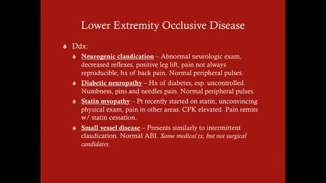
Claudication, which is defined as reproducible ischemic muscle pain, is one of the most common manifestations of peripheral arterial occlusive disease (PAOD) caused by atherosclerosis. Claudication occurs during physical activity and is relieved after a short rest. Pain develops because of inadequate blood flow. Examination of a patient with claudication should include a complete lower-extremity evaluation and pulse examination, including measuring segmental pressures. Attempt to palpate pulses from the abdominal aorta to the foot, with auscultation for bruits in the abdominal and pelvic regions. When palpable pulses are not present, a handheld Doppler device may be used to assess circulation.
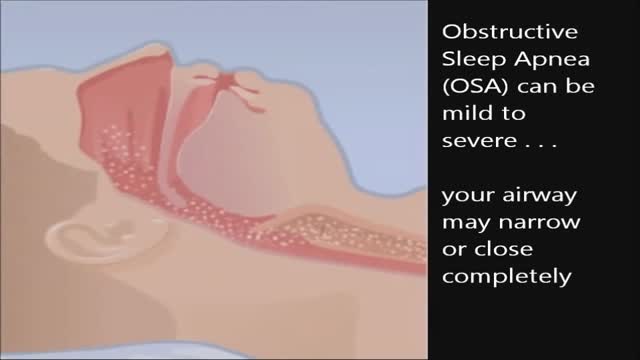
CPAP, or continuous positive airway pressure, is a treatment that uses mild air pressure to keep the airways open. CPAP typically is used by people who have breathing problems, such as sleep apnea. CPAP also may be used to treat preterm infants whose lungs have not fully developed.
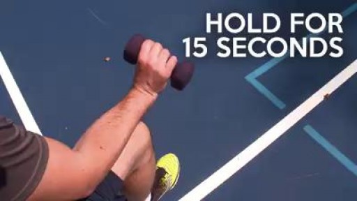
You don't have to play tennis to get tennis elbow. These easy exercises can help:
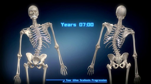
If you look at someone’s back, you’ll see that the spine runs straight down the middle. When a person has scoliosis, their backbone curves to the side. The angle of the curve may be small, large or somewhere in between. But anything that measures more than 10 degrees is considered scoliosis. Doctors may use the letters “C” and “S” to describe the curve of the backbone. You probably don’t look directly at too many spines, but what you might notice about someone with scoliosis is the way they stand. They may lean a little or have shoulders or hips that look uneven. What Causes Scoliosis? In as many as 80% of cases, doctors don’t find the exact reason for a curved spine. Scoliosis without a known cause is what doctors call “idiopathic.” Some kinds of scoliosis do have clear causes. Doctors divide those curves into two types -- structural and nonstructural. In nonstructural scoliosis, the spine works normally, but looks curved. Why does this happen? There are a number of reasons, such as one leg’s being longer than the other, muscle spasms, and inflammations like appendicitis. When these problems are treated, this type of scoliosis often goes away. In structural scoliosis, the curve of the spine is rigid and can’t be reversed

SSFTV is the official YouTube channel of the Seattle Science Foundation. Subscribe now to be updated on the latest videos: tinyurl.com/yt8kt8mg.
The Seattle Science Foundation is a not for profit organization dedicated to advancing the quality of patient care through education, research, innovation and technology. As a physician driven organization, we have created a trusted community of nationally recognized experts from the world’s best medical and academic institutions.
To join our upcoming meeting for CME credit, visit https://www.ssfcme.org.
Get Social With SSF:
On Instagram: https://www.instagram.com/seattlesciencefoundation
On Facebook: https://www.facebook.com/SeattleScienceFoundation
On Twitter: https://twitter.com/seattlescifdtn
On LinkedIn: https://www.linkedin.com/company/756824
On YouTube: http://www.ssfyoutube.org
Learn More at http://www.seattlesciencefoundation.org
Dr. Rod J. Oskouian, is a neurosurgeon who specializes in the diagnosis and treatment of complex spinal disorders. Dr. Oskouian is currently the Chief of Spine at the Swedish Neuroscience Institute and President and CEO of the Seattle Science Foundation. His research and clinical focus is on scoliosis, spinal deformities and anomalies, osteoporosis, spinal cord injury, degenerative disc disease, spinal oncology, stereotactic spinal radiosurgery, and minimally invasive spinal surgery. He has published in numerous medical journals and textbooks, including Neuroscience, Neurosurgery, Neurosurgical Clinics of North America, the Journal of Neurosurgery, Neurosurgical Focus and Spine.
How do you know if you have pneumonia? They may include: Cough. You will likely cough up mucus (sputum) from your lungs. ... Fever. Fast breathing and feeling short of breath. Shaking and "teeth-chattering" chills. Chest pain that often feels worse when you cough or breathe in. Fast heartbeat. Feeling very tired or very weak. Nausea and vomiting.

Thought a snake in your boot was bad? That old 19th-century idiom is nothing compared to one in your ear.
Shocking footage captured the alleged moment that a “surgeon” tried to remove a live snake that infiltrated a woman’s ear. Video of the herpetological surgery has racked up more than 125,000 views as viewers speculate whether or not the squirm-inducing footage is authentic.
“The snake has gone in the ear,” reads the caption to the bizarre Facebook clip, which was posted Sept. 1 by an India-based social media star named Chandan Singh to his 20,126 followers. However, it’s unclear where, when or how this unfortunate event transpired, local outlet the Economic Times reported.
In the nearly four-minute clip, an alleged medical practitioner can be seen using tweezers in a desperate attempt to extract a black and yellow serpent that’s peeking its head out from a female patient’s ear.
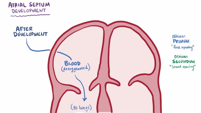
An atrial septal defect (ASD) is a hole in the wall between the two upper chambers of your heart (atria). The condition is present from birth (congenital). Small atrial septal defects may close on their own during infancy or early childhood. Large and long-standing atrial septal defects can damage your heart and lungs. Small defects may never cause a problem and may be found incidentally. An adult who has had an undetected atrial septal defect for decades may have a shortened life span from heart failure or high blood pressure that affects the arteries in the lungs (pulmonary hypertension). Surgery may be necessary to repair atrial septal defects to prevent complications.
