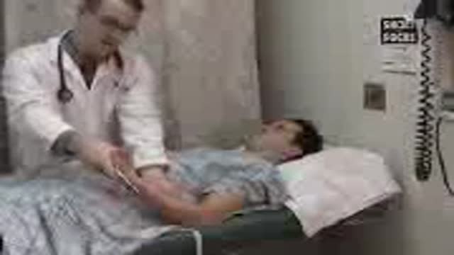
- Physical Examination
- Surgical Examination
- Ophthalmology
- Clinical Skills
- Orthopedics
- Surgery Videos
- Laparoscopy
- Pediatrics
- Funny Videos
- Cardiothoracic Surgery
- Nursing Videos
- Plastic Surgery
- Otorhinolaryngology
- Histology and Histopathology
- Neurosurgery
- Dermatology
- Pediatric Surgery
- Urology
- Dentistry
- Oncology and Cancers
- Anatomy Videos
- Health and Fitness
- Radiology
- Anaesthesia
- Physical Therapy
- Pharmacology
- Interventional Radiology
- Cardiology
- Endocrinology
- Gynecology
- Emergency Medicine
- Psychiatry and Psychology
- Childbirth Videos
- General Medical Videos
- Nephrology
- Physiology
- Diet and Food Health
- Diabetes Mellitus
- Neurology
- Women Health
- Osteoporosis
- Gastroenterology
- Pulmonology
- Hematology
- Rheumatology
- Toxicology
- Nuclear Medicine
- Infectious Diseases
- Vascular Disease
- Reproductive Health
- Burns and Wound Healing
- Other
Top videos

A DMC patient suffering from an abdominal aortic aneurysm receives an endovascular graft to alleviate the potentially deadly problem, performed by DMC cardiac specialist Dr. Ali Kafi. ~ Detroit Medical Center
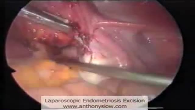
Laparoscopic excision of endometriosis
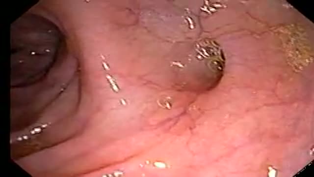
Small colon polyp (redish bump)and many diverticuli (small outpouches in wall of the colon)

One Handed Surgical Knot

Ophthalmoscopic exam

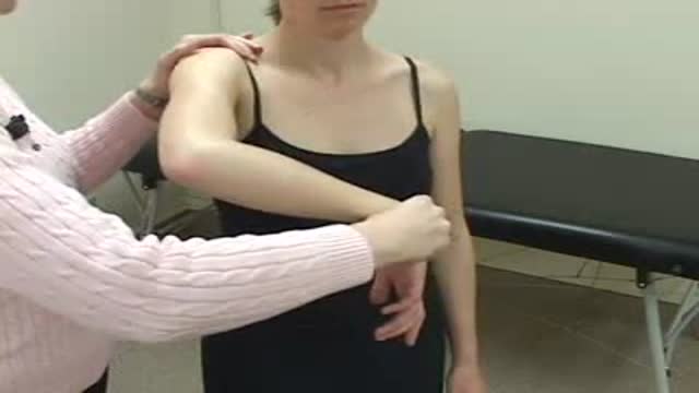
Examination of the cervical spines
Mitral valve repair of anterior leaflet perforation and ruptured chordae

Watch that Real Human Body Decomposing Process On Video
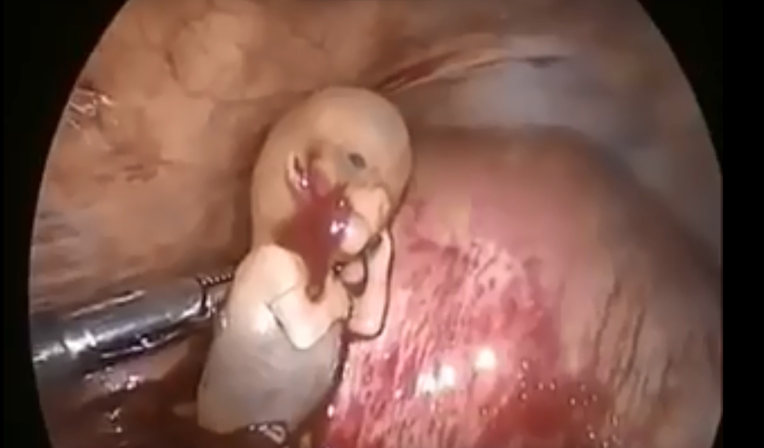
Watch that Ectopic Pregnancy Abortion Surgery
Continuous Lumbar Epidural
Hawkin's Test
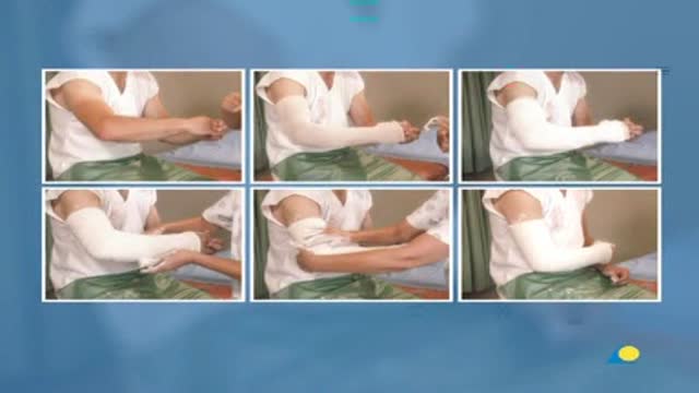
indicated in Radius and Ulna Fractures

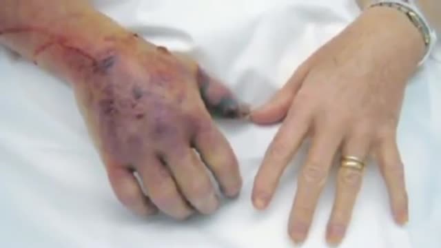
Watch that video of Unbelievable Mutations and Medical Conditions

Total Laparoscopic Hysterectomy

Watch that Female Foley Catheter Insertion Procedure
Sensory and reflexes exam of the upper limb from the USMLE collection Sensory and reflexes exam of the upper limb
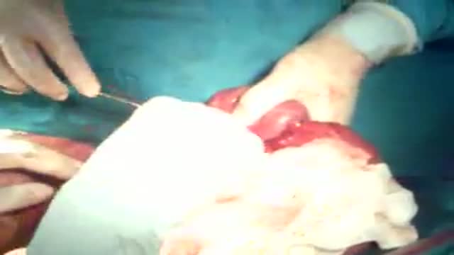
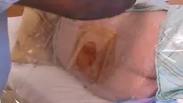
The operation was done by cut opening the abdomen for resection anastamoses of intestine. You can see all intestines. The patient unfortunately died of sepsis. He was just 15 yrs old
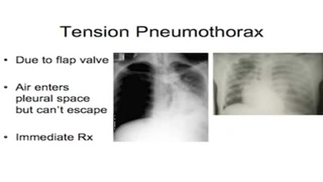
Intercostal Tube Insertion in case of pneumothorax
Two types of clinically distinct necrotizing fasciitis have been described. The most common form (type II) usually occurs in individuals with no concurrent medical illness. Many patients report a history of laceration, blunt trauma, or a surgical procedure as a predisposing factor. It is typically caused by group A Streptococcus (Streptococcus pyogenes). In contrast, type I is usually seen in patients with underlying diabetes and peripheral vascular disease. It is generally a polymicrobial infection; some commonly isolated organisms include Staphylococcus aureus, Bacteroides tragi/is, Escherichia coli, group A Streptococcus, and Pre vote/fa species. Crepitus is more common if anaerobic organisms, such as Clostridium perfringens or 8 tragi/is, are involved.

Orgasm is the sudden discharge of accumulated sexual excitement during the sexual response cycle, resulting in rhythmic muscular contractions in the pelvic region characterized by sexual pleasure
