أهم مقاطع الفيديو

This multi award winning video talks about a time of increased demands on our healthcare system and healthcare providers, ensuring that each and every patient and their family members are provided with compassionate care is a massive goal, but one that the staff at the Royal Alexandra Hospital are pursuing every day. Good quality care is always important, but caring for our patients is what they will really remember.

Dr. Ailawadi, M.D., the Chair of Cardiac Surgery at Michigan Medicine, specializes in minimally invasive valve surgery as well as complex cardiac operations. This video shows step by step footage of a Coronary Artery Bypass Graft (CABG) in a complex patient. In this case, CABG was performed through a sternotomy (through the breast bone) using the internal thoracic artery and saphenous leg veins to bypass obstructed coronary arteries. In this video, Dr. Ailawadi will perform a triple vessel bypass (CABG) which has been shown to minimize the risk of future heart attack and help patients live longer in the setting of complex coronary artery disease.
To learn more about cardiac surgery at Michigan Medicine, visit: https://medicine.umich.edu/dept/cardiac-surgery
To learn more about Frankel Cardiovascular Center, visit: https://www.umcvc.org/
To watch the full playlist, visit: https://www.youtube.com/playli....st?list=PLNxqP-XbH8B
-------------------------------------------------------
Subscribe to Michigan Medicine’s YouTube channel for upcoming videos and future live streams featuring our experts answering your questions.
-------------------------------------------------------
Follow Michigan Medicine on Social:
Twitter: https://twitter.com/umichmedicine
Instagram: https://www.instagram.com/umichmedicine/
Facebook: https://www.facebook.com/MichiganMedicine/
Follow the U-M Frankel Cardiovascular Center on Social:
Twitter: https://twitter.com/umichcvc
Facebook: https://www.facebook.com/Unive....rsityofMichiganCardi
#MichiganMedicine #MedEd #CardiacSurgery #UniversityOfMichiganHealth #FrankelCardiovascularCenter #Cardiology #CardiacSurgeon

Warning: This video contains actual surgical footage, which may not be suitable for all viewers.
To learn more about coronary artery bypass surgery, please visit http://cle.clinic/3b7dqpE
Cardiothoracic surgeon Faisal Bakaeen, MD, discusses how he does single and bilateral internal mammary arteries, and the benefits of doing this type of coronary artery bypass.
If you liked the video hit like and subscribe for more!

Follow along on a typical day with UCSF Medical Center's chief of cardiothoracic surgery Dr. Tom Nguyen. Take a walk on rounds with his team as they check on patients who are recovering or preparing for heart valve surgeries to treat conditions such as mitral valve prolapse and mitral regurgitation. Get a glimpse into the operating room as Dr. Nguyen and his team use the latest non-invasive techniques to help patients achieve the best outcomes.
0:00 Surgeon begins day with morning report
0:53 Meet with fellows and visit patients
1:28 Surgeon thoughts on his practice
Minimally Invasive Surgeries
2:09 Mitral valve replacement for mitral stenosis
3:11 Mitral valve repair for AFib and mitral regurgitation
3:36 Stopping the heart
4:15 Culture 1 - Everyone's voice matters
4:45 Mitral valve repair for heart murmur
5:12 Culture 2 - Patient first
To view more UCSF videos relating to Mitral Regurgitation Treatment and Aortic Stenosis Treatment view:
Mitral Regurgitation Treatment Options https://youtu.be/7nUUOMx4tJ0
Aortic Stenosis Treatment Options https://youtu.be/A2rZK0oFWcc
If you want to learn more about the Cardiac Surgery clinic and to request an appointment visit: https://www.ucsfhealth.org/cli....nics/cardiac-surgery
#dayinthelife #heartsurgeon #heartsurgery #CardiacSurgery #Cardiology #ucsf #drnguyen#ucsfhealth #Cardiothoracic

Elizabeth Stephens, MD joined the Department of Cardiovascular Surgery at Mayo Clinic Rochester, Minnesota in 2019. To learn more about Dr. Stephens’ practice: https://www.mayoclinic.org/bio....graphies/stephens-el
Elizabeth H. Stephens, M.D., Ph.D., is an Assistant Professor of Surgery in Cardiovascular Surgery specializing in congenital cardiac surgery. She received her medical degree from Baylor College of Medicine and Ph.D in Bioengineering from Rice University focusing on tissue engineering heart valves. Her adult cardiothoracic training was completed at Columbia University and congenital training at Lurie Children's Hospital in Chicago. Her clinical areas of expertise include the treatment of:
• Neonates, infants, and children with complex congenital heart disease
• Adult patients with congenital heart disease, including patients previously repaired
• Valve disease, including Ebstein's anomaly
• Pediatric patients with heart failure, including mechanical circulatory support and heart transplantation
• Patients with vascular rings and tracheal stenosis
In addition to her clinical areas of expertise, Dr. Stephens is active in outcomes research relative to congenital heart disease and is extensively published on various cardiac surgery conditions. She has a particular interest in education, including serving on national committees and mentoring trainees of all levels.

If you notice a patient beginning to fall, follow these steps to help lower them safely to floor. Always stay with the patient and call for additional help.
Download the CNA Mastery app: https://onelink.to/cnamastery
Download the My Mastery nursing app: https://mynursingmastery.com/get-started

Ellis demonstrates how to administer an intradermal, subcutaneous, and intramuscular injection.
Our Critical Nursing Skills video tutorial series is taught by Ellis Parker MSN, RN-BC, CNE, CHS and intended to help RN and PN nursing students study for your nursing school exams, including the ATI, HESI and NCLEX.
#NCLEX #ClinicalSkills #injections #HESI #Kaplan #ATI #NursingSchool #NursingStudent #Nurse #RN #PN #Education #LVN #LPN #nurseeducator
00:00 What to expect
00:20 Intradermal injections
00:35 Cleaning site
00:54 Explaining bevel up
1:40 Inserting needle
2:00 Injecting medication
2:16 Withdrawing needle
2:29 Subcutaneous Injections
2:39 Selecting site for subcutaneous injections
3:08 Cleaning subcutaneous injections site
3:18 Pinching subcutaneous injections site
3:30 Inserting needle subcutaneous injections
4:13 Injecting medication subcutaneous injections
4:23 Post injection
4:36 Intramuscular injection
4:54 Locating intramuscular injection site
5:18 Cleaning intramuscular injection site
5:38 Inserting needle intramuscular injection
6:28 Anchoring needle intramuscular injection
6:44 Injecting medication intramuscular injection
6:55 Withdrawing needle intramuscular injection
7:05 Disposing of needle
7:43 Cleaning site
8:00 Displacing with Z-track
8:10 Inserting needle
8:23 Releasing tissue
🚨 Reminder: shipping deadlines are looming 👀
🎁 Regular Shipping: Order by Friday, December 15
🚀 Expedited Shipping: Order by Monday, December 18
🔍 Still searching for last-minute gifts? Consider a Level Up RN Gift Card! 💌 It’s not only a thoughtful present but also the perfect way to share treasures like Pharmacology Flashcards OR digital treasures like Flashables Digital Nursing Flashcards & the Level Up RN membership. Give the gift of knowledge this holiday season! 🧠⚡️💖 bit.ly/LevelUpRNGC
🚪 Access our Cram Courses, Quizzes and Videos all in one ad free space with Level Up RN Membership https://bit.ly/LevelUpRNMembership
Want more ways to MASTER Clinical Skills? Check out our flashcards & videos!
👇👇👇👇👇👇👇👇👇👇
👉 https://bit.ly/clinicalnursingskills 👈
☝️👆☝️👆☝️👆☝️👆☝️👆
This is your one-stop-shop for materials to help you LEARN & REVIEW so you can PASS Nursing School.
🤔🤔🤔 DO YOU WANT TO PASS your classes, proctored exams and the NCLEX? 🤔🤔🤔 Our resources are the best you can buy. They are built with a single goal: help you pass with no fluff. Everything you need, and nothing you don’t. Don’t take our word for it, though! Check out our hundreds of ⭐️⭐️⭐️⭐️⭐️ reviews from nurses who passed their exams and the NCLEX with Level Up RN.
🗂️ Our Ultimate Nursing School Survival kit is your number 1 resource to get through nursing school and to pass the NCLEX. Whether you're just starting school or you’re already prepping for the NCLEX, this bundle of flashcards is the best you can buy. It covers all the information you need to know to pass all your exams and it has FREE shipping!
➡️ https://bit.ly/TUNSSK ⬅️
L👀king for EVEN MORE resources to survive Nursing School? Make your Nursing School experience your own! Life’s difficult enough—learning shouldn’t be.
🪅 Games https://nursesquad.com
💻 Digital resources https://bit.ly/NursingStudyCourses
📅 Organizational tools https://bit.ly/OrganizingSchool
✨Want perks? Join our channel!
https://youtube.com/leveluprn/join
🏷 Head to https://leveluprn.com/specials for all our latest deals!🥳️
📧 LOOKING FOR FREE RESOURCES TO HELP WITH YOUR EXAMS? Get exclusive tips, latest video releases and more delivered to your email!
➡️ https://leveluprn.com/signup ⬅️
⚕ 👩 LEVEL UP NURSE SQUAD 👩⚕️
All of the nurses at Level Up RN are here to help! Cathy Parkes started helping her fellow classmates back when she was in nursing school, tutoring so they could pass their exams and graduate. After she got her BSN and started working as an RN at Scripps Encinitas Hospital, she started this YouTube channel to help nursing students around the world. Since then she has built a team of top-notch dedicated nurses and nurse educators who are focused on improving nursing education and supporting career advancement for nurses everywhere. With flashcards, videos, courses, organizational tools and more, we are singularly focused on helping students and nurses Level Up on their exams and nursing careers.

Ellis demonstrates how to set up an intravenous piggyback medication (i.e., secondary).
Our Critical Nursing Skills video tutorial series is taught by Ellis Parker MSN, RN-BC, CNE, CHS and intended to help RN and PN nursing students study for your nursing school exams, including the ATI, HESI and NCLEX.
#NCLEX #ClinicalSkills #IVPush #IVpiggyback #HESI #Kaplan #ATI #NursingSchool #NursingStudent #Nurse #RN #PN #Education #LVN #LPN
00:00 What to expect from IV Piggyback
00:32 Ejecting air, saline flush for IV Piggyback
1:11 Saline lock
2:28 Clamping tubing
2:38 Spiking bag
2:50 Hanging bag
3:07 Priming the tubing
3:50 Attaching to pump port
4:04 Unclamping tubing
4:45 Lowering the primary
5:08 Setting the pump
🚨 Reminder: shipping deadlines are looming 👀
🎁 Regular Shipping: Order by Friday, December 15
🚀 Expedited Shipping: Order by Monday, December 18
🔍 Still searching for last-minute gifts? Consider a Level Up RN Gift Card! 💌 It’s not only a thoughtful present but also the perfect way to share treasures like Pharmacology Flashcards OR digital treasures like Flashables Digital Nursing Flashcards & the Level Up RN membership. Give the gift of knowledge this holiday season! 🧠⚡️💖 bit.ly/LevelUpRNGC
🚪 Access our Cram Courses, Quizzes and Videos all in one ad free space with Level Up RN Membership https://bit.ly/LevelUpRNMembership
Want more ways to MASTER Clinical Skills? Check out our flashcards & videos!
👇👇👇👇👇👇👇👇👇👇
👉 https://bit.ly/clinicalnursingskills 👈
☝️👆☝️👆☝️👆☝️👆☝️👆
This is your one-stop-shop for materials to help you LEARN & REVIEW so you can PASS Nursing School.
🤔🤔🤔 DO YOU WANT TO PASS your classes, proctored exams and the NCLEX? 🤔🤔🤔 Our resources are the best you can buy. They are built with a single goal: help you pass with no fluff. Everything you need, and nothing you don’t. Don’t take our word for it, though! Check out our hundreds of ⭐️⭐️⭐️⭐️⭐️ reviews from nurses who passed their exams and the NCLEX with Level Up RN.
🗂️ Our Ultimate Nursing School Survival kit is your number 1 resource to get through nursing school and to pass the NCLEX. Whether you're just starting school or you’re already prepping for the NCLEX, this bundle of flashcards is the best you can buy. It covers all the information you need to know to pass all your exams and it has FREE shipping!
➡️ https://bit.ly/TUNSSK ⬅️
L👀king for EVEN MORE resources to survive Nursing School? Make your Nursing School experience your own! Life’s difficult enough—learning shouldn’t be.
🪅 Games https://nursesquad.com
💻 Digital resources https://bit.ly/NursingStudyCourses
📅 Organizational tools https://bit.ly/OrganizingSchool
✨Want perks? Join our channel!
https://youtube.com/leveluprn/join
🏷 Head to https://leveluprn.com/specials for all our latest deals!🥳️
📧 LOOKING FOR FREE RESOURCES TO HELP WITH YOUR EXAMS? Get exclusive tips, latest video releases and more delivered to your email!
➡️ https://leveluprn.com/signup ⬅️
⚕ 👩 LEVEL UP NURSE SQUAD 👩⚕️
All of the nurses at Level Up RN are here to help! Cathy Parkes started helping her fellow classmates back when she was in nursing school, tutoring so they could pass their exams and graduate. After she got her BSN and started working as an RN at Scripps Encinitas Hospital, she started this YouTube channel to help nursing students around the world. Since then she has built a team of top-notch dedicated nurses and nurse educators who are focused on improving nursing education and supporting career advancement for nurses everywhere. With flashcards, videos, courses, organizational tools and more, we are singularly focused on helping students and nurses Level Up on their exams and nursing careers.

Ellis Parker MSN, RN-BC, CNE, CHSE covers Incentive Spirometry. The Critical Nursing Skills - Shorts series is intended to help RN and PN nursing students study for nursing school exams, including the ATI, HESI and NCLEX.
#NCLEX #HESI #Kaplan #ATI #NursingSchool #NursingStudent #Nurse #RN #PN #Education #LVN #LPN #clinicalskills #safety
Comments? Suggestions? Please share! Your feedback can help inform our future videos and study resources. 🙂
🤔🤔🤔 DO YOU WANT TO PASS your classes, proctored exams and the NCLEX? 🤔🤔🤔 Our flashcards are the best you can buy. They are built with a single goal: help you pass with no fluff. Everything you need, and nothing you don’t. Don’t take our word for it, though! Check out our hundreds of 5-star reviews from nurses who passed their exams and the NCLEX with Level Up RN.
Our #Clinical Nursing Skills Flashcards are available at
➡️ https://bit.ly/clinicalnursingskills
👇SHOP ALL OUR FLASHCARDS👇
http://bit.ly/allstudycards
🗂️ Our Ultimate Nursing School Survival kit is your number 1 resource to get through nursing school and to pass the NCLEX. Whether you're just starting school or you’re already prepping for the NCLEX, this bundle of flashcards is the best you can buy. It covers all the information you need to know to pass all your exams and it has FREE shipping!
➡️ https://bit.ly/TUNSSK ⬅️
📧 LOOKING FOR FREE RESOURCES TO HELP WITH YOUR EXAMS? Get exclusive tips, latest video releases and more delivered to your email!
➡️ https://www.leveluprn.com/signup ⬅️
Want perks? Join our channel!
➡️ https://www.youtube.com/leveluprn/join ⬅️
👩⚕️ LEVEL UP NURSE SQUAD 👩⚕️
All of the nurses at Level Up RN are here to help! Cathy Parkes started helping her fellow classmates back when she was in nursing school, tutoring so they could pass their exams and graduate. After she got her BSN and started working as an RN at Scripps Encinitas Hospital, she started this YouTube channel to help nursing students around the world. Since then she has built a team of top-notch dedicated nurses and nurse educators who are focused on improving nursing education and supporting career advancement for nurses everywhere. With flashcards, videos, courses, organizational tools and more, we are singularly focused on helping students and nurses Level Up on their exams and nursing careers.
👋 STAY CONNECTED 👋
TikTok: https://tiktok.com/@leveluprn
Instagram: https://www.instagram.com/leveluprn/
Facebook: https://fb.me/LevelUpRN
Pinterest: https://www.pinterest.com/leveluprn/

You can now test your knowledge with a free lesson quiz on NURSING.com!
Click here for your free quiz: https://bit.ly/3HwJr8t
Stoma Care- Changing a Colostomy Bag (Nursing Skills)
FREE Nursing School Cheat Sheets at: http://www.NURSING.com
Get the full lesson on Stoma Care here:
05.01 Stoma Care (Colostomy bag) | NURSING.com
Check out our new Nurse Care Plan Lessons here:
https://bit.ly/3BPRfPL
Watch the Nursing Skills Course Introduction here:
https://nursing.com/lesson/ski....lls-00-01-course-int
Get Access to Thousands of Lessons here:
https://nursing.com/courses/
Welcome to the NURSING Family, we call it the most supportive nursing cohort on the planet.
At NURSING.com, we want to help you remove the stress and overwhelm of nursing school so that you can focus on becoming an amazing nurse.
Check out our freebies and learn more at: (http://www.nursing.com)
Stoma Care- Changing a Colostomy Bag (Nursing Skills)
In this video, we’re going to talk about stoma care. Now, the wafer and bag for an ostomy only NEEDS to be changed every 3 days, or if it’s leaking. But, you still need to be able to assess the stoma itself. In this case we’re going to show you how to replace the bag and clean and assess the stoma. Start by putting a towel under the patient on the side of the stoma. We love you guys! Go out and be your best selves today! And, as always, happy nursing!
Bookmarks:
0.05 Introduction to Stoma Care
0:20 Assessing the stoma
0:47 Cleaning the stoma
1:12 Inspecting the stoma
1:25 Measuring and cutting the stoma
2:00 Applying and sealing the bag
2:35 Documentation
2:41 Outro
Visit us at https://nursing.com/medical-disclaimer/ for disclaimer information.
NCLEX®, NCLEX-RN® are registered trademarks of the National Council of State Boards of Nursing, INC. and hold no affiliation with NURSING.com.

You can now test your knowledge with a free lesson quiz on NURSING.com!
Click here for your free quiz: https://bit.ly/3GF9I3h
NG (Nasogastric) Tube Insertion Techniques (Nursing Skills)
FREE Nursing School Cheat Sheets at: http://www.NURSING.com
Get the full lesson on NG Tube Insertion here:
https://nursing.com/lesson/ski....lls-04-01-inserting-
Get Access to Thousands of Lessons here:
https://nursing.com/courses/
Welcome to the NURSING Family, we call it the most supportive nursing cohort on the planet.
At NURSING.com, we want to help you remove the stress and overwhelm of nursing school so that you can focus on becoming an amazing nurse.
Check out our freebies and learn more at: (http://www.nursing.com)
NG (Nasogastric) Tube Insertion Techniques (Nursing Skills)
In this video we’re going to show you the correct technique for insertion of an NG tube or Nasogastric tube). We’ll also give you a few tips and tricks we use. Of course, before you get started, make sure you’ve determined which nare is more patent and that the patient doesn’t have a deviated septum. Before you start, lay a towel across the patient’s chest – I’m telling you I’ve had patients throw up on me – this step is WORTH IT!! We love you guys! Go out and be your best selves today! And, as always, happy nursing!
Bookmarks:
0.05 Introduction to NG Tube Insertion techniques
0.25 Towel placement
0.32 Measuring NG tube length
1.04 Tape preparation
1.27 Give patient water
1.34 NG Tube lubrication
1.42 NG Tube insertion technique
2.25 Securing the NG tube
2.36 Checking placement/ aspiration
2.55 Assessing pH
3.08 Confirming placement
3.22 Waiting for abdominal X-ray
3.35 Supply clean-up
3.48 NG Tube insertion outro
Visit us at https://nursing.com/medical-disclaimer/ for disclaimer information.
NCLEX®, NCLEX-RN® are registered trademarks of the National Council of State Boards of Nursing, INC. and hold no affiliation with NURSING.com.
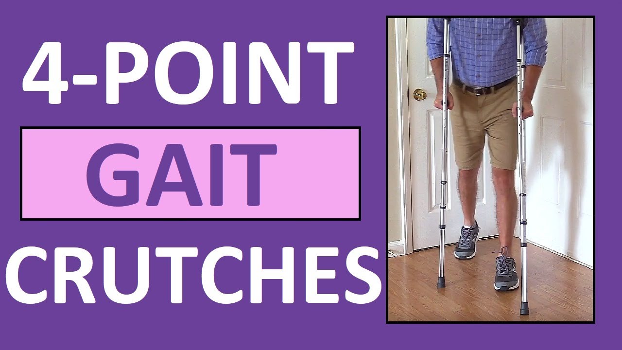
Four-point gait crutches walking pattern demonstration review for
NCLEX assistive devices and nurses.
One of the gaits that you'll have to learn for crutches is the 4-point gait. An example of a four point gait crutch pattern would be the patient moving the right crutch first (on the injured side), followed by the left foot, then the left crutch, and then the right foot. Then, you'll repeat this pattern.
In addition to this video, we have an entire compilation that features the various crutch gait patterns, as well as walkers and canes:
https://www.youtube.com/watch?v=k2-w3LZlCVk
#crutches
#nclex
#nursing
#nurse
Website: https://www.registerednursern.com/
More Videos: https://www.youtube.com/watch?v=R2XMro13dD0&list=UUPyMN8DzkFl2__xnTEiGZ1w
Nursing Gear: https://teespring.com/stores/registerednursern
Instagram: https://www.instagram.com/registerednursern_com/
Facebook: https://www.facebook.com/RegisteredNurseRNs
Twitter: https://twitter.com/NursesRN
Popular Playlists:
NCLEX Reviews: https://www.youtube.com/playli....st?list=PLQrdx7rRsKf
Fluid & Electrolytes: https://www.youtube.com/playli....st?list=PLQrdx7rRsKf
Nursing Skills: https://www.youtube.com/playli....st?list=PLQrdx7rRsKf

WATCH MORE NURSING SKILLS HERE: https://nursing.com/course/nursing-skills/?utm_source=youtube&utm_medium=social
In our Nursing Skills course, we show you the most common and most important skills you will use as a nurse! We included everything from bed baths, to inserting a foley, to advanced skills like chest tube management.
Welcome to the NURSING Family, we call it the most supportive nursing cohort on the planet.
At NURSING.com, we want to help you remove the stress and overwhelm of nursing school so that you can focus on becoming an amazing nurse.
Check out our freebies and learn more at: (http://www.nursing.com)
Visit us at http://www.nursing.com/medical....-information-disclai for disclaimer information.
NCLEX®, NCLEX-RN® are registered trademarks of the National Council of State Boards of Nursing, INC. and hold no affiliation with NURSING.

Glass ampules are often used to store medication, and as a nurse, you'll need to know how to use them.
In this video, I demonstrate how to clean an ampule using alcohol prep, how to open (or break) an ampule, as well as how to dispose of the ampule.
In addition, I show how to use an ample filter straw while drawing up (withdrawing) medication, how to use the syringe, and how to remove the air bubbles in the syringe.
This is another video in our series on clinical nursing skills.
Notes: https://www.registerednursern.....com/how-to-withdraw-
Website: https://www.registerednursern.com/
More Videos: https://www.youtube.com/watch?v=R2XMro13dD0&list=UUPyMN8DzkFl2__xnTEiGZ1w
Nursing Gear: https://teespring.com/stores/registerednursern
Instagram: https://www.instagram.com/registerednursern_com/
Facebook: https://www.facebook.com/RegisteredNurseRNs
Twitter: https://twitter.com/NursesRN
Popular Playlists:
NCLEX Reviews: https://www.youtube.com/playli....st?list=PLQrdx7rRsKf
Fluid & Electrolytes: https://www.youtube.com/playli....st?list=PLQrdx7rRsKf
Nursing Skills: https://www.youtube.com/playli....st?list=PLQrdx7rRsKf
FREE Nursing School Cheat Sheets at: http://www.NURSING.com
Get the full lesson on IM Injections here:
https://nursing.com/lesson/ski....lls-06-01-pill-crush
Check out our new Nurse Care Plan Lessons here:
https://bit.ly/3BPRfPL
Get Access to Thousands of Lessons here:
https://nursing.com/courses/
Welcome to the NURSING Family, we call it the most supportive nursing cohort on the planet.
At NURSING.com, we want to help you remove the stress and overwhelm of nursing school so that you can focus on becoming an amazing nurse.
Check out our freebies and learn more at: (http://www.nursing.com)
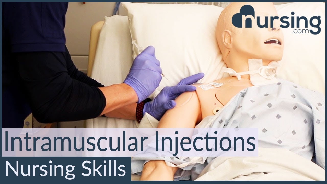
Intramuscular Injection Techniques (Nursing Skills)
In this video, we’re going to look at proper administration techniques for intramuscular medication administration. Of course, always follow your 5 rights and calculate the correct volume for administration. We love you guys! Go out and be your best selves today! And, as always, happy nursing!
Bookmarks:
0.05 Introduction to Intramuscular injections
0.16 site and needle selection
0.35 site sterilization
0.43 Z track method
0.58 needle insertion
1.10 medication injection
1.14 needle removal
1.25 bandaging and needle disposal
1.30 documentation and patient monitoring
1.35 Outro
Visit us at https://nursing.com/medical-disclaimer/ for disclaimer information.
NCLEX®, NCLEX-RN® are registered trademarks of the National Council of State Boards of Nursing, INC. and hold no affiliation with NURSING.com.

Tummy Tuck ( Classic Method ) : Surgery | 3D Animation
How long does tummy tuck last?
Tummy tuck results are considered permanent, insofar that the fat cells and skin removed during an abdominoplasty cannot grow back. Likewise, the internal sutures placed to repair abdominal muscles are designed to remain in place indefinitely.
What is tummy tuck surgery?
A tummy tuck — also known as abdominoplasty — is a cosmetic surgical procedure to improve the shape and appearance of the abdomen. During a tummy tuck, excess skin and fat are removed from the abdomen. Connective tissue in the abdomen (fascia) usually is tightened with sutures as well.
How much does tummy tuck cost?
How much does it cost? It can cost from about £5,000 to £10,000 to have an abdominoplasty in the UK, plus the cost of any consultations or follow-up care.
How painful is a tummy tuck?
A tummy tuck requires significant downtime
At the beginning, you will be fatigued, swollen and sore. It is normal to have moderate pain during these first several days, although this will steadily improve. It is vital to allow yourself time to focus on rest and healing.
What is the disadvantage of tummy tuck?
The cons of a tummy tuck include: A full abdominoplasty is a major operation with a considerable recovery. Expect to postpone strenuous activities for at least 6 weeks. Results take time.
Is tummy tuck more painful than C section?
That's something many women want to know. While patients have different experiences, most plastic surgeons would agree that a cesarean section is more painful than most tummy tucks.
- Tummy tuck
- Abdominoplasty
- Abdominal tuck
- Tummy tuck procedure
- Tummy tuck process
- Tummy tuck surgery
- Tummy tuck operation
- Tummy tuck video
- Tummy tuck recovery
- Tummy tuck before and after
- Abdominoplasty surgery
- Abdominal contouring surgery
- Postpartum tummy tuck
- Post pregnancy tummy tuck
- Mini tummy tuck
- Tummy tuck cost
- Tummy tuck risks
- Tummy tuck complications
- How long does a tummy tuck take
- Tummy tuck scarring
- Tummy tuck skin removal
- Tummy tuck muscle tightening
#tummytuck
#abdominoplasty
#plastic_surgery
#cosmetic_surgery
#body_contouring
#tummy_tuck_surgery
#surgery
#cosmetic_procedure
#beauty
#health
#fitness
#medical_animation
#3d_animation
#medical_video
#explainer_video
#education
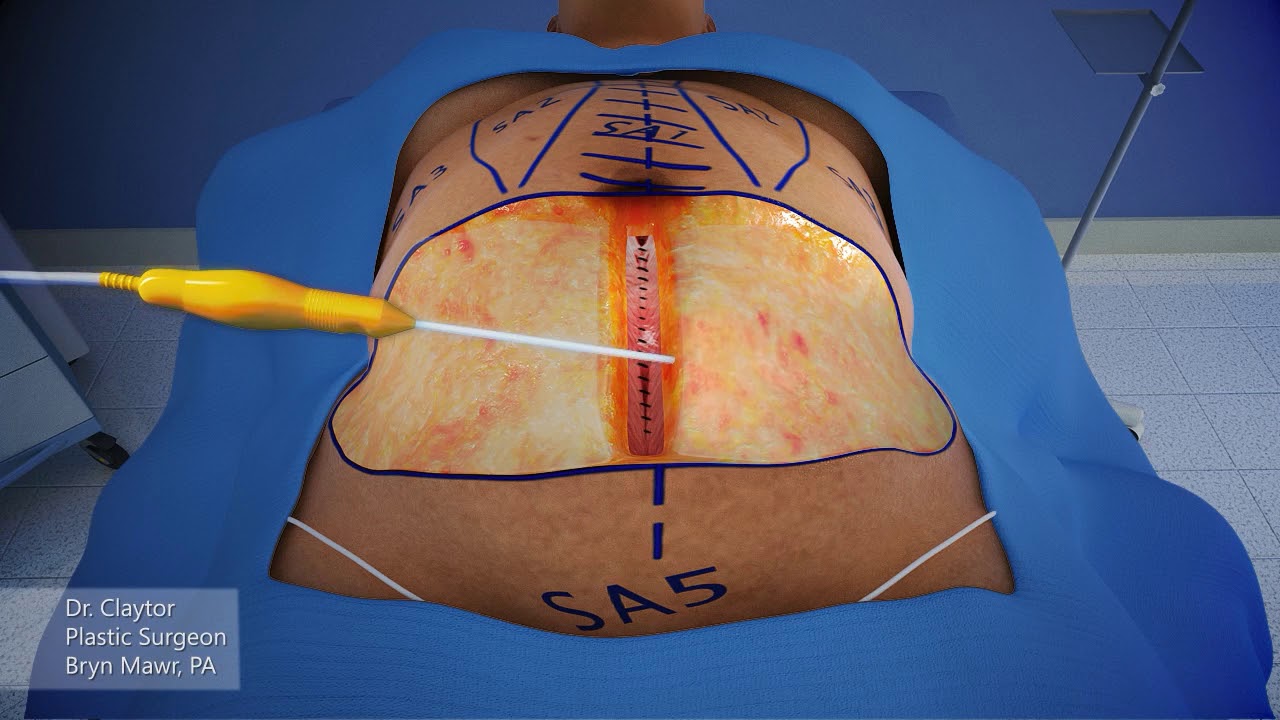
Dr. Claytor uses a 3-D animation to demonstrate how a drainless tummy tuck combined with liposuction can effectively reduce excess skin and fat on the abdomen WITHOUT the need for drains during post-op recovery!
Learn more about Dr. Claytor’s drainless tummy tucks here: https://www.cnplasticsurgery.c....om/procedures/body/t
R. Brannon Claytor, MD, FACS is a renowned double board-certified plastic surgeon and director of Claytor Noone Plastic Surgery, a premium plastic surgery practice in Bryn Mawr, PA that proudly serves the Philadelphia, Main Line, and surrounding areas. Dr. Claytor’s superb skill and results have been recognized for over a decade, earning him numerous awards in both local and national publications, including Philadelphia Magazine, Main Line Today, and Newsweek.
Together, Dr. Claytor and his experienced aesthetics team provide a variety of surgical and non-surgical procedures for the face, breasts, and body to help you look and feel your best. To learn more about how Dr. Claytor and our entire staff can help you reach your goals, please visit our website or give us a call at 610-527-4833.
About Dr. Claytor: https://www.cnplasticsurgery.c....om/our-practice/dr-r
Claytor Noone Plastic Surgery: https://www.cnplasticsurgery.com/
Essential guide to plastic surgery (procedures, costs, planning and more): https://www.cnplasticsurgery.c....om/our-practice/esse
Questions? Contact us online: https://www.cnplasticsurgery.com/contact-us/

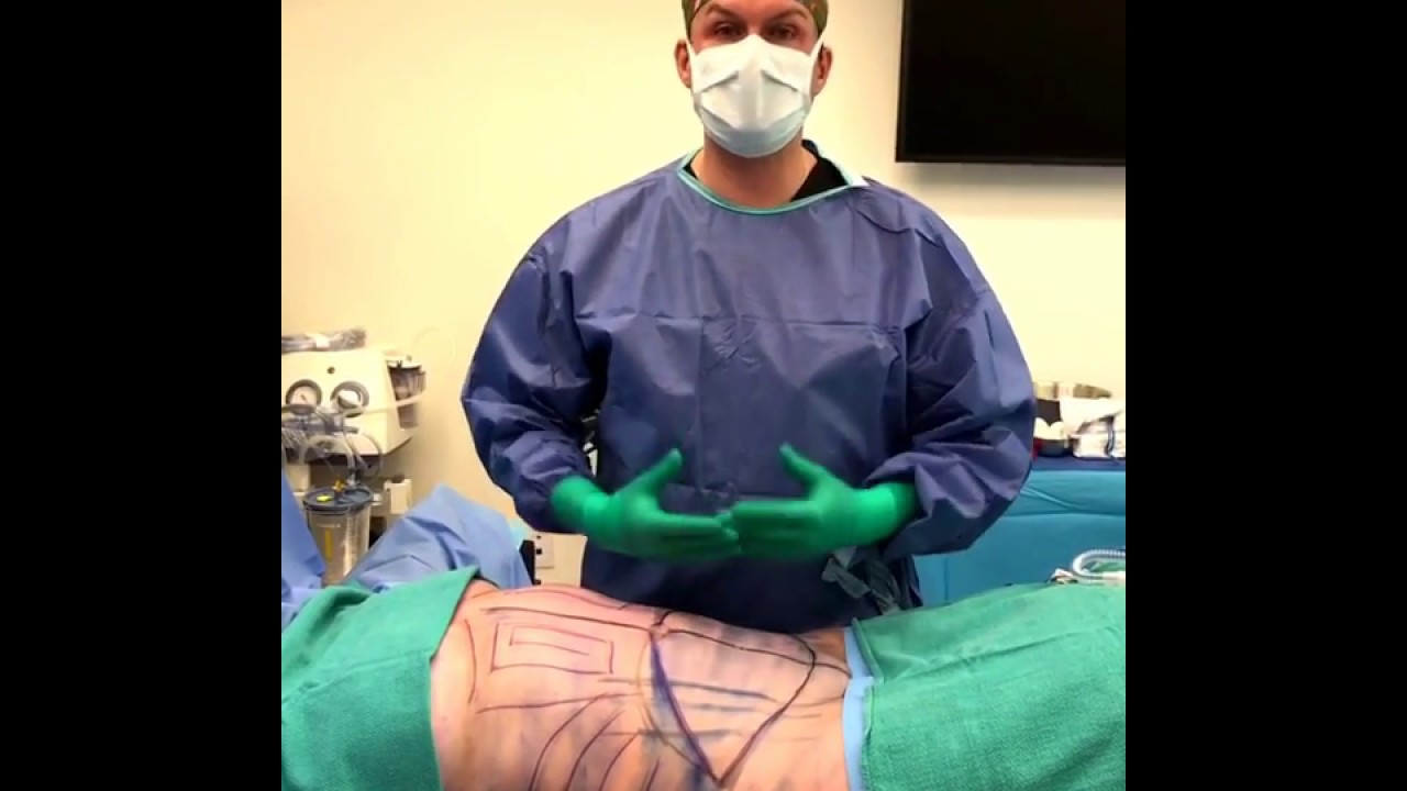
TUMMY TUCK 🤩 Immediate OR Results
This patient wanted to get her abs back, but unfortunately NO diet or workout can tighten muscles that have been stretched apart from carrying a baby 👀 But we can fix that at Lemmon Avenue Plastic Surgery & Laser Center!
To learn more about the #tummytuck click here: https://drdeuber.com/procedures/tummy-tuck/
For #mommymakeover, click here: https://drdeuber.com/procedures/mommy-makeover/
👙
#MarkDeuberMD

A tummy tuck is a surgical process that removes excess fat and skin. Learn more about the procedure by watching this video!
Looking to book a consultation? Call Zuri Plastic Surgery now at 786-804-1603 or DM us today to schedule a complimentary consultation with Dr. Z.
Un tummy tuck es un procedimiento quirúrgico que elimina el exceso de grasa y piel. ¡Aprenda más sobre este procedimiento viendo este video!
¿Quiere agendar una consulta? Llame a Zuri Plastic Surgery ahora al 786-804-1603 o envíenos un DM hoy para programar una consulta gratuita con el Dr. Z.
WARNING: Explicit and Educational Surgical Content.
Visage Clinic's Dr. Marc DuPéré - located in Toronto, Ontario, Canada discusses Liposuction (upper bra, back rolls, lower back rolls, love handles & abdomen) and "Tummy Tuck" (Abdominoplasty): Skin excision, muscle repair and umbilicoplasty.
For more info and to book a consultation visit www.VisageClinic.com/cosmetic-....surgery/mommy-makeov or call (416) 929-9800.
