
- Physical Examination
- Surgical Examination
- Ophthalmology
- Clinical Skills
- Orthopedics
- Surgery Videos
- Laparoscopy
- Pediatrics
- Funny Videos
- Cardiothoracic Surgery
- Nursing Videos
- Plastic Surgery
- Otorhinolaryngology
- Histology and Histopathology
- Neurosurgery
- Dermatology
- Pediatric Surgery
- Urology
- Dentistry
- Oncology and Cancers
- Anatomy Videos
- Health and Fitness
- Radiology
- Anaesthesia
- Physical Therapy
- Pharmacology
- Interventional Radiology
- Cardiology
- Endocrinology
- Gynecology
- Emergency Medicine
- Psychiatry and Psychology
- Childbirth Videos
- General Medical Videos
- Nephrology
- Physiology
- Diet and Food Health
- Diabetes Mellitus
- Neurology
- Women Health
- Osteoporosis
- Gastroenterology
- Pulmonology
- Hematology
- Rheumatology
- Toxicology
- Nuclear Medicine
- Infectious Diseases
- Vascular Disease
- Reproductive Health
- Burns and Wound Healing
- Other
Top videos
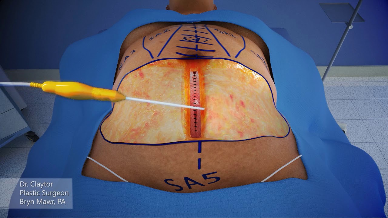

Dr. Claytor uses a 3-D animation to demonstrate how a drainless tummy tuck combined with liposuction can effectively reduce excess skin and fat on the abdomen WITHOUT the need for drains during post-op recovery!
Learn more about Dr. Claytor’s drainless tummy tucks here: https://www.cnplasticsurgery.c....om/procedures/body/t
R. Brannon Claytor, MD, FACS is a renowned double board-certified plastic surgeon and director of Claytor Noone Plastic Surgery, a premium plastic surgery practice in Bryn Mawr, PA that proudly serves the Philadelphia, Main Line, and surrounding areas. Dr. Claytor’s superb skill and results have been recognized for over a decade, earning him numerous awards in both local and national publications, including Philadelphia Magazine, Main Line Today, and Newsweek.
Together, Dr. Claytor and his experienced aesthetics team provide a variety of surgical and non-surgical procedures for the face, breasts, and body to help you look and feel your best. To learn more about how Dr. Claytor and our entire staff can help you reach your goals, please visit our website or give us a call at 610-527-4833.
About Dr. Claytor: https://www.cnplasticsurgery.c....om/our-practice/dr-r
Claytor Noone Plastic Surgery: https://www.cnplasticsurgery.com/
Essential guide to plastic surgery (procedures, costs, planning and more): https://www.cnplasticsurgery.c....om/our-practice/esse
Questions? Contact us online: https://www.cnplasticsurgery.com/contact-us/

It’s not tummy tuck procedure.. it’s liposuction only.. don’t get confused with both procedure..
#beforeandafter #kmc #nose #aesthetic #antiaging #beauty #drhabibhairtransplant #peshawar #nose #islamabad #swat #kohat #nowshehra #karakin #mardan
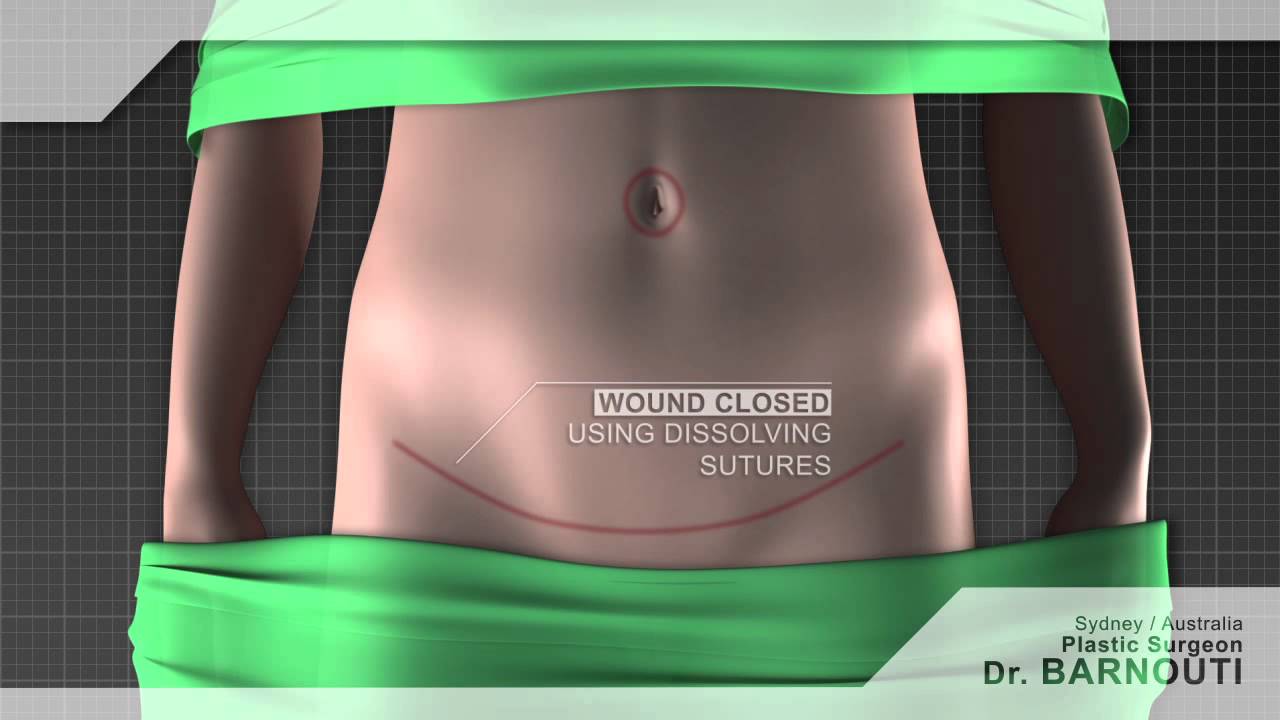
Tummy tuck Sydney Dr Barnouti. Call us on 02-9561 0222 or 1300 002 006
Broadway, Chatswood, Burwood NSW Australia
email:drbarnouti@australiaplasticsurgery.com.au
https://www.plasticsurgery-syd....ney.com.au/abdominop
What is a tummy tuck?
A tummy tuck operation is also known as abdominoplasty. It involves removing excess skin and fat from the stomach area, mainly the lower part of the tummy through surgical procedure. A tummy tuck operation is intended to leave the patient with a flatter tummy and to remove any signs of an 'apron' stomach or an overhang which is sometimes visible above underwear. The skin on this area tends to be stretched and of poor quality. A tummy tuck operation will usually focus on the lower part of the stomach, below the belly button and may require the belly button to be repositioned in some cases. The procedure is often carried out on women or men who have suffered from stretched skin in the stomach area after pregnancy, giving birth, excess fat deposition or weight loss.
What happens during a tummy tuck?
During a tummy tuck procedure the aim of the surgeon is to cut away fat and excess skin. To do this Dr Barnouti will make in incision on the lowest part of the stomach, where a fold will be visible above the pubic bone. He will take out as much excess fat as can be removed and will then cut the skin to fit back over the place where the fat has been removed from. It is important to have realistic expectations of a tummy tuck. Taking too much fat and skin away can result in folds at each end of the resulting scar which are sometimes referred to as "dog ears". Dr Barnouti will make sure you will not have this problem.
Who should have a tummy tuck?
Tummy tucks are recommended for either men or women who have an excess of fat and skin around their abdomen which cannot be removed by weight loss, exercise or liposuction. Tummy tuck operations in women are usually reserved for those who are not likely to have children as it is inadvisable to get pregnant again after having skin removed, this can cause the wound to stretch and scar.
The cost of a tummy tuck in Sydney Australia
The total cost is $7,900 if the patient's health fund cover the hospital's fees. In case the health fund does not cover the hospital's fee, the total cost will be around $12,000 inclusive of the Surgeon, assistant surgeon, Anaesthetist, hospital, operating theatre and follow ups visit.
Payment plans are alos available from Dr Barnouti's office in Chatswood, Burwood or Broadway.
A tummy tuck is a cosmetic procedure that removes excess skin and fatty tissue in order to give a flatter appearance to the stomach. Tummy tucks, also known as abdominoplasties, are ideal for patients who are not excessively overweight but suffer from an overhang of skin around the abdomen.
Performed under general anaesthetic, tummy tucks involve a horizontal incision being made just above the pubic area between the hip bones. Skin and fatty tissue is separated from the muscle and the area is tightened, with the excess skin and fatty tissues then being pulled downwards and removed.
Following your tummy tuck, there will be a scar present across the lower abdomen, but this will gradually fade. You may experience moderate tissue swelling for several months, but this will disappear with time. There may also be a sensation reduction just above the pubic area.
Once your tummy tuck recovery is complete however, you'll benefit from a more attractive figure and the ability to wear a wider selection of clothes.
After MacKenzie Walker lost 100 pounds, her "after" picture remained elusive. So she asked plastic surgeon Dr. Anthony Youn to perform an abdominoplasty.
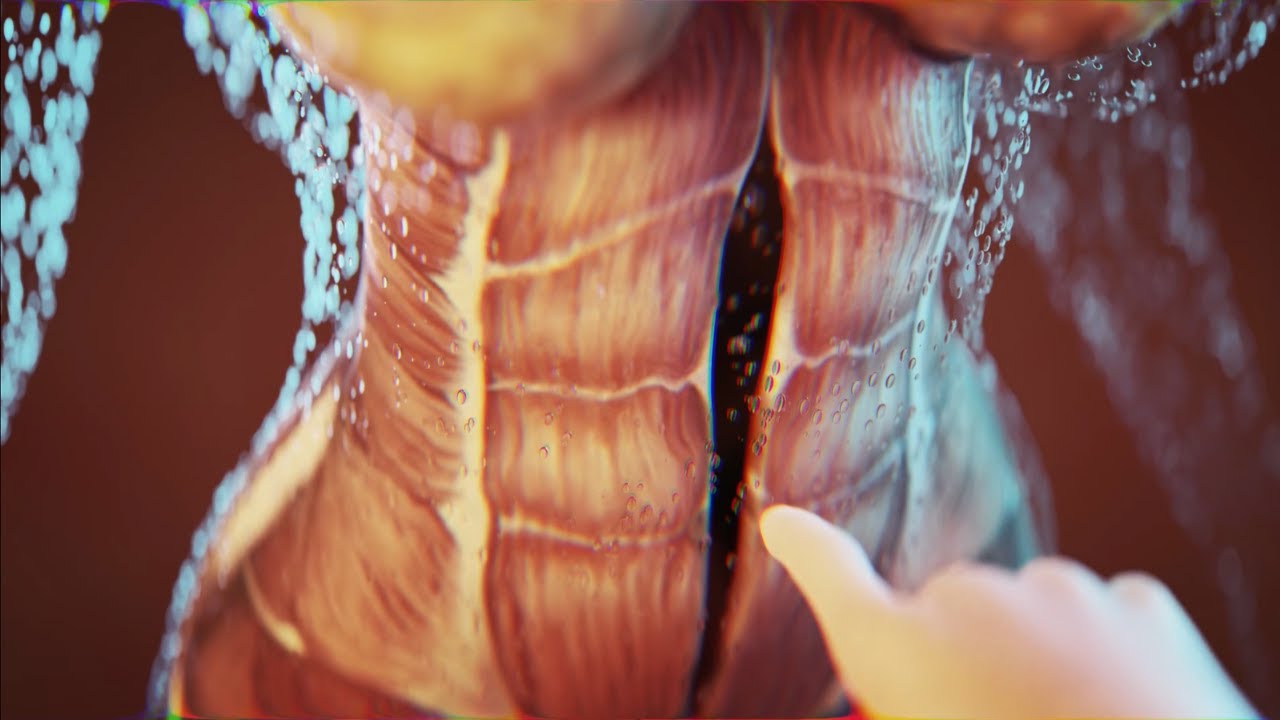
Today I'm using the best 3D animation to explain WHAT IS DIASTASIS RECTI and what you need to know about diastasis recti after pregnancy! Grab the Complete Diastasis Recti Healing Guide: https://landing.mailerlite.com..../webforms/landing/n0
If you are't sure what video to start with and you just want step-by-step daily instructions you can start my 30-day core healing program. You get a new 10-min core healing video daily for 30 days. https://pregnancyandpostpartum....tv.thinkific.com/cou
How I healed my 4-finger diastasis recti gap:
Jessica Pumple is a registered dietitian, and pre & postnatal fitness instructor and certified pregnancy and postpartum core exercise specialist (CPES). She helps pregnant women stay fit, have healthy babies, and easier labors. She helps new moms with postpartum recovery, to heal and strengthen their core and feel energized after pregnancy!
If you enjoy our content subscribe to our channel, hit the bell button, leave a comment and share with your friends so I can make you more of the videos you enjoy!
Disclaimer: This is general postnatal fitness only. Please check with your doctor or health care provider to see if this video is safe for you. Wait until you get clearance (usually 4-6 weeks or 6-8 weeks after a c-section).You are responsible for your own safety. Don’t do anything that feels unsafe for you or baby. Stop if you have any pain or discomfort, bleeding, chest pain or shortness of breath, dizziness or if you feel unwell. P&P Health Inc., Pregnancy and Postpartum TV and Jessica Pumple are not liable in any way for any injury, loss, damages, costs or expenses suffered by you in relation to this video or its content.
Copyright 2023 P&P Health Inc. All rights reserved
#diastasisrecti #whatisdiastasisrecti #3danimation
Music: Epidemic Sound

A complete organized library of all my videos, digital slides, pics, & sample pathology reports is available here: https://kikoxp.com/posts/5084 (dermpath) & https://kikoxp.com/posts/5083 (bone/soft tissue sarcoma pathology)
Topics discussed:
Epidermis:
Layers of epidermis: 0:10
Melanocytes vs Keratinocytes: 5:16
Langerhans cells: 10:10 & 33:30 & 57:30
Dermis:
Papillary and reticular dermis: 11:50
Three types of white empty spaces on a slide: vessels, glands/ducts/cysts, or artifact: 15:25
Blood vessels & nerves: 18:24 & 48:50 & 58:59
Arrector pili & other dermal smooth muscle: 20:00
Adnexal:
Sebaceous gland: 21:10
Hair follicle 23:14
Eccrine sweat glands and ducts 24:45 & 50:00
Gland/duct vs blood vessel 27:20 & 48:50
Apocrine glands: this video https://kikoxp.com/posts/7837 (at 12:30)
Acrosyringium: this video https://kikoxp.com/posts/7837 (at 10:00)
Three types of pink bundles: smooth muscle, nerve, dense connective tissue: 27:50
Acral skin (palm sole) with contact dermatitis 29:37
Parakeratosis 30:00
Perivascular lymphocytes 30:40
Eosinophils vs neutrophils 31:20
Spongiosis with desmosome keratinocyte spines 32:10
Spongiotic vesicles with Langerhans cells 33:30
Normal acral skin (palm & sole) with stratum lucidum 34:20
Normal glomus body/apparatus (canal of Sucquet-Hoyer) 35:40
Nerve 36:46 & 51:50
Adipose tissue (white fat cells) in subcutis with Lochkern 37:55
Normal scalp skin with large anagen hair follicles: 39:30
Hair follicle anatomy (bulb/matrix, inner root sheath, outer root sheath, hair shaft, isthmus, infundibulum): 40:55 (labeled images):
https://kikoxp.com/posts/3661 & https://kikoxp.com/posts/7899
Pacinian corpuscle 50:40
Meissner corpuscle 1:02:28
Dense regular connective tissue (Fascia/Tendon/Ligament) vs Smooth Muscle 53:00
Basic Normal Skin Immunohistochemistry:
-cytokeratin in epidermis: 55:33
-S100 in melanocytes and Langerhans cells and adipocytes: 57:30
-Desmin in smooth muscle (arrector pili and blood vessels): 58:59
-CD31 in endothelial cells of blood vessels: 59:33
-SOX-10 in melanocytes: 1:00:40
Digit/Finger/Toe histology (amputation for subungual acral melanoma) 1:04:10 & 1:08:30
-bone 1:05:40
-glomus body 1:05:15
-tendon/ligament 1:06:10
-artery 1:06:58
-fingernail/toenail 1:08:54
-acrosyringium 1:10:45
Solar elastosis (what wrinkles look like microscopically!) 1:11:50
Other videos you might like:
Tendon vs Nerve Histology Made Simple with the Ramen Noodle Sign (of Fulton) video: https://kikoxp.com/posts/4466
Melanocytes vs Keratinocytes made easy video: https://kikoxp.com/posts/3802
Blood Vessel vs Gland vs Artifact Made Easy video: https://kikoxp.com/posts/4808
The basic normal structures of the skin discussed and described by a dermatopathologist. This material is intended for use by medical students, junior pathology or dermatology residents, or for anyone else studying normal human histology. Special thanks to two of my medical students at UAMS for helping make this video possible. Miki Lindsey convinced me that I really needed to sit down and record this video. Akash Patel took time to edit the video and make it ready for YouTube. My sincere thanks to both of them for helping me overcome procrastination.
Huge thanks to Abigail Cline, a medical student at Medical College of Georgia, for volunteering to type a transcript of this ENTIRE video (over 14,000 words!) so that I could provide closed caption subtitles for those with hearing impairments and for those who may need assistance in understanding spoken English (particularly given how quickly I speak!). You can access a text version of her transcript of my video here: https://kikoxp.com/posts/5390
Correction - I made a mistake in the video. I said that sebaceous gland secretions are turned into smelly substances by bacteria and that this makes body odor. That is incorrect. That is actually true of APOCRINE gland secretions not sebaceous secretions.
Also, in the past I used "keratinocyte" and "squamous cell" interchangeably (this is because in dermatopathology, we see and talk about squamous cell carcinomas all the time, and those tumors are composed of keratinocytes). But technically, in normal skin histology, "squamous cell" refers only to the flattened keratinocytes in the superficial epidermis. Thankfully, a histology PhD colleague pointed this out to me and corrected my lazy nomenclature!
Please check out my Soft Tissue Pathology & Dermatopathology survival guide textbooks: http://bit.ly/2Te2haB
This video is geared towards medical students, pathology or dermatology residents, or practicing pathologists or dermatologists. Of course, this video is for educational purposes only and is not formal medical advice or consultation.
Presented by Jerad M. Gardner, MD. Please subscribe to my channel to be notified of new pathology teaching videos.
Follow me on:
Snapchat: JMGardnerMD
Twitter: @JMGardnerMD
Instagram: @JMGardnerMD
Facebook: https://www.facebook.com/JMGardnerMD/

How to approach histology for Human Anatomy students. Using a key will help get you through it! Add some penguin fairy dust will help too!
Please note: I mis-spoke and said "striated" instead of "stratified epithelium" a couple of times... apologies!
There are lots of histology keys out there, but the one I showed in the video is here: http://www.penguinprof.com/upl....oads/8/4/3/1/8431323
Want more?
Subscribe: http://www.youtube.com/user/ThePenguinProf
FB Page: https://www.facebook.com/ThePenguinProf
Twitter: https://twitter.com/penguinprof
Web: http://www.penguinprof.com/
---------------------------------------------------------------------------------------------------
Details:
Tissue in the human body:
Epithelial: Is made of cells arranged in a continuous sheet with one or more layers, has apical & basal surfaces.
A basement membrane is the attachment between the basal surface of the cell & the underlying connective tissue.
Two types of epithelial tissues: (1) Covering & lining epithelia and (2) Glandular Epithelium.
The number of cell layers & the shape of the cells in the top layer can classify epithelium.
Simple Epithelium - one cell layer
Stratified epithelium - two or more cell layers
Pseudostratified Columnar Epithelium - When cells of an epithelial tissue are all anchored to the basement Membrane but not all cells reach the apical surface.
Glandular Epithelium -- (1) Endocrine: Release hormones directly into the blood stream and (2) Exocrine - Secrete into ducts.
Connective: contains many different cell types including: fibroblasts, macrophages, mast cells, and adipocytes. Connective Tissue Matrix is made of two materials: ground substance - proteins and polysaccharides, fiber -- reticular, collagen and elastic.
Classification of Connective Tissue:
Loose Connective - fibers & many cell types in gelatinous matrix, found in skin, & surrounding blood vessels, nerves, and organs.
Dense Connective - Bundles of parallel collagen fibers& fibroblasts, found in tendons& ligaments.
Cartilage - Cartilage is made of collagen & elastin fibers embedded in a matrix glycoprotein & cells called chondrocytes, which was found in small spaces.
Cartilage has three subtypes:
Hyaline cartilage -- Weakest, most abundant type, Found at end of long bones, & structures like the ear and nose,
Elastic cartilage- maintains shape, branching elastic fibers distinguish it from hyaline and
Fibrous Cartilage - Strongest type, has dense collagen & little matrix, found in pelvis, skull & vertebral discs.
Muscle: is divided into 3 categories, skeletal, cardiac and smooth.
Skeletal Muscle -- voluntary, striated, striations perpendicular to the muscle fibers and it is mainly found attached to bones.
Cardiac Muscle -- involuntary, striated, branched and has intercalated discs
Smooth Muscle -- involuntary, nonstriated, spindle shaped and is found in blood vessels & the GI tract.
Nervous: Consists of only two cell types in the central nervous system (CNS) & peripheral nervous system (PNS):
Neurons - Cells that convert stimuli into electrical impulses to the brain, and Neuroglia -- supportive cells.
Neurons -- are made up of cell body, axon and dendrites. There are 3 types of neurons:
Motor Neuron -- carry impulses from CNS to muscles and glands,
Interneuron - interpret input from sensory neurons and end responses to motor neurons
Sensory Neuron -- receive information from environment and transmit to CNS.
Neuroglia -- is made up of astrocytes, oligodendrocytes, ependymal cells and microglia in the CNS, and schwann cells and satellite cells in the PNS.

► Sign up here and try our FREE content: http://lectur.io/freecontentyt
► If you’re an medical educator or faculty member, visit: http://lectur.io/medytb2u
This video “Epithelium” is part of the Lecturio course “Histology” ► WATCH the complete course on http://lectur.io/epithelium
► LEARN ABOUT:
- Introduction: Epithelial Tissue
- Summary of structure and function
- Epithelium
- Three characteristics of any Epithelium
- Epithelium: Classification
- Epithelium: Epithelial Cells Exhibit Polarity
- Epithelium: How Are Epithelial Cells Joined Together?
- Epithelium: Surfaces of Epithelial Cells
- Epithelium: Basement Membrane
► THE PROF: Your lecturer is Professor Geoff Meyer. He is currently teaching at the School of Anatomy, Physiology and Human Biology at the University of Western Australia (UWA). As a leading anatomy and histology expert he is also coordinating the Federative International Program for Anatomical Terminologies (FIPAT) of the International Federation of Associations of Anatomists (IFAA). Besides medical research on the ovarian function, steroidogenesis, corpus luteum, angiogenesis, and microcirculation, Geoff Meyer’s research activities also focus on developing innovative, computer-aided learning and teaching tools. For his inventiveness, Geoff Meyer has received a number of awards, including the Australian University Teaching Award.
► LECTURIO is your single-point resource for medical school:
Study for your classes, USMLE Step 1, USMLE Step 2, MCAT or MBBS with video lectures by world-class professors, recall & USMLE-style questions and textbook articles. Create your free account now: http://lectur.io/epithelium
► INSTALL our free Lecturio app
iTunes Store: https://app.adjust.com/z21zrf
Play Store: https://app.adjust.com/b01fak
► READ TEXTBOOK ARTICLES related to this video:
Epithelium — Functions and Types of Epithelial Tissue
http://lectur.io/epitheliumarticle
► SUBSCRIBE to our YouTube channel: http://lectur.io/subscribe
► WATCH MORE ON YOUTUBE: http://lectur.io/playlists
► LET’S CONNECT:
• Facebook: https://www.facebook.com/lectu....rio.medical.educatio
• Instagram: https://www.instagram.com/lecturio_medical_videos
• Pinterest: https://www.pinterest.de/lecturiomedical
• LinkedIn: https://www.linkedin.com/company/lecturio-medical/


Join the Amoeba Sisters a they explore different muscle tissues and then focus on the sliding filament theory in skeletal muscle! This video also briefly talks about muscle naming, some vocabulary (such as agonists and antagonists) before focusing on the sliding filament model. Video also mentions general roles of tropomyosin and troponin.
---------------------------------------------------------
Table of Contents:
00:00 Intro
0:39 Muscle Tissue Types
1:58 Muscle Characteristics
2:33 Skeletal Muscle Naming and Arrangement
3:26 Actin Myosin and Sarcomere
4:32 Sliding Filament Model
6:55 Tropomyosin an Troponin
---------------------------------------------------------
Factual References:
Betts, J. Gordon, et al. “10.3 Muscle Fiber Contraction and Relaxation - Anatomy and Physiology 2e | OpenStax.” Openstax.org, 20 Apr. 2022, openstax.org/books/anatomy-and-physiology-2e/pages/10-3-muscle-fiber-contraction-and-relaxation.
Urry, Lisa A, et al. Campbell Biology. 11th ed., New York, Ny, Pearson Education, Inc, 2017.
---------------------------------------------------------
Further Reading Recommendations:
What about I and A bands? What actually initiates the power stroke? How does calcium get released and from where? Remember, there is a lot more detail! We recommend this page from Openstax to learn more:
https://openstax.org/books/bio....logy-2e/pages/38-4-m
-----------------------------------------------
The Amoeba Sisters videos demystify science with humor and relevance. The videos center on Pinky's certification and experience in teaching biology at the high school level. Amoeba Sisters videos only cover concepts that Pinky is certified to teach, and they focus on her specialty: secondary life science. Learn more about our videos here: https://www.amoebasisters.com/our-videos
Support Us? https://www.amoebasisters.com/support-us
Our Resources and Handouts: https://www.amoebasisters.com/handouts
Biology Video Playlist: https://www.youtube.com/playli....st?list=PLwL0Myd7Dk1
GIFs: https://www.amoebasisters.com/gifs.html
Comics: https://www.amoebasisters.com/....parameciumparlorcomi
Unlectured Series: https://www.amoebasisters.com/unlectured
Connect with us!
Website: https://www.AmoebaSisters.com
Twitter: https://www.twitter.com/AmoebaSisters
Facebook: https://www.facebook.com/AmoebaSisters
Tumblr: https://www.amoebasisters.tumblr.com
Pinterest: https://www.pinterest.com/AmoebaSisters
Webtoon: https://www.webtoons.com/en/challenge/amoeba-sisters-sisterhood/list?title_no=289479&page=1
Instagram: https://www.instagram.com/amoebasistersofficial/
TikTok: https://www.tiktok.com/@amoebasistersofficial
Visit our Redbubble store at https://www.amoebasisters.com/store
TIPS FOR VIEWING EDU YOUTUBE VIDEOS:
Want to learn tips for viewing edu YouTube videos including changing the speed, language, viewing the transcript, etc? https://www.amoebasisters.com/....pinkys-ed-tech-favor
MUSIC:
Our intro music designed and performed by Jeremiah Cheshire.
End music in this video is listed free to use/no attribution required from the YouTube audio library.
COMMUNITY:
We take pride in our AWESOME community, and we welcome feedback and discussion. However, please remember that this is an education channel. See YouTube's community guidelines and how YouTube handles comments that are reported by the community. We also reserve the right to remove comments.
TRANSLATIONS:
Spanish Subtitles Translated by Jeremy García
Hindi Subtitles: Translated by Alisha Aggarwal
We gladly accept subtitle translations from our community. Learn more here: https://www.amoebasisters.com/....pinkys-ed-tech-favor We want to thank our amazing community for the generosity of their time in continuing to create translated subtitles.
We also have videos dubbed in Spanish and Portuguese using an artificial voice via https://aloud.area120.google.com to increase accessibility. See our Amoeba Sisters en Español channel https://www.youtube.com/channe....l/UC1Njo3LBy53cOPngz and Amoeba Sisters em Português https://www.youtube.com/channe....l/UCYTQPX2X_mXe0ZMPi


#GIT #Esophagus #histology

Classification of epithelium, discussion on lining epithelium of 3 major system (GIT, Urogenital and Respiratory system


Website : https://www.udemy.com/course/h....istology/?referralCo
Human Histology is one of the basic subject in a Medical Student career. By learning Histology in a proper way, this will help you to get a Visual memory of the Human body. Using this Visual memory, you can Learn any other subjects with little effort.
This Course is very well organized with lot of Histology images, Line diagrams, simple presentations and clear Explanations. This course has 33 videos, 19 chapters, 6 hours long covering all topics. Every topic is made Simple and Complete. Dr Ram has a great teaching style and has a good experience in teaching medical subjects to students.
After finishing this course, you will be better in your basics, with ability to visualize the human body and this will create an intense thirst to learn more. We give 100% guarantee that you will have a complete and in-depth understanding in short time, You will start to enjoy Learning Medicine because of the visualization of human body you get from this course and you will be ready to face any Medical exams in world.
Course features:
- Complete Histology lectures covering all chapters
- 19 chapters | 33 Videos | 6 Hours
- Clear Histology images
- Line diagrams for easy understanding
- Lot of memory tips
- High quality audio and Videos
- Can be viewed in Pc, or Phones or TV
Course content: ( 19 Chapters, 33 videos )
I The Cell - 3 Lessons
1. Nucleus
2. Cytoplasm
3. Cell Junctions
II Tissues - 11 Lessons
4. Epithelial tissues
5. Connective tissues
6. Muscular tissues
7. Nervous tissues
8. Bones
9. Cartilage
10. Lymphoid tissues
III Organ systems - 19 Lessons
11. Cardiovascular system
12. Respiratory system
13. Gastrointestinal system
14. Liver and Exocrine pancreas
15. Endocrine system
16. Urinary system
17. male reproductive system
18. Female reproductive system
19. The skin
Instructor : Dr Ram , Med Madness

✨This video is on the structure and functions of the three types of cartilage (Hyaline, Elastic and Fibrocartilage). I hope it helps! ☀️
🌟What's in this video?
0:00 - Intro
0:07 - Connective Tissue Recap
1:16 - Structure of Connective Tissue
1:57 - Structural Components of Cartilage
3:38 - Types of Cartilage
3:49 - Hyaline Cartilage
8:05 - Elastic Cartilage
8:55 - Fibrocartilage
✨ Other videos you may need:
🔅 Connective Tissue : https://youtu.be/xw_ALdt5n-A
🔅 Collagen : https://youtu.be/3e2JYMNS_W4
🔅 Ossification: https://youtu.be/86V9SNWD_No
🔅Histology: https://www.youtube.com/playli....st?list=PL1rG930trF2
💫 For more videos like this, subscribe to my channel!
Byte Size Med: https://youtube.com/channel/UC....ZghvlgylH3r_CWfA18eF
📚Factual References & for Further Reading:
- DiFiore's Atlas of Histology
- Junqueira's Basic Histology
- Gartner's Concise Histology
- Openstax Anatomy and Physiology
https://openstax.org/details/b....ooks/anatomy-and-phy
- Openstax Biology
https://openstax.org/details/books/biology-2e
(The last two are links to open-source references. They are NOT affiliate links)
🌤 Note:
These are just a collection of my notes. So use them the way you would use borrowed notes from a friend. 📝
The images in this video are hand-drawn for illustration and explanation only.✍️ Hence, they may not be anatomically accurate. I am just one person making these videos. If there are any errors, that is unintentional. I try super hard to avoid them. Please let me know if you find any, so it gets clarified for other viewers. Science constantly evolves and changes. New discoveries are made everyday. So some of the information in these videos may become outdated. If you notice that, please let me know so I can update them.
⚡️Disclaimer:
These videos are NOT a substitute for a medical textbook. Textbooks are written by experts (which I do not claim to be), edited, proofread and referenced. Please use them.
The information has been sourced from multiple references as mentioned above. I draw all the pictures myself. But if I have inadvertently infringed on any copyright, that is completely unintentional. I only make these videos to impart education. If I have accidentally violated copyright in any way, do let me know so I can make the necessary changes or give credit to anyone who is owed the same.
These videos are NOT intended for patient education. They are NOT a substitute for diagnosis and treatment by a licensed medical professional. Always seek the advice of a qualified health care provider for any questions you may have regarding any medical condition, so that they can address your individual needs.
🔅They are ONLY meant to help students of medicine and health sciences with studying, and should be used for just that purpose and absolutely nothing else.
Byte Size Med. All Rights Reserved.
![Histology of Exocrine Gland [Epithelium 7 of 7]](https://i.ytimg.com/vi/NkU7YJ7eYd0/maxresdefault.jpg)
Histological features and cellular biology of exocrine glands. This video is a part of our Histology Video Course (https://youtube.com/playlist?l....ist=PLnr1l7WuQdDynxT
Additional YouTube Content
Biochemistry videos: https://youtube.com/playlist?l....ist=PLnr1l7WuQdDzCUC
Anatomy Videos: https://youtube.com/playlist?l....ist=PLnr1l7WuQdDz2dK
DaVinci Cases Videos: https://youtube.com/playlist?l....ist=PLnr1l7WuQdDyJUl
The DaVinci Hour Podcast: https://youtube.com/playlist?l....ist=PLnr1l7WuQdDwSm9
DaVinci Academy Website: https://www.dviacademy.com/

**PLEASE READ FULLY
Purpose of the video is to help Esthetician’s review chapters in their text book to better prepare for State Bord testing, by simply reading and going over some of the material, it’s not intended to replace any teaching from any Beauty College. Every instructor does things different, Keep in mind I am in the state of Texas, also keep in mind that when in school students are to follow guidelines and might be required to do things a bit different, I teach my students the text book because that is where the state board questions come from and the goal is for them to pass their board exams. I also teach them and go over real working situations they might come across in the salon or spa.
* I am not affiliated with TDLR or PSI in any way
PSI Bulletin Link
https://candidate.psiexams.com/bulletin/display_bulletin.jsp?ro=yes&actionname=83&bulletinid=173&bulletinurl=.pdf
Glymed store: https://glymedplus.io/home/index?store=0011298
email: glamandbeyondinfo@gmail.com

http://www.highimpact.com - This brain surgery animation was used to demonstrate a young girl's craniotomy, cranioplasty, and reconstructive skull surgery after her vehicle was struck by a tractor-trailer. The procedures included the evacuation of a large epidural hematoma, the draining of the epidural space, and the reassembly of bone fragments to repair the skull.
More Brain Surgery Animations: https://tinyurl.com/y6m4lkdf
WHAT HAPPENED
A teenage girl was riding home with her parents and boyfriend from a Wednesday night church service when a tractor-trailer struck the back driver’s side of their car as they were traveling through an intersection. The impact sent the car spinning into oncoming traffic where it struck another vehicle. When paramedics arrived, the 17-year-old was unresponsive with bleeding from her left ear and a laceration from behind her left ear.
She was rushed to the hospital where she underwent a series of CT scans that showed a severely comminuted open skull fracture with an underlying 1.1 cm subdural hematoma. She was taken to the operating room where an emergency craniotomy was performed to evacuate the hematoma and reassemble the skull fragments. The patient gradually began to wake up and was discharged six days later, after she showed she could maneuver up and down the hallway.
The biggest challenge in a traumatic brain injury case like this - where most of the damages are deeply underlying and undetectable on the surface - is that the only visual evidence is in the form of 2D black-and-white radiographic films. This can look ambiguous to the typical juror because it’s often difficult to discern where these snapshots are located inside the person’s skull. Tony Seaton, Esq., and Robert Bates, Esq., needed to reinforce this 2D radiographic evidence with maximum 3D context.
We equipped them with a custom Diagnostic Slice Chooser: an interactive presentation that presents radiographic slides within a three-dimensional model of the patient’s head. We also designed the model accurately to the patient’s likeness and colorized the films to highlight key areas of damage. The attorneys could show the complete depth and magnitude of his client’s injuries at every level both before and after the surgery. After establishing the full extent of damages, we also created an animation to walk viewers through the surgical experience the patient would undergo as a result of her injuries.
The visual presentation helped jurors understand the destructive impact this collision had on this young teenager’s life, and Mr. Seaton and Mr. Bates, Esq., were able to acquire a $4.5M settlement for his client.
Read the Full Case Study: https://tinyurl.com/yy4v2dyh

“Neurosurgery necessitates a very high level of detail involving complex procedures. I’m a very intense person inside the hospital and I feel like neurosurgery matched that level of intensity.”
It’s that intensity that made Dr. Jonathan Pindrik want to become a neurosurgeon. But it’s his certainty and skill inside the operating room that make him one of the best pediatric neurosurgeons in the country.
Dr. Pindrik is a neurosurgeon at Nationwide Children’s Hospital. While he performs multiple complex brain and spinal procedures each week, he also specializes in surgical intervention for children with epilepsy. Dr. Pindrik serves as co-director of the Epilepsy Surgery Program at Nationwide Children’s Epilepsy Center. It’s level-four accreditation means we offer the highest level of epilepsy care including advanced epilepsy surgery.
Connect with a specialist: http://bit.ly/2qdhDj7
Our team of neurosurgeons: http://bit.ly/2qcvxSl
Nationwide Children's Epilepsy Center: http://bit.ly/2qcGtj1
Learn more about Nationwide Children’s Level 4 Epilepsy Center: http://bit.ly/2qcGtj1
Meet our Chief of Neurosurgery: https://bit.ly/2GJSuYm
