- Physical Examination
- Surgical Examination
- Ophthalmology
- Clinical Skills
- Orthopedics
- Surgery Videos
- Laparoscopy
- Pediatrics
- Funny Videos
- Cardiothoracic Surgery
- Nursing Videos
- Plastic Surgery
- Otorhinolaryngology
- Histology and Histopathology
- Neurosurgery
- Dermatology
- Pediatric Surgery
- Urology
- Dentistry
- Oncology and Cancers
- Anatomy Videos
- Health and Fitness
- Radiology
- Anaesthesia
- Physical Therapy
- Pharmacology
- Interventional Radiology
- Cardiology
- Endocrinology
- Gynecology
- Emergency Medicine
- Psychiatry and Psychology
- Childbirth Videos
- General Medical Videos
- Nephrology
- Physiology
- Diet and Food Health
- Diabetes Mellitus
- Neurology
- Women Health
- Osteoporosis
- Gastroenterology
- Pulmonology
- Hematology
- Rheumatology
- Toxicology
- Nuclear Medicine
- Infectious Diseases
- Vascular Disease
- Reproductive Health
- Burns and Wound Healing
- Other
Top videos
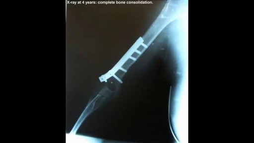
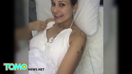
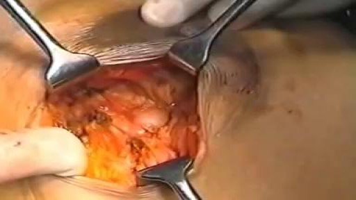
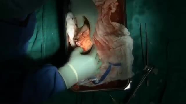
A case of replantation of a completely amputated arm
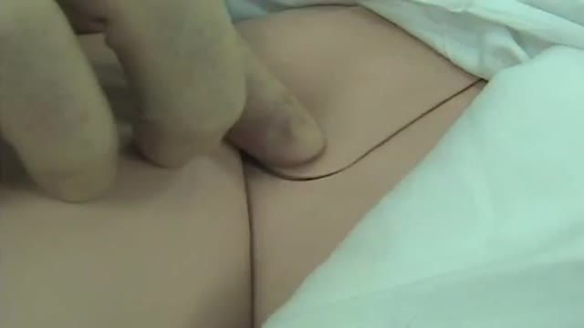
Femoral Venous Line Placement
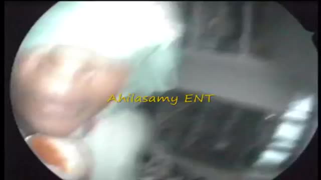
Results Sinusitis was characterized as acute in 26 patients, subacute in 5 (including 1 pyocele), and chronic in 8 (including 2 fungal infections). No tumors were found. Isolated sinus cysts were excluded from the study. Headache, the main symptom in 32 patients (82%), was localized most commonly on the vertex. Other common complaints were rhinitis, dizziness, eye symptoms, and fever. In 2 patients, the finding was occult. Eight patients (21%) presented with cranial nerve deficits, and 1 patient had an intracranial complication. Sinus irrigation was performed in 16 patients (41%) and sphenoidotomy was performed in 10 (26%). Fifteen patients (38%) were treated with antibiotic drugs alone. Within 3 months, 31 (84%) of 37 patients had recovered from the illness; 5 still experienced headaches despite having normalized radiographic findings; and 1 had permanent unilateral visual loss. Two patients were lost to follow-up.
دكتور مصطفى ياقوت فى حوار على القناة الفضائية المصرية عن الطب البديل و الطب الفرعونى فى علاج الأرق
TV interview with Dr. Mostafa Yakoot about Alternative medicine
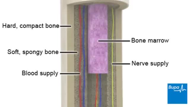
Osteoporosis is a disease in which bones become brittle and fragile due to low bone mass and bone tissue loss. It's the most common type of bone disease, according to the National Institutes of Health (NIH), and increases your risk of fractures, particularly of the hips, spine, and wrists. Prevalence In the United States, nearly 54 million people ages 50 and older were living with osteoporosis or osteopenia (low bone mass ) in 2010, according to a 2014 article in the Journal of Bone and Mineral Research. More specifically, 10.2 million adults had osteoporosis, and 43.4 million adults had osteopenia, which puts a person at high risk for osteoporosis.
Hipertension Arterial Pdf, Hipertension Esencial, Hipertension Pulmonar Tratamiento
http://bajar-presion-arterial.good-info.co
Lo Que Todos Necesitan Saber Sobre La Presión Arterial Alta
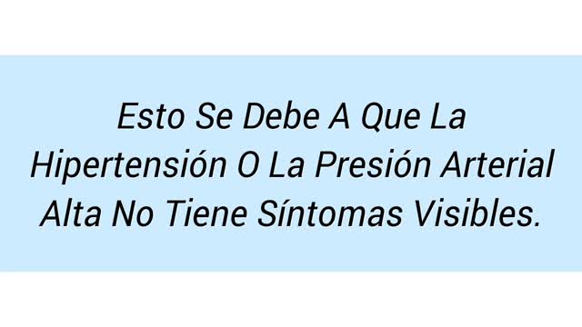
Si le han dicho que tiene presión arterial alta, usted puede decir, "Pero me siento muy bien!" Esto se debe a que la hipertensión o la presión arterial alta no tiene síntomas visibles. Es por eso que se la describe a menudo como el "asesino silencioso". No hay achaques o limitaciones físicas sólo porque tiene la presión arterial alta. Entonces, por qué siquiera preocuparse?
La hipertensión afecta a uno de cada tres adultos estadounidenses, y muchas de estas personas ni siquiera saben que la tienen. Además, aquellos con presión arterial alta tienen también un mayor riesgo de tener el colesterol alto.
Haga clic en el enlace de abajo para comprobar que funciona
http://bajar-presion-arterial.good-info.co
Suscríbete a nuestro canal
http://bajar-presion-arterial.blogspot.com/
https://www.youtube.com/watch?v=wQU4dgC1FM8
Hipertension Arterial Pdf, Hipertension Esencial, Hipertension Pulmonar Tratamiento,
hipertension renal,
hipertension en embarazo,
hipertension alimentacion,
hipertension arterial diagnostico,
hipertension arterial sistolica,
hipertension remedios naturales,
hipertension pulmonar leve,
hipertension arterial prevencion,
embarazo e hipertension,
diagnostico de hipertension,
sintomas de tension arterial alta,
remedios caseros hipertension
The goal of breast cancer surgery is to remove the entire tumor from the breast. Some of the lymph nodes in the underarm area (axillary nodes) may also be removed to see if cancer cells are present.
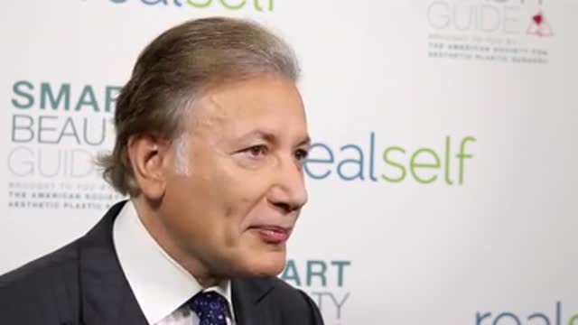
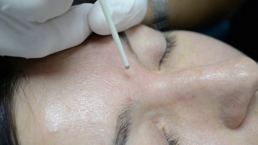
Dr. Arthur Handal discusses how injectable fillers can be used to restore a patient's youth.
Alcoholic hepatitis can occur in people who drink heavily for many years. Symptoms include yellow skin and eyes along with increasing belly size due to fluid accumulation. Treatment involves hydration, nutritional care, and stopping alcohol use. Steroid drugs can help reduce liver inflammation.
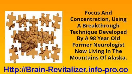
How To Increase Brain Power, How To Increase Brain Activity, How To Boost Your Concentration--- http://brain-revitalizer.info-pro.co --- Improve Concentration, Ask anyone today what they have a hard time with and more often than not they are likely to cite trouble focusing or concentrating. With laptops, smart phones, television, GPS systems and social media, it is hard to block out all the distractions when you need to focus on something specific. Attention deficit disorder may be one of the most popular excuses for our lack of concentration, but it's not an entirely accurate diagnosis since many of us simply aren't training our brains the right way to improve our focus. Concentration is necessary just to make it through a typical workday. From driving and working to shopping and cooking, focus is essential in order to get tasks accomplished. Unfortunately many of us have a hard time zoning in on a specific project and seeing it through to completion. Being easily distracted is not necessarily a disorder but it will keep you from managing your time wisely and getting done the important things that need your attention. There are several ways people can improve their concentration, including strategic brain games and exercises, along with guided meditation and brainwave entrainment. Research has shown that the more a person trains their brain in specific skill sets, the better one can get at cognitive thinking, problem solving and memorization. Using computer generated pulses, or isochronic tones, during brain entrainment meditation is another way to synchronize your brain's frequencies so you can operate on a fast or slow frequency that will assist in achieving states of productivity or relaxation. As we all know, when we are relaxed our concentration and decision making are much better than if we are under stress or hurried. Being able to tap in to specific Alpha, Theta and Delta waves allows us to synchronize our brainwaves with sound pulses so we can be more focused on specific projects and tasks. But YOU can be different! You can use Genius Brain Power to empower your brain so that you come alive with more energy, learn quicker, think more creatively, focus on your work like never before and drastically reduce stress with amazingly deep states of relaxation and meditation. click here: http://brain-revitalizer.info-pro.co

Vesicoureteral (ves-ih-koe-yoo-REE-tur-ul) reflux is the abnormal flow of urine from your bladder back up the tubes (ureters) that connect your kidneys to your bladder. Normally, urine flows only down from your kidneys to your bladder. Vesicoureteral reflux is usually diagnosed in infants and children. The disorder increases the risk of urinary tract infections, which, if left untreated, can lead to kidney damage. Vesicoureteral reflux can be primary or secondary. Children with primary vesicoureteral reflux are born with a defect in the valve that normally prevents urine from flowing backward from the bladder into the ureters. Secondary vesicoureteral reflux is due to a urinary tract malfunction, often caused by infection. Children may outgrow primary vesicoureteral reflux. Treatment, which includes medication or surgery, aims at preventing kidney damage.
When you get a kidney transplant, a healthy kidney is placed inside your body to do the work your own kidneys can no longer do. On the plus side, there are fewer limits on what you can eat and drink, but you should follow a heart-healthy diet. Your health and energy should improve. In fact, a successful kidney transplant may allow you to live the kind of life you were living before you got kidney disease. Studies show that people with kidney transplants live longer than those who remain on dialysis. On the minus side, there are the risks of surgery. You will also need to take anti-rejection medicines for as long as your new kidney is working, which can have side effects. You will have a higher risk for infections and certain types of cancer.
Vertigo is a sensation of spinning. If you have these dizzy spells, you might feel like you are spinning or that the world around you is spinning. Causes of Vertigo Vertigo is often caused by an inner ear problem. Some of the most common causes include: BPPV. These initials stand for benign paroxysmal positional vertigo. BPPV occurs when tiny calcium particles (canaliths) clump up in canals of the inner ear. The inner ear sends signals to the brain about head and body movements relative to gravity. It helps you keep your balance. BPPV can occur for no known reason and may be associated with age. Meniere's disease. This is an inner ear disorder thought to be caused by a buildup of fluid and changing pressure in the ear. It can cause episodes of vertigo along with ringing in the ears (tinnitus) and hearing loss. Vestibular neuritis or labyrinthitis. This is an inner ear problem usually related to infection (usually viral). The infection causes inflammation in the inner ear around nerves that are important for helping the body sense balance
How To Breastfeed - Deep Latch Technique
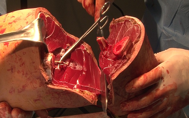
Watch that Above Knee Amputation Surgery video

A seizure occurs when there’s abnormal electrical activity in the brain. Seizures may go virtually unnoticed. Or, in severe cases, they may produce a change or loss of consciousness and involuntary muscle spasms called convulsions. Seizures usually come on suddenly and vary in duration and severity. A seizure may be a one-time event, or you may have seizures repeatedly. Recurrent seizures are called epilepsy, or a seizure disorder. Less than one in 10 people who has a seizure develops epilepsy. Experts classify seizures into two general categories and many subtypes based on the pattern of the attack. Generalized seizures involve both sides of the brain from the start of the attack. Common subtypes include tonic-clonic (grand mal) and absence seizures (petit mal). Febrile and infantile spasms are two types of generalized seizures that occur almost exclusively in young children. Partial (or focal) seizures are the second major seizure type. These begin in a specific area of the brain and may be contained there. Or they may spread to the entire brain. With simple partial seizures, the person remains conscious. Complex partial seizures involve impaired consciousness. What Causes Seizures? Often the cause of a seizure is unknown. Many conditions can provoke seizures, including: Stroke Brain tumors Head injuries Electrolyte imbalance Very low blood sugar Repetitive sounds or flashing lights, such as in video games Medications, such as antipsychotics and some asthma drugs Withdrawal from medications, such as Xanax, narcotics, or alcohol Use of drugs such as cocaine and heroin Cancer Brain infections, such as meningitis
Chronic obstructive pulmonary disease (COPD) is defined as progressive, chronic airflow obstruction due to chronic bronchitis, emphysema, or both. The majority of patients have components of both, although one of these entities will frequently dominate the clinical picture. Emphysema�airspace enlargement distal to the terminal bronchioles due to destruction of alveolar septa. Chronic bronchitis�chronic airway inflammation and bronchospasm. Clinically defined as productive cough lasting for at least 3 mo over 2 consecutive years. Although COPD is irreversible, patients with acute exacerbations do have reversible bronchospastic and inflammatory components.
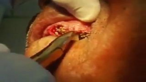
Watch that video of Removing Hundreds of Worms Living Inside Teeth