- Physical Examination
- Surgical Examination
- Ophthalmology
- Clinical Skills
- Orthopedics
- Surgery Videos
- Laparoscopy
- Pediatrics
- Funny Videos
- Cardiothoracic Surgery
- Nursing Videos
- Plastic Surgery
- Otorhinolaryngology
- Histology and Histopathology
- Neurosurgery
- Dermatology
- Pediatric Surgery
- Urology
- Dentistry
- Oncology and Cancers
- Anatomy Videos
- Health and Fitness
- Radiology
- Anaesthesia
- Physical Therapy
- Pharmacology
- Interventional Radiology
- Cardiology
- Endocrinology
- Gynecology
- Emergency Medicine
- Psychiatry and Psychology
- Childbirth Videos
- General Medical Videos
- Nephrology
- Physiology
- Diet and Food Health
- Diabetes Mellitus
- Neurology
- Women Health
- Osteoporosis
- Gastroenterology
- Pulmonology
- Hematology
- Rheumatology
- Toxicology
- Nuclear Medicine
- Infectious Diseases
- Vascular Disease
- Reproductive Health
- Burns and Wound Healing
- Other
Top videos
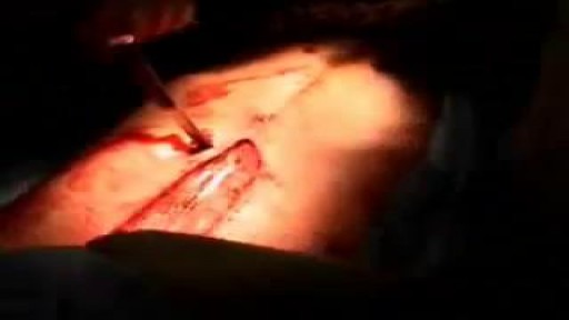
Watch that video of a Knife Stabbed Inside Chest Removing Surgery
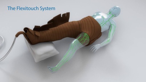
Treating Lymphedema -
Plastic Surgery New York Dr. Carlin Vickery of 5th Avenue Aesthetics Surgery in Manhattan
(http://www.5thavesurgery.com) speaks at a Fab Over 50 event on having great breasts after the age
of 50. In this presentation Dr. Carlin shares patient results by providing before and after pictures from
different types of breast surgeries including breast lifts, implants and reductions.
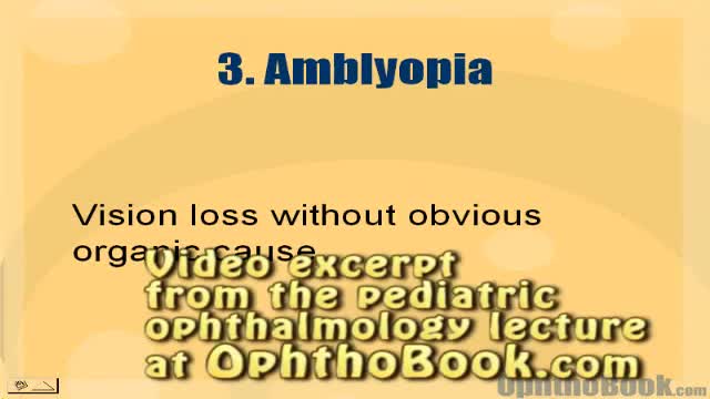
How amblyopia develops in children. Basically, if one eye doesn't see well from an early age, the wiring never forms correctly back to the occipital cortex.
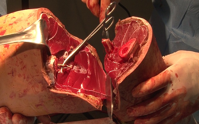
Watch that Above Knee Amputation Surgery video
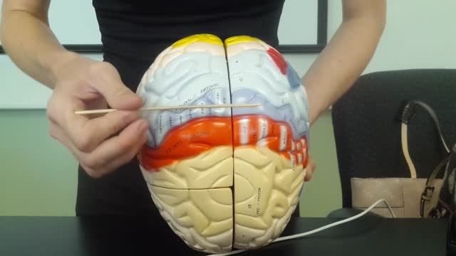
The brain is that part of the CNS contained within the cranial cavity (figure 13.1). It is the control center for many of the body's functions. The brain is much like a complex central computer but with additional functions that no computer can as yet match. Indeed, one goal in computer technology is to make computers that can function more like the human brain. The brain consists of the brainstem, the cerebellum, the diencephalon, and the cerebrum (table 13.1). The brainstem includes the medulla oblongata, pons, midbrain, and reticular formation. The structure of the brain is described in this chapter. Its functions are primarily discussed in chapter 14. Twelve pairs of cranial nerves, which are part of the PNS, arise directly from the brain. Two pairs arise from the cerebrum, nine pairs arise from the brainstem, and one pair arises from the spinal cord.
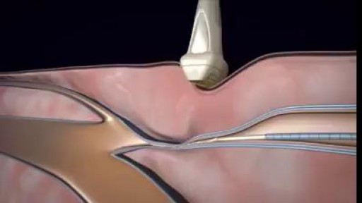
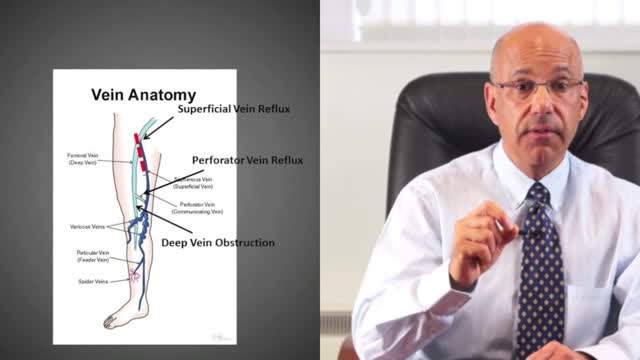
3D animation video of Varicose Veins Sclerotherapy Treatment

Lichen sclerosus is a skin condition that mainly affects the genital skin (vulva) in women and the penis in men. It most commonly occurs in middle-aged women. Symptoms may include itch, soreness, and changes in the appearance of affected skin.
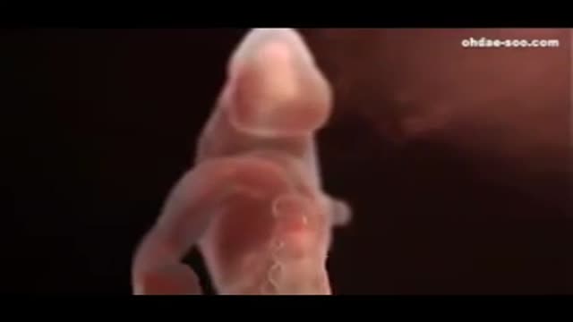
0 to 9 Months Journey In The Womb
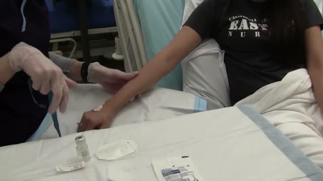
IV cannulation is a skill that has scared a lot of student nurses and even professionals. Perhaps it’s because IV insertion is an invasive procedure, and nurses are too worried that they might hurt their patients. Or maybe it’s because they are just clueless about IV therapy do’s and don’ts–things that one can only fully understand through constant practice.
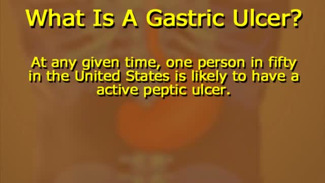
What Causes Ulcers? No single cause has been found for ulcers. However, it is now clear that an ulcer is the end result of an imbalance between digestive fluids in the stomach and duodenum. Most ulcers are caused by an infection with a type of bacteria called Helicobacter pylori (H. pylori). Factors that can increase your risk for ulcers include: Use of painkillers called nonsteroidal anti-inflammatory drugs (NSAIDs), such as aspirin, naproxen (Aleve, Anaprox, Naprosyn, and others), ibuprofen (Motrin, Advil, some types of Midol, and others), and many others available by prescription; even safety-coated aspirin and aspirin in powered form can frequently cause ulcers. Excess acid production from gastrinomas, tumors of the acid producing cells of the stomach that increases acid output (seen in Zollinger-Ellison syndrome) Excessive drinking of alcohol Smoking or chewing tobacco Serious illness Radiation treatment to the area What Are the Symptoms of an Ulcer? An ulcer may or may not have symptoms. When symptoms occur, they may include: A gnawing or burning pain in the middle or upper stomach between meals or at night Bloating Heartburn Nausea or vomiting In severe cases, symptoms can include: Dark or black stool (due to bleeding) Vomiting blood (that can look like "coffee-grounds") Weight loss Severe pain in the mid to upper abdomen
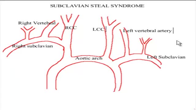
The term subclavian steal describes retrograde blood flow in the vertebral artery associated with proximal ipsilateral subclavian artery stenosis or occlusion, usually in the setting of subclavian artery occlusion or stenosis proximal to the origin of the vertebral artery. Alternatively, innominate artery disease has also been associated with retrograde flow in the ipsilateral vertebral artery, particularly where the subclavian artery origin is involved. Subclavian steal is frequently asymptomatic and may be discovered incidentally on ultrasound or angiographic examination for other indications, or it may be prompted by a clinical examination finding of reduced unilateral upper limb pulse or blood pressure. In some cases, patients may develop upper limb ischemic symptoms due to reduced arterial flow in the setting of subclavian artery occlusion, or they may develop neurologic symptoms due to posterior circulation ischemia associated with exercise of the ipsilateral arm.[1] Treatment has traditionally consisted of open subclavian artery revascularization, typically via carotid-subclavian bypass or subclavian artery transposition, which are generally durable procedures. Newer, less invasive options include endovascular intervention with recanalization as appropriate and angioplasty and stenting if required. The clinical relevance of subclavian steal was described in 1961 by Reivich, Holling and Roberts; however, the recognition of retrograde vertebral artery flow dates back another 100 years to Harrison and Smyth. Some papers, including a previous version of this article, advocate restricting the term subclavian steal to patients with neurologic symptoms only, but this is incorrect in view of the substantial literature using this term to describe the hemodynamic scenario of retrograde vertebral flow and proximal subclavian artery disease.
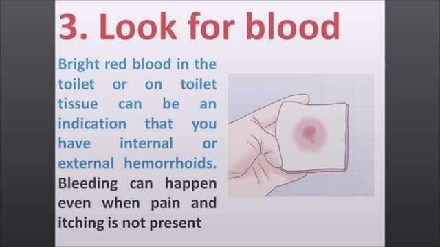
Most of the time, treatment for hemorrhoids involves steps that you can take on your own, such as lifestyle modifications. But sometimes medications or surgical procedures are necessary. Medications If your hemorrhoids produce only mild discomfort, your doctor may suggest over-the-counter creams, ointments, suppositories or pads. These products contain ingredients, such as witch hazel or hydrocortisone, that can relieve pain and itching, at least temporarily. Don't use an over-the-counter cream or other product for more than a week unless directed by your doctor. These products can cause side effects, such as skin rash, inflammation and skin thinning. Minimally invasive procedures If a blood clot has formed within an external hemorrhoid, your doctor can remove the clot with a simple incision, which may provide prompt relief. For persistent bleeding or painful hemorrhoids, your doctor may recommend another minimally invasive procedure. These treatments can be done in your doctor's office or other outpatient setting. Rubber band ligation. Your doctor places one or two tiny rubber bands around the base of an internal hemorrhoid to cut off its circulation. The hemorrhoid withers and falls off within a week. This procedure — called rubber band ligation — is effective for many people. Hemorrhoid banding can be uncomfortable and may cause bleeding, which might begin two to four days after the procedure but is rarely severe. Injection (sclerotherapy). In this procedure, your doctor injects a chemical solution into the hemorrhoid tissue to shrink it. While the injection causes little or no pain, it may be less effective than rubber band ligation. Coagulation (infrared, laser or bipolar). Coagulation techniques use laser or infrared light or heat. They cause small, bleeding, internal hemorrhoids to harden and shrivel. While coagulation has few side effects, it's associated with a higher rate of hemorrhoids coming back (recurrence) than is the rubber band treatment. Surgical procedures If other procedures haven't been successful or you have large hemorrhoids, your doctor may recommend a surgical procedure. Surgery can be performed on an outpatient basis or you may need to stay in the hospital overnight. Hemorrhoid removal. During a hemorrhoidectomy, your surgeon removes excessive tissue that causes bleeding. Various techniques may be used. The surgery may be done with a local anesthetic combined with sedation, a spinal anesthetic or a general anesthetic. Hemorrhoidectomy is the most effective and complete way to treat severe or recurring hemorrhoids. Complications may include temporary difficulty emptying your bladder and urinary tract infections associated with this problem. Most people experience some pain after the procedure. Medications can relieve your pain. Soaking in a warm bath also may help. Hemorrhoid stapling. This procedure, called stapled hemorrhoidectomy or stapled hemorrhoidopexy, blocks blood flow to hemorrhoidal tissue. Stapling generally involves less pain than hemorrhoidectomy and allows an earlier return to regular activities. Compared with hemorrhoidectomy, however, stapling has been associated with a greater risk of recurrence and rectal prolapse, in which part of the rectum protrudes from the anus. Talk with your doctor about what might be the best option for you.
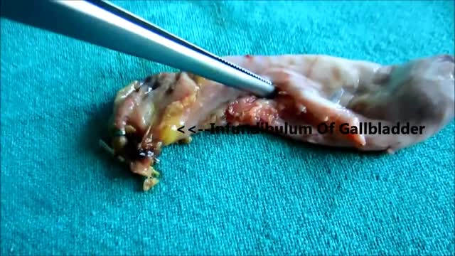
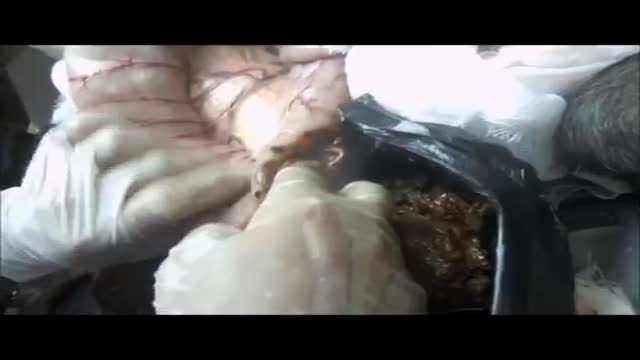
Watch Inside Of Gall bladder - Stone Removed
A leg ulcer is simply a break in the skin of the leg, which allows air and bacteria to get into the underlying tissue. This is usually caused by an injury, often a minor one that breaks the skin. In most people such an injury will heal up without difficulty within a week or two. However, when there is an underlying problem the skin does not heal and the area of breakdown can increase in size. This is a chronic leg ulcer.
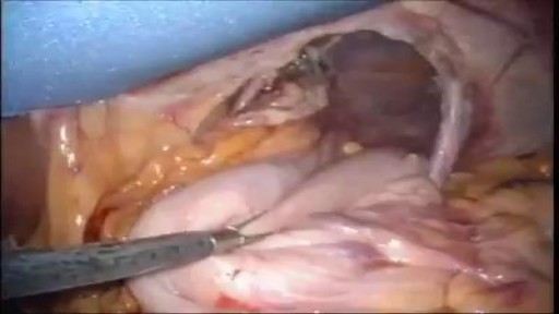
A hiatal hernia occurs when the upper part of the stomach pushes through an opening in the diaphragm and into the chest cavity. The diaphragm is the thin muscle wall that separates the chest cavity from the abdomen. The opening in the diaphragm is where the esophagus and stomach join.
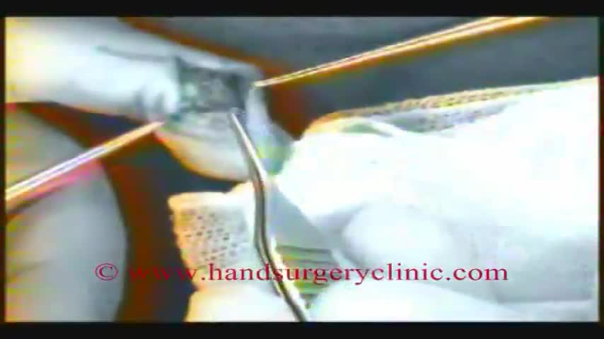
Glomus tumors are rare soft tissue neoplasms that typically present in adults (ages 20-40 years) as small, blue-red papules or nodules of the distal extremities, with most cases involving subungual sites. These tumors are typically painful, often causing paroxysmal pain in response to temperature changes or pressure. Glomus tumors are thought to arise from the glomus body, a thermoregulatory shunt concentrated in the fingers and toes. Most lesions are solitary and localized to cutaneous sites; however, generalized glomuvenous malformations, or multiple glomangiomas, have also been described, and may have extracutaneous involvement.
Megacolon, as well as megarectum, is a descriptive term. It denotes dilatation of the colon that is not caused by mechanical obstruction.[1, 2] Although the definition of megacolon has varied in the literature, most researchers use the measurement of greater than 12 cm for the cecum as the standard. Because the diameter of the large intestine varies, the following definitions would also be considered: greater than 6.5 cm in the rectosigmoid region and greater than 8 cm for the ascending colon. Megacolon can be divided into the following 3 categories: Acute megacolon ( pseudo-obstruction) Chronic megacolon, which includes congenital, acquired, and idiopathic causes Toxic megacolon
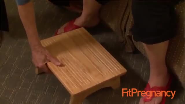
How To Breastfeed - Deep Latch Technique

#dialysis #hemodialysis #kidneys
Follow us: https://www.instagram.com/7activestudio/
For more information:
www.7activestudio.com
7activestudio@gmail.com
Contact: +91- 9700061777, 040-66564777
7 Active Technology Solutions Pvt.Ltd. is an educational 3D digital content provider for K-12. We also customise the content as per your requirement for companies platform providers colleges etc . 7 Active driving force "The Joy of Happy Learning" -- is what makes difference from other digital content providers. We consider Student needs, Lecturer needs and College needs in designing the 3D & 2D Animated Video Lectures. We are carrying a huge 3D Digital Library ready to use.
Voice Over Credit: Muls N Ravs Entertainment Pvt. Ltd. (www.mulsnravs.com)
