Principais vídeos
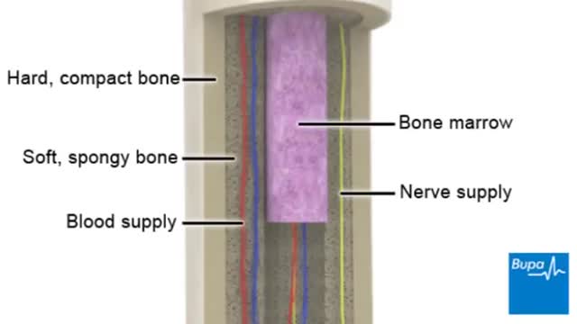
Osteoporosis is a disease in which bones become brittle and fragile due to low bone mass and bone tissue loss. It's the most common type of bone disease, according to the National Institutes of Health (NIH), and increases your risk of fractures, particularly of the hips, spine, and wrists. Prevalence In the United States, nearly 54 million people ages 50 and older were living with osteoporosis or osteopenia (low bone mass ) in 2010, according to a 2014 article in the Journal of Bone and Mineral Research. More specifically, 10.2 million adults had osteoporosis, and 43.4 million adults had osteopenia, which puts a person at high risk for osteoporosis.

Femoral Venous Line Placement

دكتور مصطفى ياقوت فى حوار على القناة الفضائية المصرية عن الطب البديل و الطب الفرعونى فى علاج الأرق
TV interview with Dr. Mostafa Yakoot about Alternative medicine
Learn one of the hidden reasons why you still have a thyroid symptoms. If your lab results are "normal"--then why do you still have thyroid symptoms like: * Tired, sluggish * Can't lose weight even with exercise * Feel cold—hands, feet, or all over * Require excessive amounts of sleep to function properly * Increase in weight gain even with low-calorie diet * Gain weight easily * Difficult, infrequent bowel movements * Depression, lack of motivation * Morning headaches that wear off as the day progresses * Outer third of eyebrow thins * Thinning of hair on scalp, face or genitals or hair loss * Dryness of skin and/or scalp * Mental sluggishness * Nervousness and emotional * Insomnia * Night sweats
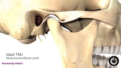
What Causes TMD? We don’t know what causes TMD. Dentists believe symptoms arise from problems with the muscles of your jaw or with the parts of the joint itself. Injury to your jaw, the joint, or the muscles of your head and neck -- like from a heavy blow or whiplash -- can lead to TMD. Other causes include: Grinding or clenching your teeth, which puts a lot of pressure on the joint Movement of the soft cushion or disc between the ball and socket of the joint Arthritis in the joint Stress, which can cause you to tighten facial and jaw muscles or clench the teeth

In most people, post-concussion syndrome symptoms occur within the first seven to 10 days and go away within three months, though they can persist for a year or more. Post-concussion syndrome treatments are aimed at easing specific symptoms.

Diabetic retinopathy involves changes to retinal blood vessels that can cause them to bleed or leak fluid, distorting vision. Diabetic retinopathy is the most common cause of vision loss among people with diabetes and a leading cause of blindness among working-age adults.

How surgeons remove kidney stones كيف يقوم الجراحون بإزالة حصى الكلى see more http://www.kidneymy.com/
Hipertension Arterial Pdf, Hipertension Esencial, Hipertension Pulmonar Tratamiento
http://bajar-presion-arterial.good-info.co
Lo Que Todos Necesitan Saber Sobre La Presión Arterial Alta
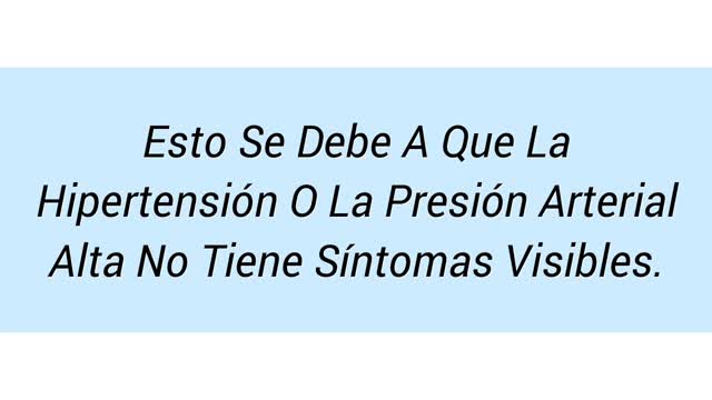
Si le han dicho que tiene presión arterial alta, usted puede decir, "Pero me siento muy bien!" Esto se debe a que la hipertensión o la presión arterial alta no tiene síntomas visibles. Es por eso que se la describe a menudo como el "asesino silencioso". No hay achaques o limitaciones físicas sólo porque tiene la presión arterial alta. Entonces, por qué siquiera preocuparse?
La hipertensión afecta a uno de cada tres adultos estadounidenses, y muchas de estas personas ni siquiera saben que la tienen. Además, aquellos con presión arterial alta tienen también un mayor riesgo de tener el colesterol alto.
Haga clic en el enlace de abajo para comprobar que funciona
http://bajar-presion-arterial.good-info.co
Suscríbete a nuestro canal
http://bajar-presion-arterial.blogspot.com/
https://www.youtube.com/watch?v=wQU4dgC1FM8
Hipertension Arterial Pdf, Hipertension Esencial, Hipertension Pulmonar Tratamiento,
hipertension renal,
hipertension en embarazo,
hipertension alimentacion,
hipertension arterial diagnostico,
hipertension arterial sistolica,
hipertension remedios naturales,
hipertension pulmonar leve,
hipertension arterial prevencion,
embarazo e hipertension,
diagnostico de hipertension,
sintomas de tension arterial alta,
remedios caseros hipertension
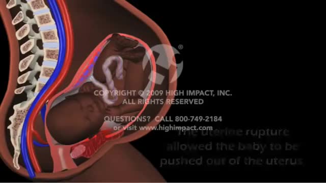
Uterine rupture is usually when the scar from your previous caesarean section tears open. Though it's uncommon, you should be aware of this risk, particularly if you're thinking about giving birth vaginally next time. It's possible for your scar to gape slightly while you're pregnant (scar dehiscence).
Watch that Above Knee Amputation Surgery video

Dr. Arthur Handal discusses how injectable fillers can be used to restore a patient's youth.
When you get a kidney transplant, a healthy kidney is placed inside your body to do the work your own kidneys can no longer do. On the plus side, there are fewer limits on what you can eat and drink, but you should follow a heart-healthy diet. Your health and energy should improve. In fact, a successful kidney transplant may allow you to live the kind of life you were living before you got kidney disease. Studies show that people with kidney transplants live longer than those who remain on dialysis. On the minus side, there are the risks of surgery. You will also need to take anti-rejection medicines for as long as your new kidney is working, which can have side effects. You will have a higher risk for infections and certain types of cancer.

Vesicoureteral (ves-ih-koe-yoo-REE-tur-ul) reflux is the abnormal flow of urine from your bladder back up the tubes (ureters) that connect your kidneys to your bladder. Normally, urine flows only down from your kidneys to your bladder. Vesicoureteral reflux is usually diagnosed in infants and children. The disorder increases the risk of urinary tract infections, which, if left untreated, can lead to kidney damage. Vesicoureteral reflux can be primary or secondary. Children with primary vesicoureteral reflux are born with a defect in the valve that normally prevents urine from flowing backward from the bladder into the ureters. Secondary vesicoureteral reflux is due to a urinary tract malfunction, often caused by infection. Children may outgrow primary vesicoureteral reflux. Treatment, which includes medication or surgery, aims at preventing kidney damage.

Vertigo is a sensation of spinning. If you have these dizzy spells, you might feel like you are spinning or that the world around you is spinning. Causes of Vertigo Vertigo is often caused by an inner ear problem. Some of the most common causes include: BPPV. These initials stand for benign paroxysmal positional vertigo. BPPV occurs when tiny calcium particles (canaliths) clump up in canals of the inner ear. The inner ear sends signals to the brain about head and body movements relative to gravity. It helps you keep your balance. BPPV can occur for no known reason and may be associated with age. Meniere's disease. This is an inner ear disorder thought to be caused by a buildup of fluid and changing pressure in the ear. It can cause episodes of vertigo along with ringing in the ears (tinnitus) and hearing loss. Vestibular neuritis or labyrinthitis. This is an inner ear problem usually related to infection (usually viral). The infection causes inflammation in the inner ear around nerves that are important for helping the body sense balance
How To Breastfeed - Deep Latch Technique

A seizure occurs when there’s abnormal electrical activity in the brain. Seizures may go virtually unnoticed. Or, in severe cases, they may produce a change or loss of consciousness and involuntary muscle spasms called convulsions. Seizures usually come on suddenly and vary in duration and severity. A seizure may be a one-time event, or you may have seizures repeatedly. Recurrent seizures are called epilepsy, or a seizure disorder. Less than one in 10 people who has a seizure develops epilepsy. Experts classify seizures into two general categories and many subtypes based on the pattern of the attack. Generalized seizures involve both sides of the brain from the start of the attack. Common subtypes include tonic-clonic (grand mal) and absence seizures (petit mal). Febrile and infantile spasms are two types of generalized seizures that occur almost exclusively in young children. Partial (or focal) seizures are the second major seizure type. These begin in a specific area of the brain and may be contained there. Or they may spread to the entire brain. With simple partial seizures, the person remains conscious. Complex partial seizures involve impaired consciousness. What Causes Seizures? Often the cause of a seizure is unknown. Many conditions can provoke seizures, including: Stroke Brain tumors Head injuries Electrolyte imbalance Very low blood sugar Repetitive sounds or flashing lights, such as in video games Medications, such as antipsychotics and some asthma drugs Withdrawal from medications, such as Xanax, narcotics, or alcohol Use of drugs such as cocaine and heroin Cancer Brain infections, such as meningitis
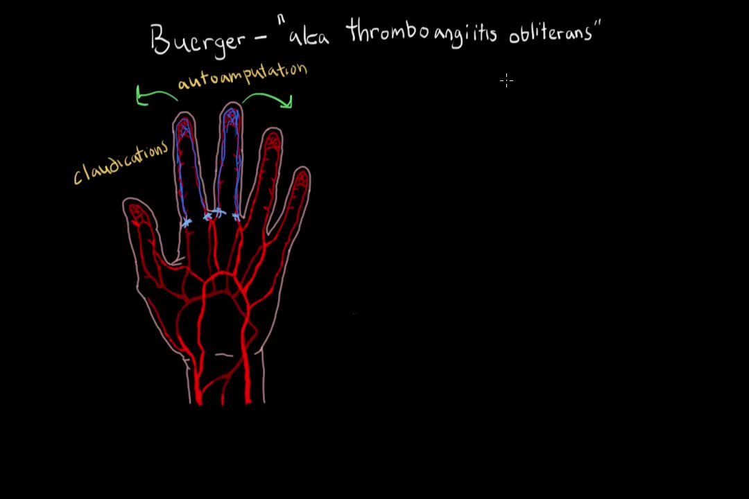
Buerger's disease (thromboangiitis obliterans) is a rare disease of the arteries and veins in the arms and legs. In Buerger's disease, your blood vessels become inflamed, swell and can become blocked with blood clots (thrombi). This eventually damages or destroys skin tissues and may lead to infection and gangrene. Buerger's disease usually first shows in your hands and feet and may eventually affect larger areas of your arms and legs. Virtually everyone diagnosed with Buerger's disease smokes cigarettes or uses other forms of tobacco, such as chewing tobacco. Quitting all forms of tobacco is the only way to stop Buerger's disease. For those who don't quit, amputation of all or part of a limb is sometimes necessary.

Iron is a mineral that plays a vital role in health and well-being. Without it, many bodily functions would malfunction. “The primary role of iron is to carry oxygen in the blood to every cell in the body,” says Beth Thayer, RDN, MS, director of the Center for Health Promotion and Disease Prevention at Henry Ford Health System in Detroit. Iron is an important component of hemoglobin, the protein in red blood cells that carries oxygen from the lungs and transports it throughout the body.

Gunshot wounds have become increasing common in urban cities and many such cases can lead to undesirable outcomes. While gunshot wounds to the head are considered most lethal, gunshot wounds to the chest too may be dangerous. Gunshot wound to the chest is challenging owing to the presence of vital organs like lungs, heart and their surrounding structures including major blood vessels. Gunshot wound is caused by penetration of the bullet, which travels through a projectile path after being shot from a firearm. The bullet, on hitting the chest, punctures the tissue it first encounters with, the bones or the muscular chest wall. The extent and severity of the injury depends on the characteristics of the bullet and the firearm, the position and the distance of the victim, the projectile path and the nature of the tissue penetrated.
