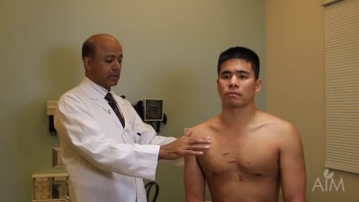
- Physical Examination
- Surgical Examination
- Ophthalmology
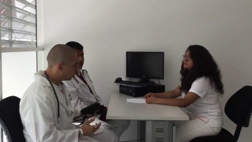
- Clinical Skills
- Orthopedics
- Surgery Videos
- Laparoscopy
- Pediatrics
- Funny Videos
- Cardiothoracic Surgery
- Nursing Videos
- Plastic Surgery
- Otorhinolaryngology
- Histology and Histopathology
- Neurosurgery
- Dermatology
- Pediatric Surgery
- Urology
- Dentistry
- Oncology and Cancers
- Anatomy Videos
- Health and Fitness
- Radiology
- Anaesthesia
- Physical Therapy
- Pharmacology
- Interventional Radiology
- Cardiology
- Endocrinology
- Gynecology
- Emergency Medicine
- Psychiatry and Psychology
- Childbirth Videos
- General Medical Videos
- Nephrology
- Physiology
- Diet and Food Health
- Diabetes Mellitus
- Neurology
- Women Health
- Osteoporosis
- Gastroenterology
- Pulmonology
- Hematology
- Rheumatology
- Toxicology
- Nuclear Medicine
- Infectious Diseases
- Vascular Disease
- Reproductive Health
- Burns and Wound Healing
- Other
Top videos

Dr. Arthur Handal discusses how injectable fillers can be used to restore a patient's youth.

Iron is a mineral that plays a vital role in health and well-being. Without it, many bodily functions would malfunction. “The primary role of iron is to carry oxygen in the blood to every cell in the body,” says Beth Thayer, RDN, MS, director of the Center for Health Promotion and Disease Prevention at Henry Ford Health System in Detroit. Iron is an important component of hemoglobin, the protein in red blood cells that carries oxygen from the lungs and transports it throughout the body.
Chronic obstructive pulmonary disease (COPD) is defined as progressive, chronic airflow obstruction due to chronic bronchitis, emphysema, or both. The majority of patients have components of both, although one of these entities will frequently dominate the clinical picture. Emphysema�airspace enlargement distal to the terminal bronchioles due to destruction of alveolar septa. Chronic bronchitis�chronic airway inflammation and bronchospasm. Clinically defined as productive cough lasting for at least 3 mo over 2 consecutive years. Although COPD is irreversible, patients with acute exacerbations do have reversible bronchospastic and inflammatory components.

Vesicoureteral (ves-ih-koe-yoo-REE-tur-ul) reflux is the abnormal flow of urine from your bladder back up the tubes (ureters) that connect your kidneys to your bladder. Normally, urine flows only down from your kidneys to your bladder. Vesicoureteral reflux is usually diagnosed in infants and children. The disorder increases the risk of urinary tract infections, which, if left untreated, can lead to kidney damage. Vesicoureteral reflux can be primary or secondary. Children with primary vesicoureteral reflux are born with a defect in the valve that normally prevents urine from flowing backward from the bladder into the ureters. Secondary vesicoureteral reflux is due to a urinary tract malfunction, often caused by infection. Children may outgrow primary vesicoureteral reflux. Treatment, which includes medication or surgery, aims at preventing kidney damage.

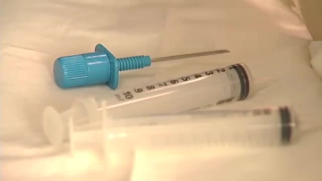
Thoracentesis is a procedure to remove fluid or air from around the lungs. A needle is put through the chest wall into the pleural space. The pleural space is the thin gap between the pleura. The pleura are a double layer of membranes that surrounds the lungs.
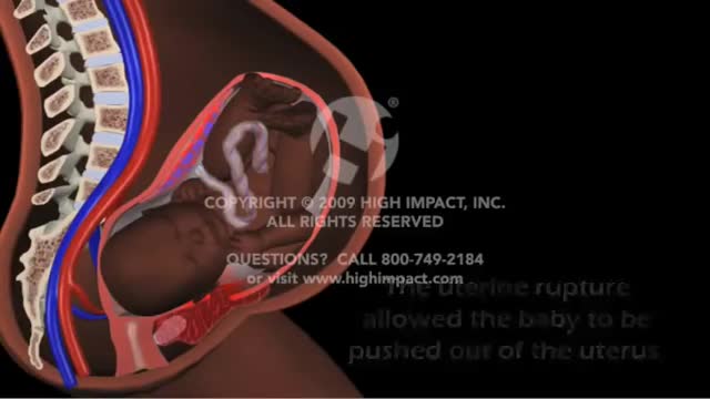
Uterine rupture is usually when the scar from your previous caesarean section tears open. Though it's uncommon, you should be aware of this risk, particularly if you're thinking about giving birth vaginally next time. It's possible for your scar to gape slightly while you're pregnant (scar dehiscence).
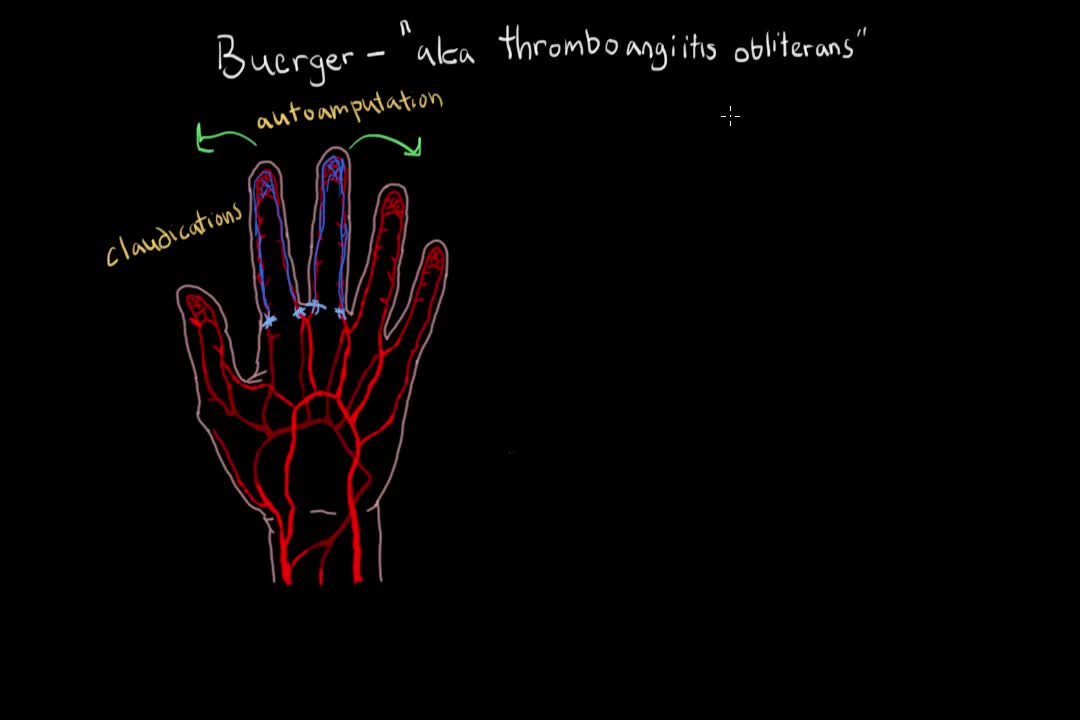
Buerger's disease (thromboangiitis obliterans) is a rare disease of the arteries and veins in the arms and legs. In Buerger's disease, your blood vessels become inflamed, swell and can become blocked with blood clots (thrombi). This eventually damages or destroys skin tissues and may lead to infection and gangrene. Buerger's disease usually first shows in your hands and feet and may eventually affect larger areas of your arms and legs. Virtually everyone diagnosed with Buerger's disease smokes cigarettes or uses other forms of tobacco, such as chewing tobacco. Quitting all forms of tobacco is the only way to stop Buerger's disease. For those who don't quit, amputation of all or part of a limb is sometimes necessary.

Vertigo is a sensation of spinning. If you have these dizzy spells, you might feel like you are spinning or that the world around you is spinning. Causes of Vertigo Vertigo is often caused by an inner ear problem. Some of the most common causes include: BPPV. These initials stand for benign paroxysmal positional vertigo. BPPV occurs when tiny calcium particles (canaliths) clump up in canals of the inner ear. The inner ear sends signals to the brain about head and body movements relative to gravity. It helps you keep your balance. BPPV can occur for no known reason and may be associated with age. Meniere's disease. This is an inner ear disorder thought to be caused by a buildup of fluid and changing pressure in the ear. It can cause episodes of vertigo along with ringing in the ears (tinnitus) and hearing loss. Vestibular neuritis or labyrinthitis. This is an inner ear problem usually related to infection (usually viral). The infection causes inflammation in the inner ear around nerves that are important for helping the body sense balance
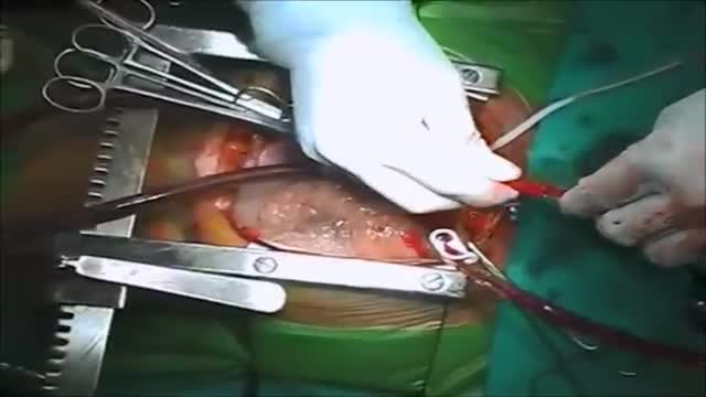
Ventricular septal rupture (VSR) is a rare but lethal complication of myocardial infarction (MI). The event occurs 2-8 days after an infarction and often precipitates cardiogenic shock. [1] The differential diagnosis of postinfarction cardiogenic shock should exclude free ventricular wall rupture and rupture of the papillary muscles. (See the image below.)

How To Breastfeed - Deep Latch Technique

A seizure occurs when there’s abnormal electrical activity in the brain. Seizures may go virtually unnoticed. Or, in severe cases, they may produce a change or loss of consciousness and involuntary muscle spasms called convulsions. Seizures usually come on suddenly and vary in duration and severity. A seizure may be a one-time event, or you may have seizures repeatedly. Recurrent seizures are called epilepsy, or a seizure disorder. Less than one in 10 people who has a seizure develops epilepsy. Experts classify seizures into two general categories and many subtypes based on the pattern of the attack. Generalized seizures involve both sides of the brain from the start of the attack. Common subtypes include tonic-clonic (grand mal) and absence seizures (petit mal). Febrile and infantile spasms are two types of generalized seizures that occur almost exclusively in young children. Partial (or focal) seizures are the second major seizure type. These begin in a specific area of the brain and may be contained there. Or they may spread to the entire brain. With simple partial seizures, the person remains conscious. Complex partial seizures involve impaired consciousness. What Causes Seizures? Often the cause of a seizure is unknown. Many conditions can provoke seizures, including: Stroke Brain tumors Head injuries Electrolyte imbalance Very low blood sugar Repetitive sounds or flashing lights, such as in video games Medications, such as antipsychotics and some asthma drugs Withdrawal from medications, such as Xanax, narcotics, or alcohol Use of drugs such as cocaine and heroin Cancer Brain infections, such as meningitis

Gunshot wounds have become increasing common in urban cities and many such cases can lead to undesirable outcomes. While gunshot wounds to the head are considered most lethal, gunshot wounds to the chest too may be dangerous. Gunshot wound to the chest is challenging owing to the presence of vital organs like lungs, heart and their surrounding structures including major blood vessels. Gunshot wound is caused by penetration of the bullet, which travels through a projectile path after being shot from a firearm. The bullet, on hitting the chest, punctures the tissue it first encounters with, the bones or the muscular chest wall. The extent and severity of the injury depends on the characteristics of the bullet and the firearm, the position and the distance of the victim, the projectile path and the nature of the tissue penetrated.
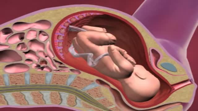
If you have placenta previa, it means that your placenta is lying unusually low in your uterus, next to or covering your cervix. The placenta is the pancake-shaped organ – normally located near the top of the uterus – that supplies your baby with nutrients through the umbilical cord.
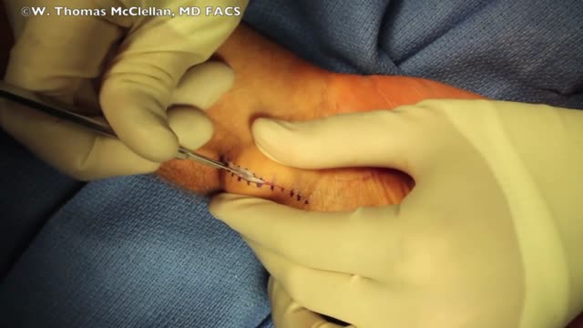
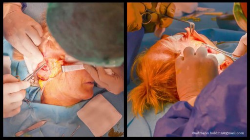
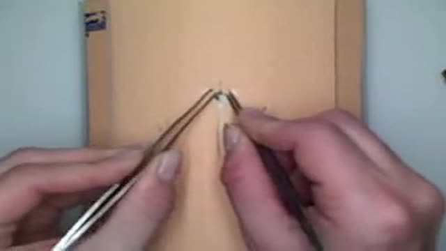
Ganglion Cyst Surgical Removal
A bone marrow biopsy is part of a bone marrow test that takes a sample of your solid bone tissue. This test looks for abnormalities in your blood cells and signs of any diseases. You can request anesthesia or a sedative before the biopsy, and manage any pain afterward with over-the-counter medications.
Demonstration of staple insertion and removal technique for laceration repair or wound closure in the operating room.

AZT Mechanism of Antiviral Activity

New device claims to stimulate brain for depression treatment
