- Physical Examination
- Surgical Examination
- Ophthalmology
- Clinical Skills
- Orthopedics
- Surgery Videos
- Laparoscopy
- Pediatrics
- Funny Videos
- Cardiothoracic Surgery
- Nursing Videos
- Plastic Surgery
- Otorhinolaryngology
- Histology and Histopathology
- Neurosurgery
- Dermatology
- Pediatric Surgery
- Urology
- Dentistry
- Oncology and Cancers
- Anatomy Videos
- Health and Fitness
- Radiology
- Anaesthesia
- Physical Therapy
- Pharmacology
- Interventional Radiology
- Cardiology
- Endocrinology
- Gynecology
- Emergency Medicine
- Psychiatry and Psychology
- Childbirth Videos
- General Medical Videos
- Nephrology
- Physiology
- Diet and Food Health
- Diabetes Mellitus
- Neurology
- Women Health
- Osteoporosis
- Gastroenterology
- Pulmonology
- Hematology
- Rheumatology
- Toxicology
- Nuclear Medicine
- Infectious Diseases
- Vascular Disease
- Reproductive Health
- Burns and Wound Healing
- Other
Top videos
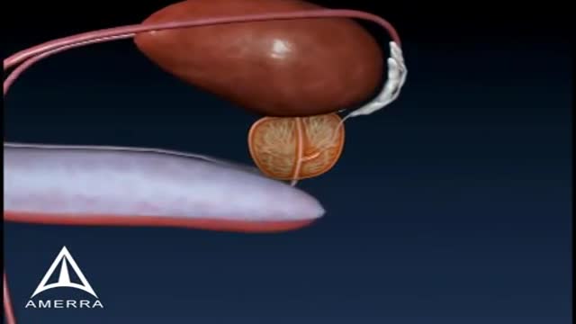
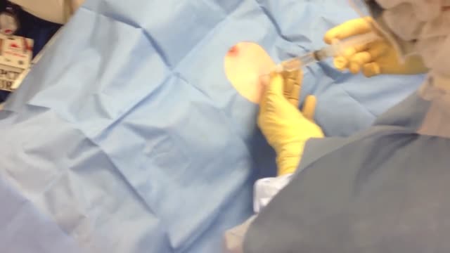
Prostate biopsy is a procedure in which small hollow needle-core samples are removed from a man's prostate gland to be examined microscopically for the presence of cancer. It is typically performed when the result from a PSA blood test rises to a level that is associated with the possible presence of prostate cancer.
Sialorrhea or excessive drooling is a major issue in children with cerebral palsy and adults with neurodegenerative disorders. In this review, we describe the clinical features, anatomy and physiology of sialorrhea, as well as a review of the world literature on medical treatment using Yale University’s search engine; including but not limited to Medline and Erasmus. Level of drug efficacy is defined according to the guidelines of American Academy of Neurology. Current medical management is unsatisfactory. Topical agents (scopolamine and tropicamide) and oral agents (glyccopyrolate) combined render a level B evidence (probably effective); however, this treatment is associated with troublesome side effects. Double-blind and placebo-controlled studies of botulinum toxin (BoNT) provide a level A evidence for type B (two class I studies; effective and established) and both overall and individual B level of evidence for OnabotulinumtoxinA (A/Ona) and AbobotulinumtoxinA (A/Abo); these are probably effective. For IncobotulinumtoxinA (A/Inco), the level of evidence is U (insufficient) due to lack of blinded studies. Side effects are uncommon; transient and comparable between the two types of toxin. A clinical note at the end of this review comments on fine clinical points. Administration of BoNTs into salivary glands is currently the most effective way of treating sialorrhea.
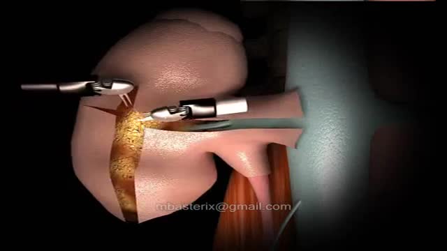
The robotic approach to renal surgery, particularly partial nephrectomy, has some inherent challenges, and some familiarity with the da Vinci robotic system is necessary. The surgeon must gain an understanding of the robotic arm movements and range of motion, especially in relation to the clutch and camera. The advent of robotically assisted prostatectomy in 2001 [23] paved the way for widespread accessibility to the da Vinci robotic unit and its application to renal surgery. Since that time, at least one multi-institutional survey has demonstrated superiority of the robotic approach when compared to laparoscopic for outcomes of blood loss, hospital stay and a substantially shorter warm ischemia time, while maintaining equivalence in positive margin rate, operative time and complications. [11] A transperitoneal approach is most commonly used. Prior abdominal operation is not necessarily a contraindication to this procedure, but access should be approached with regard for previous operation(s) by an experienced team.
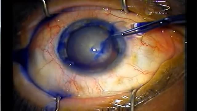
In cataract surgery, the lens inside your eye that has become cloudy is removed and replaced with an artificial lens (called an intraocular lens, or IOL) to restore clear vision. The procedure typically is performed on an outpatient basis and does not require an overnight stay in a hospital or other care facility.

Preeclampsia is a pregnancy complication characterized by high blood pressure and signs of damage to another organ system, often the kidneys. Preeclampsia usually begins after 20 weeks of pregnancy in a woman whose blood pressure had been normal. Even a slight rise in blood pressure may be a sign of preeclampsia. Left untreated, preeclampsia can lead to serious — even fatal — complications for both you and your baby. If you have preeclampsia, the only cure is delivery of your baby. If you're diagnosed with preeclampsia too early in your pregnancy to deliver your baby, you and your doctor face a challenging task. Your baby needs more time to mature, but you need to avoid putting yourself or your baby at risk of serious complications.
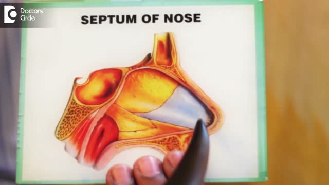
A deviated septum occurs when the thin wall (nasal septum) between your nasal passages is displaced to one side. In many people, the nasal septum is displaced — or deviated — making one nasal passage smaller. When a deviated septum is severe, it can block one side of your nose and reduce airflow, causing difficulty breathing. The additional exposure of a deviated septum to the drying effect of airflow through the nose may sometimes contribute to crusting or bleeding in certain individuals. Nasal obstruction can occur from a deviated nasal septum, from swelling of the tissues lining the nose, or from both. Treatment of nasal obstruction may include medications to reduce the swelling or nasal dilators that help open the nasal passages. To correct a deviated septum, surgery is necessary.
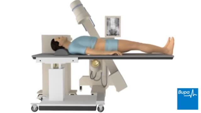
Extracorporeal shock wave lithotripsy (ESWL) uses shock waves to break a kidney stone into small pieces that can more easily travel through the urinary tract camera.gif and pass from the body. See a picture of ESWL camera.gif. You lie on a water-filled cushion, and the surgeon uses X-rays or ultrasound tests to precisely locate the stone. High-energy sound waves pass through your body without injuring it and break the stone into small pieces. These small pieces move through the urinary tract and out of the body more easily than a large stone. The process takes about an hour. You may receive sedatives or local anesthesia. Your surgeon may use a stent if you have a large stone. A stent is a small, short tube of flexible plastic mesh that holds the ureter open. This helps the small stone pieces to pass without blocking the ureter.
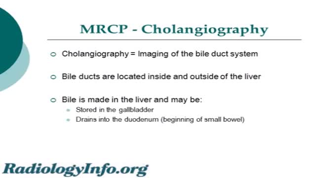
An MRCP scan is a scan that uses magnetic resonance imaging (MRI) to produce pictures of the liver, bile ducts, gallbladder and pancreas. Note: the information below is a general guide only. The arrangements,and the way tests are performed, may vary between different hospitals.
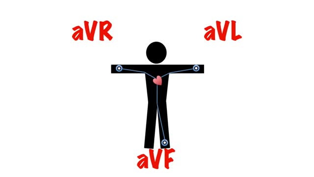
It then spreads down the bundle of his and then purkinje fibres to cause ventricular contraction. So when viewing the heart from the front, the direction of depolarisation is 11 o'clock to 5 o'clock. The general direction of depolarisation is known as the cardiac axis.
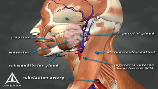
The external jugular vein receives the greater part of the blood from the exterior of the cranium and the deep parts of the face, being formed by the junction of the posterior division of the retromandibular vein with the posterior auricular vein.
A pneumothorax is usually caused by an injury to the chest, such as a broken rib or puncture wound. It may also occur suddenly without an injury. A pneumothorax can result from damage to the lungs caused by conditions such as chronic obstructive pulmonary disease (COPD), asthma, cystic fibrosis, and pneumonia.
Treatment may not be needed for an eschar if it is part of the natural healing process. However, if an eschar looks like it may have a wound infection – symptoms can include oozing fluid such as pus or blood, your clinician will likely recommend topical treatment or debridement to help control and remove the infection.
Results Sinusitis was characterized as acute in 26 patients, subacute in 5 (including 1 pyocele), and chronic in 8 (including 2 fungal infections). No tumors were found. Isolated sinus cysts were excluded from the study. Headache, the main symptom in 32 patients (82%), was localized most commonly on the vertex. Other common complaints were rhinitis, dizziness, eye symptoms, and fever. In 2 patients, the finding was occult. Eight patients (21%) presented with cranial nerve deficits, and 1 patient had an intracranial complication. Sinus irrigation was performed in 16 patients (41%) and sphenoidotomy was performed in 10 (26%). Fifteen patients (38%) were treated with antibiotic drugs alone. Within 3 months, 31 (84%) of 37 patients had recovered from the illness; 5 still experienced headaches despite having normalized radiographic findings; and 1 had permanent unilateral visual loss. Two patients were lost to follow-up.
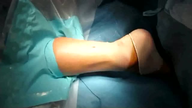
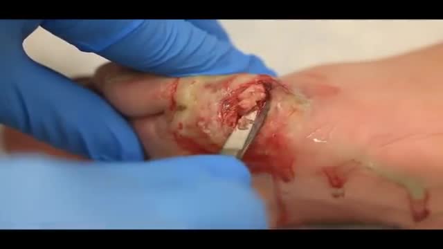
an incision made on the back of the lower leg starting just above the heel bone. After the surgeon finds the two ends of the ruptured tendon, these ends are sewn together with sutures. The incision is then closed. Another repair method makes a small incision on the back of the lower leg at the site of the rupture.
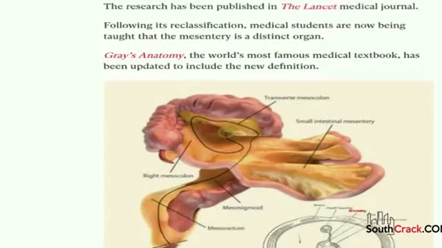
an. 4, 2017 -- Scientists say they've identified a new organ in the body -- a swath of tissue dubbed the mesentery that connects the intestine to the abdomen and holds everything in place. For years, anatomical experts have thought the organ was composed of several different segments of tissue, as opposed to being one single structure, according to Discover magazine. Since an organ must be one structure that performs a vital function, it was not deemed worthy of organ status. But recent research from doctors at the University Hospital Limerick in Ireland shows that the mesentery is actually one single band of tissue, the magazine reported Tuesday. It begins at the pancreas and wraps around the small intestine and colon. Its purpose: to hold these organs in position so they can perform their respective functions. "Without it you can't live," lead researcher Dr. J. Calvin Coffey, a colorectal surgeon at Limerick, told the magazine. "There are no reported instances of a Homo sapien living without a mesentery." "Understanding how and why our digestive system is arranged the way it is could be crucial to our understanding of diseases like Crohn's and irritable bowel syndrome," Coffey added. "There are a lot of diseases that we are stalled on, and we need to refresh our approach to these diseases," Coffey said. "Now that we've clarified its [the mesentery's] structure, we can systematically examine it. We're at a very exciting place right now." The discovery was published recently in the Lancet Gastroenterology & Hepatology journal.
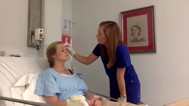
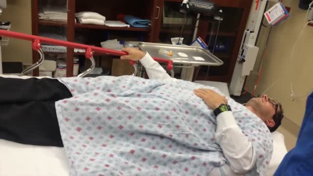
Physical assessment is taking an educated, systematic look at all aspects of an individual’s health status utilizing knowledge, skills and tools of health history and physical exam. To collect data- information about the client’s health, including physiological, psychological, sociocultural and spiritual aspects To establish actual and potential problems To establish the nurse-client relationship Method: The history is done first, then the physical examination focuses on finding data associated with the history. Health History- obtained through interview and record review. Physical exam- accomplished by tools and techniques ** A complete assessment is not necessarily carried out each time. A comprehensive assessment is part of a health screening examination. On admission, you will do an admission assessment (not necessarily including everything presented here) and document it on the admission form. You will do a daily shift assessment (patient systems review). And, if client has a specific problem, you may assess only that part of the body (focused). Data Collection: Information is organized into objective and subjective data: Subjective: Apparent only to person affected; includes client’s perceptions, feelings, thoughts, and expectations. It cannot be directly observed and can be discovered only asking questions. Objective: Detectable by an observer or can be tested against an acceptable standard; tangible, observable facts; includes observation of client behavior, medical records, lab and diagnostic tests, data collected by physical exam. ** To obtain data for the nursing health history, you must utilize good interview techniques and communications skills. Record accurately. DO NOT ASSUME. D. Frameworks for Health Assessment There are two main frameworks utilized in health assessment: Head to Toe- systematic collection of data starting with the head and working downward. Functional Health Assessment- Gordon’s 11 functional health patterns that address the behaviors a person uses to maintain health. PERSON is the ACC-ADN framework for assessment. It is similar to Gordon's functional health patterns.
Head to Toe Assesment
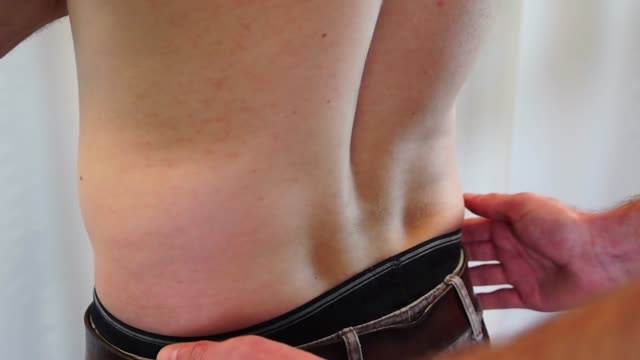
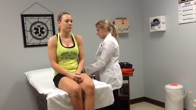
Clinical Examination - Gait, Arms, Legs, Spine
What's helping me become a better doctor
Sex easily falls to the wayside during pregnancy. Research shows that good sex has a significant impact upon not just the relationship, but also a woman’s ability to have an easeful and even joyful birth. Unfortunately, sex during pregnancy can be quite complicated for a variety of physical and emotional reasons. This week’s video will outline how to overcome these hurdles and make sure you continue to enjoy the wonders of sex as you embark on the first steps of parenthood.
