- Physical Examination
- Surgical Examination
- Ophthalmology
- Clinical Skills
- Orthopedics
- Surgery Videos
- Laparoscopy
- Pediatrics
- Funny Videos
- Cardiothoracic Surgery
- Nursing Videos
- Plastic Surgery
- Otorhinolaryngology
- Histology and Histopathology
- Neurosurgery
- Dermatology
- Pediatric Surgery
- Urology
- Dentistry
- Oncology and Cancers
- Anatomy Videos
- Health and Fitness
- Radiology
- Anaesthesia
- Physical Therapy
- Pharmacology
- Interventional Radiology
- Cardiology
- Endocrinology
- Gynecology
- Emergency Medicine
- Psychiatry and Psychology
- Childbirth Videos
- General Medical Videos
- Nephrology
- Physiology
- Diet and Food Health
- Diabetes Mellitus
- Neurology
- Women Health
- Osteoporosis
- Gastroenterology
- Pulmonology
- Hematology
- Rheumatology
- Toxicology
- Nuclear Medicine
- Infectious Diseases
- Vascular Disease
- Reproductive Health
- Burns and Wound Healing
- Other
Top videos
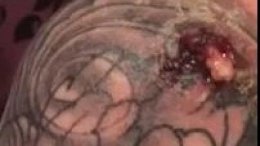
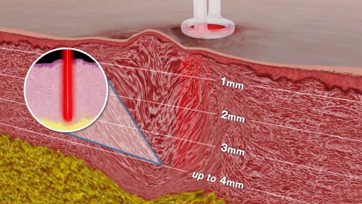
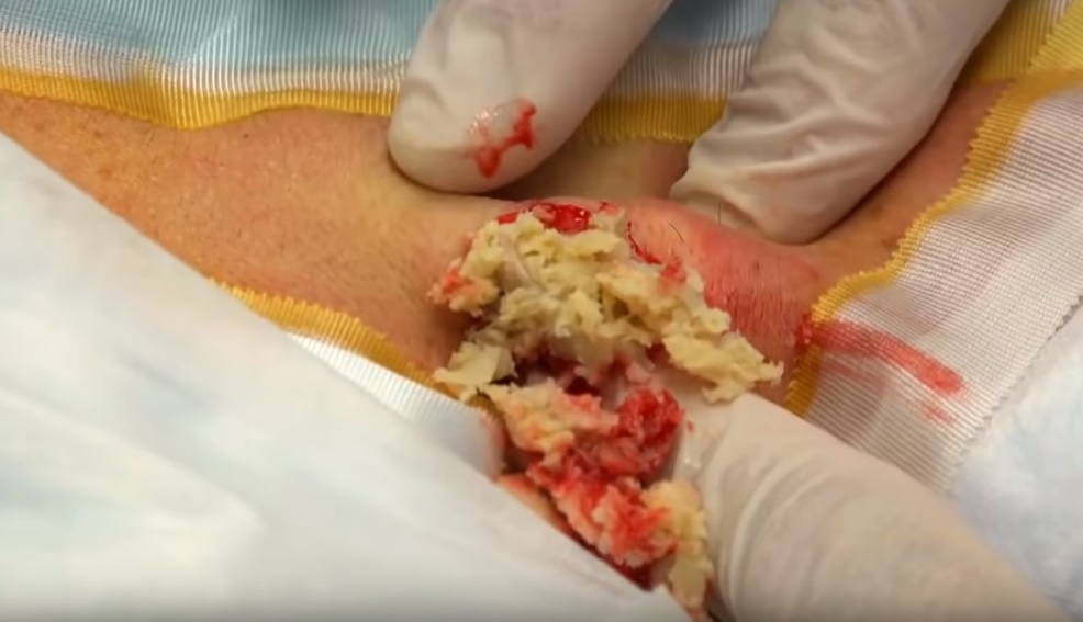
Infected Tattoo Abscess
Causes are chronic inflammation due to infection, allergies, drug sensitivity, or immune disorders. Symptoms may include a runny nose, stuffiness, or post-nasal drip. In some cases, there may be no symptoms. The condition can be treated with corticosteroids, other medications, or surgery.

Synthol, otherwise known as site enhancement oil is used by some people (including bodybuilders) to increase the apparent size of their muscles by directly injecting the oil into their muscle tissue. Users treat it as a short cut of looking like a body builder, without the actual hard work of bodybuilding training. With repeated injections, a larger volume of synthol builds up inside the muscle, expanding its size like a balloon filling up with air. Side effects of synthol can cause nerve damage, stroke, ulcers, pulmonary embolisms, and much more. Injecting synthol is very dangerous and if that doesn’t deter potential users, there is also a problem from an aesthetic standpoint; synthol use makes ones body look deformed (just see for yourself in the pictures below).
Scar revision includes techniques that improve the appearance of an unsightly scar, regardless of its size, type or age. This is typically not covered by insurance carriers and is treated as a cosmetic procedure. Though scars can never be completely removed, the appearance of scarring can be greatly diminished. Who Should Get Scar Revision? The best candidates for scar revision are in good health and have realistic expectations. Scar revision may be used to treat: Hyperpigmented scars Large or plainly visible scars Keloid scarring Raised scars Deep depression scars After scar revision, the appearance of your scar should be greatly reduced. Scar revision can improve the size, shape and color of your scar. Multiple procedures may be needed to achieve optimal results. There are several different techniques that can be used during your scar revision. During a consultation, we can discuss the best techniques and determine if you are a suitable candidate. What to Expect During Your Scar Revision Your scar revision may involve one or more of the following techniques: Topical treatments (gels, creams, external compression) can treat mild scarring or changes in pigmentation. Injectable treatments like dermal fillers are best for filling in scar depressions. These treatment options can provide long-lasting improvements, however, they are not always permanent. Surface treatments like chemical peels, dermabrasion, laser therapy and skin bleaching can improve skin tone and texture. More than one treatment may be needed to achieve optimal results. Surgical scar revision is only used in more severe cases. Reconstructive techniques like Z-plasty, tissue expansion, or skin grafting replace a prominent scar with a less noticeable scar. After Your Surgery Scar revision recovery varies depending on the procedure you have elected. Topical and injectable treatments rarely require downtime. Surface treatments and surgical removal can require several days of recovery. You may experience some temporary bruising, swelling, or discomfort. Over-the-counter or prescription medication can be used to manage pain. Topical and injectable treatments are likely to require sustained application to maintain results. The final results of surface treatments and surgical removal may not be visible for several weeks to months. It is important to protect the treatment area from direct sun exposure for several weeks. Additional details about your specific recovery will be discussed during your consultation.
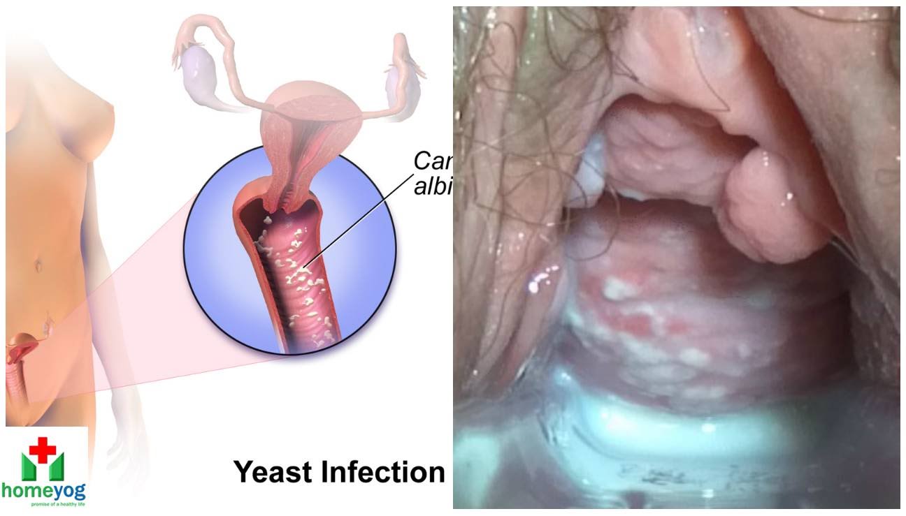
Watch that video to know the Female Genital Infections Causes and treatments.

Watch that Baby delivery Surgery video
Wisdom teeth extractions can rear their ugly head later in life. This is a video of a patient with neck pain and neck weakness. When we stimulated the nerve fibers in the area of the extracted teeth there was an immediate improvement in her ability to control her neck muscles.
Watch that video to know How To Increase Your Testosterone Levels, Naturally
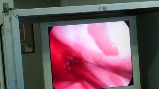
Watch that video of Popping Huge Epidermoid Cyst
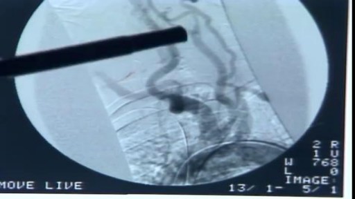
The procedure is used most often to treat a condition called supraventricular tachycardia, or SVT, which occurs because of abnormal conduction fibers in the heart. Catheter ablation is also used to help control other heart rhythm problems such as atrial flutter and atrial fibrillation.
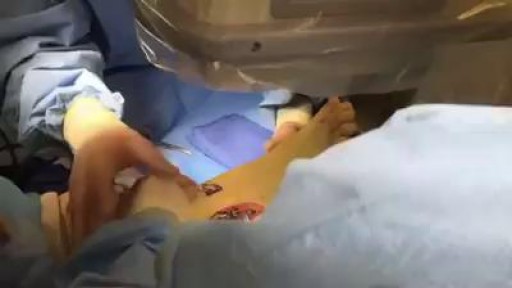
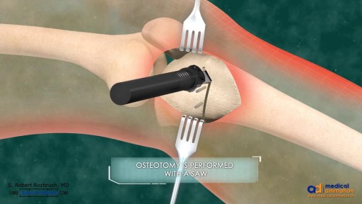
Ankle Fracture Surgery Video
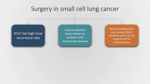
Small cell lung cancer, which occurs almost exclusively in smokers, is a malignancy characterised by rapid doubling time, high growth fraction and widespread metastasis at presentation. In this presentation, we will briefly discuss the classification of pulmonary Neuro-endocrine tumours by the World Health Organisation followed by a detailed discussion of the clinical features, lab evaluation and management of SCLC, both limited and extended stage. The frontline therapy in small cell lung cancer is etoposide and cisplatin along with thoracic radiotherapy and prophylactic cranial irradiation in patients who have a good response to therapy. Hyperfractionation of radiotherapy may provide some benefit but is also associated with increase incidence of complications. Newer agents for SCLC include Vandetanib and immunotherapy molecules, such as Iplimumab and nivolumab.
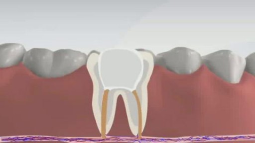
Root canal is a treatment to repair and save a badly damaged or infected tooth instead of removing it. The term "root canal" comes from cleaning of the canals inside a tooth's root. Decades ago, root canal treatments often were painful. With dental advances and local anesthetics, most people have little if any pain with a root canal. In fact, it's probably more painful living with a decayed tooth. Root canal alternatives include extracting the damaged tooth and replacing it with a dental implant, bridge or removable partial denture.

Cigarette contain tobacco that is very harmful but vaporizers does not contain tobacco. ... The most basic difference between vaping and cigarette usage is that cigarettes require combustion. You need fire to light a cigarette. On the other hand, vaping requires electricity and creates vapor.


Fibrodysplasia ossificans progressiva (FOP) is a disorder in which muscle tissue and connective tissue such as tendons and ligaments are gradually replaced by bone (ossified), forming bone outside the skeleton (extra-skeletal or heterotopic bone) that constrains movement. This process generally becomes noticeable in early childhood, starting with the neck and shoulders and proceeding down the body and into the limbs. Extra-skeletal bone formation causes progressive loss of mobility as the joints become affected. Inability to fully open the mouth may cause difficulty in speaking and eating. Over time, people with this disorder may experience malnutrition due to their eating problems. They may also have breathing difficulties as a result of extra bone formation around the rib cage that restricts expansion of the lungs.