- Physical Examination
- Surgical Examination
- Ophthalmology
- Clinical Skills
- Orthopedics
- Surgery Videos
- Laparoscopy
- Pediatrics
- Funny Videos
- Cardiothoracic Surgery
- Nursing Videos
- Plastic Surgery
- Otorhinolaryngology
- Histology and Histopathology
- Neurosurgery
- Dermatology
- Pediatric Surgery
- Urology
- Dentistry
- Oncology and Cancers
- Anatomy Videos
- Health and Fitness
- Radiology
- Anaesthesia
- Physical Therapy
- Pharmacology
- Interventional Radiology
- Cardiology
- Endocrinology
- Gynecology
- Emergency Medicine
- Psychiatry and Psychology
- Childbirth Videos
- General Medical Videos
- Nephrology
- Physiology
- Diet and Food Health
- Diabetes Mellitus
- Neurology
- Women Health
- Osteoporosis
- Gastroenterology
- Pulmonology
- Hematology
- Rheumatology
- Toxicology
- Nuclear Medicine
- Infectious Diseases
- Vascular Disease
- Reproductive Health
- Burns and Wound Healing
- Other
Top videos

What is hemodiafiltration? Hemodiafiltration, or HDF, is a renal replacement modality that combines diffusion and convection to improve removal of molecules in the middle molecular weight range versus hemodialysis.
Find our full video library only on Osmosis Prime: http://osms.it/more.
Join over 3 million current & future clinicians who learn by Osmosis, and over 130 universities around the world who partner with us to make medical and health education more engaging and efficient. We have unparalleled tools and materials to prepare you to succeed in school, on board exams, and as a future clinician. Sign up for a free trial at http://osms.it/more. If you're interested in exploring an institutional partnership, visit osmosis.org/educators to request a personalized demo.
Follow us on social:
Facebook: http://osms.it/facebook
Twitter: http://osms.it/twitter
Instagram for med: http://osms.it/instagram
Instagram for nursing: https://osms.it/ignursing
Linkedin: https://osms.it/linkedin
Our Vision: Everyone who cares for someone will learn by Osmosis.
Our Mission: To empower the world’s clinicians and caregivers with the best learning experience possible. Learn more here: http://osms.it/mission
Medical disclaimer: Knowledge Diffusion Inc (DBA Osmosis) does not provide medical advice. Osmosis and the content available on Osmosis's properties (Osmosis.org, YouTube, and other channels) do not provide a diagnosis or other recommendation for treatment and are not a substitute for the professional judgment of a healthcare professional in diagnosis and treatment of any person or animal. The determination of the need for medical services and the types of healthcare to be provided to a patient are decisions that should be made only by a physician or other licensed health care provider. Always seek the advice of a physician or other qualified healthcare provider with any questions you have regarding a medical condition. © 2023 Elsevier. All rights reserved.


Presented without captions
Starting dialysis often means creating a new normal for yourself and your family. There’s a lot to think about, from choosing a treatment option, to finding new ways to enjoy your favorite activities, to managing a new diet. The FIRST30 program is all about helping you through this period of adjustment.
Find out more at KidneyFund.org/FIRST30.

SUBSCRIBE: https://www.youtube.com/c/TVNe....phrologist?sub_confi
An animation of blood flow inside the Hemodialysis circuit.
About Dr. Rifai:
Dr. Ahmad Oussama Rifai is certified by the American Board of Internal Medicine (ABIM) in the specialty of Internal Medicine and the sub-specialty of Nephrology.
MEET DR. RIFAI
https://www.thevirtualnephrologist.com/rifai/
Follow The Virtual Nephrologist on SOCIAL MEDIA:
-Facebook: https://www.facebook.com/thevirtualnephrologist
-Instagram: https://www.instagram.com/thevirtualnephrologist/
-Twitter: https://twitter.com/VNephrologist
Schedule a virtual consult:
https://www.thevirtualnephrolo....gist.com/schedule-a-
Best wishes for great health | The Virtual Nephrologist

"Axillary Artery to Vein AV Graft for Dialysis Access"
Houston Methodist DeBakey Heart & Vascular Center, presents a cardiovascular procedure featuring Maham Rahimi, MD, M. Mujeeb Zubair, MD, and Louis Gomez, MD, as they demonstrate “Axillary Artery to Vein AV Graft for Dialysis Access".
Surgery: Maham Rahimi, MD, M. Mujeeb Zubair, MD, and Louis Gomez, MD
Narration: M. Mujeeb Zubair, MD
__________________________
FOR MORE INFORMATION
DeBakey CV Education: https://www.houstonmethodist.o....rg/education/medical
Follow Us:
Facebook: https://www.facebook.com/debakeycvedu
Twitter: https://twitter.com/DeBakeyCVedu
Livestream: https://livestream.com/debakey
Want concise, relevant reviews of the hottest topics in CV medicine? Subscribe for FREE to the Methodist DeBakey Cardiovascular Journal for quarterly, peer-reviewed issues delivered to your door.
https://journal.houstonmethodist.org/

Demystify knee pain and discover nine of the most common causes of pain in this complex joint. Join Burke Selbst PT as we work through our simple screening for the most common types of problems.
Burke is the founder and clinical director of Focus Physical Therapy in Bend Oregon.
Find him:
https://focusptbend.com
https://facebook.com/focusphysio
Intro Song Credit
Adventures by A Himitsu https://www.youtube.com/channel/UCgFw...
Creative Commons — Attribution 3.0 Unported— CC BY 3.0
http://creativecommons.org/licenses/b...
Music released by Argofox https://youtu.be/8BXNwnxaVQE
Music provided by Audio Library https://youtu.be/MkNeIUgNPQ8

Arthritis occurs when the cartilage breaks down explains Dr. Derek Papp, Sports Medicine Physician with Miami Orthopedics & Sports Medicine Institute. This it’s a very common knee injury such as the damage of the cartilage and meniscus tear.
ACL tears is another common injury especially in sports like soccer or Australian football, the specialist explains.

Runners Knee Overview:
Welcome to our Patello-Femoral Rehab video. The goal of this video is to minimize pain around the kneecap and maximize recovery. This video should not be used as a substitute for regular physical therapy visits and guidance from your physician
Visit http://www.matthewboesmd.com/p....atello-femoral-rehab for more information
![Knee Injury Rehabilitation [Early Stage] - (1st Two Weeks After Injury)](https://i.ytimg.com/vi/35zoRRUDVYo/maxresdefault.jpg)
I have shared with you in this video couple of exercises that you can follow immediately after your Knee injury.
As I promised here are 2 protocols to follow in this routine. I have also added my blog on how to strengthen your glutes and why that can help you with your knee pain.
1- Avoid Harm ( https://dublinsportsinjuryclin....ic.com/acute-injury-
2- POLICE PROTOCOL (https://dublinsportsinjuryclin....ic.com/acute-injury-
3- Read my blog and check how to strengthen your glutes (https://dublinsportsinjuryclin....ic.com/knee-injury-r
Please make sure to watch the video until the end since I'm sharing with you a couple of tips at the end of this video.
References:
1- https://www.ncbi.nlm.nih.gov/pmc/arti...
2- https://www.sciencedirect.com/science...
3- https://www.sciencedirect.com/science...
_______________________________
Music: See You
Musician: @iksonofficial
---------------------------------------------------
**MEDICAL DISCLAIMER**
All information, content, and material of this video or website are for informational and demonstration purposes only. It is not intended to serve as a substitute for the consultation, diagnosis, and/or medical treatment of a qualified physician or healthcare provider.
Don’t use this content as a replacement for treatment and advice given by your doctor or health care provider. Consult with your physical therapist or healthcare professional before doing anything contained in this content.
By watching this video, you agree to indemnify and hold harmless Dublin Sports Injury Clinic(and its representatives) for any and all losses, injuries, or damages resulting from any and all claims that arise from your use or misuse of this content. Dublin Sports Injury Clinic makes no representations about the accuracy or suitability of this content.
USE OF THIS VIDEO'S CONTENT IS AT YOUR OWN RISK.
𝐂𝐨𝐧𝐧𝐞𝐜𝐭 𝐰𝐢𝐭𝐡 𝐁𝐨𝐛 𝐚𝐭 𝐃𝐮𝐛𝐥𝐢𝐧 𝐒𝐩𝐨𝐫𝐭𝐬 𝐈𝐧𝐣𝐮𝐫𝐲 𝐂𝐥𝐢𝐧𝐢𝐜
𝐖𝐄𝐁𝐒𝐈𝐓𝐄→ https://www.dublinsportsinjuryclinic.com
𝐄𝐌𝐀𝐈𝐋 → Rehab@dublinsportsinjuryclinic.com
𝐓𝐄𝐋 → 0879276712
𝐅𝐀𝐂𝐄𝐁𝐎𝐎𝐊 → https://www.facebook.com/dublinphysicaltherapy/
𝐈𝐍𝐒𝐓𝐀𝐆𝐑𝐀𝐌 → https://www.instagram.com/dubl....in_sports_injury_cli
Tags:
Knee Injury Rehabilitation [Early Stage] - (1st Two Weeks After Injury)
Knee injury exercises, knee exercises, knee rehabilitation, Sore knee rehabilitation, Twisted knee exercises, sore kneecap exercises, runners knee injury, #kneeinjury #soreknee #runnersknee #Kneerehabilitation #kneeexercices #dublinsportsinjuryclinic
#anteriorkneepain #kneepain #kneephysio #injureknee #exerciseforknee #kneerehab #swollenknee
#runnersknee #kneeminiscus #acl #Mcl #kneeligaments#dublinsportsinjuryclinic #dublinsportsphysio #bobfiro #dulin2phyiso #bobyourphysio #bobonlinecare #Sportsinjurydublinclinic#dublinsportsinjuryclinic #dublinsportsphysio #bobfiro #dulin2phyiso #bobyourphysio #bobonlinecare #Sportsinjurydublinclinic

If you have pain on the inside of your knee, it’s likely due to an injury or arthritis. The following exercises will help strengthen and stretch your muscles to prevent further damage and improve mobility.
#kneepain #arthritis #kneepainrelief #kneeosteoarthritis
- - - - - - - - - - - - - - - - - - - - - - - - - - - - - - - - - - - - -
DISCLAIMER: This video and any related comments are not medical advice. Check with your own healthcare professional before attempting anything in this video. This information is only intended to show you the correct technique for physical therapy exercises and should not be used to self-diagnose or self-treat any medical condition. If you experience any pain or difficulty while doing these exercises, stop immediately and see your healthcare professional.
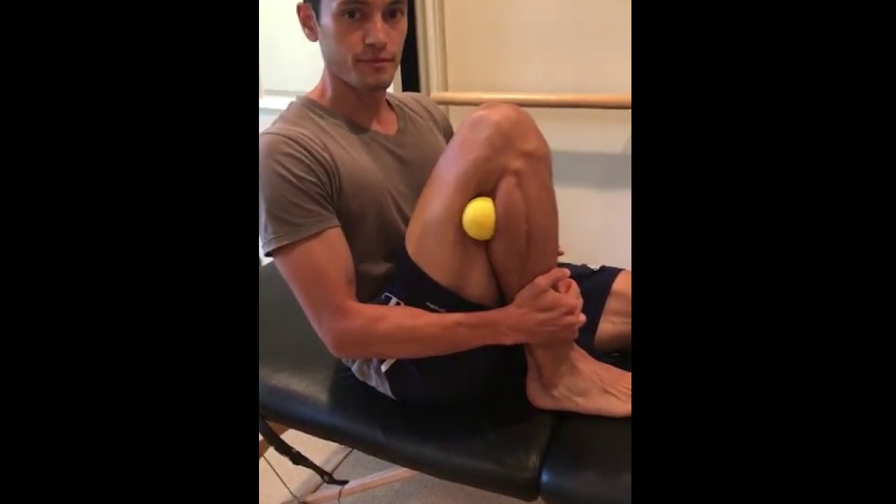
A displaced fibular head can create tightness, pain, and even numbness or tingling along the outside of your knee and down your leg. This most often occurs after a modest hyperextension knee injury, such as landing on one leg after jumping. If you have lingering knee pain and are searching for an answer, try this move
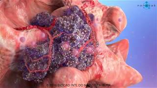
This cancer development medical video is devoted to elaborating the basics of cancer growth. We used advanced medical animation techniques to display such a complicated process.
What is happening in cancer development medical video
The fundamental abnormality described in the cancer development medical video is the nonstop unregulated multiplication of cancer cells. Being uncontrollable by body’s signals that regulate normal cell behavior; cancerous cells divide and grow populating neighboring normal tissues or even spread throughout the body. The overall lack of growth control acquired by cancer cells is due to the accumulated abnormalities in numerous cell regulatory mechanisms and is considered in some aspects of cell behavior that differs them from their healthy counterparts. The interaction of these cells is shown in our previous medical animation video.
Read full article on our webpage http://bit.ly/2LQj9ln
Follow us on Facebook https://www.facebook.com/Nanob....ot.Medical.Animation
Follow us on LinkedIn https://www.linkedin.com/compa....ny/nanobotmodels-med
Follow us on Twitter https://twitter.com/Nanobot_Studio
Follow us on Instagram https://www.instagram.com/nano....bot_medical_animatio
Follow us on Clutch https://clutch.co/profile/nano....bot-medical-animatio
Follow us on Behance https://www.behance.net/NanobotStudio
#cancer #tumor #oncology #metatastic #nanobot #visualscience #scientificcommunication #medicalanimation #animationvideo #animationdesign #animationstudio #animationmovie #nanotechnology #medicine #health #science #education #medschool #medicaleducation #animation_studio #animationstudio

Get a 60-day free trial at https://shipstation.com/doctormike. Thanks to ShipStation for sponsoring the show!
I’ll teach you how to become to media’s go-to expert in your field. Enroll in The Professional’s Media Academy now: https://www.professionalsmediaacademy.com/
Listen to my podcast, @DoctorMikeCheckup, here:
Spotify: https://go.doctormikemedia.com..../spotify/CheckUpSpot
Apple Podcasts: https://go.doctormikemedia.com..../applepodcast/AppleP
Body Bizarre is a TLC show with a name I'm not too wild about, but with stories that are nonetheless fascinating. Today we look at separating conjoined twins, a girl with ants crawling out of her ears, a man who nearly lost his hand in a factory accident, a family that all has 6 fingers, and more.
Help us continue the fight against medical misinformation and change the world through charity by becoming a Doctor Mike Resident on Patreon where every month I donate 100% of the proceeds to the charity, organization, or cause of your choice! Residents get access to bonus content, an exclusive discord community, and many other perks for just $10 a month. Become a Resident today:
https://www.patreon.com/doctormike
Let’s connect:
IG: https://go.doctormikemedia.com..../instagram/DMinstagr
Twitter: https://go.doctormikemedia.com/twitter/DMTwitter
FB: https://go.doctormikemedia.com/facebook/DMFacebook
TikTok: https://go.doctormikemedia.com/tiktok/DMTikTok
Reddit: https://go.doctormikemedia.com/reddit/DMReddit
Contact Email: DoctorMikeMedia@Gmail.com
Executive Producer: Doctor Mike
Production Director and Editor: Dan Owens
Managing Editor and Producer: Sam Bowers
Editor and Designer: Caroline Weigum
Editor: Juan Carlos Zuniga
* Select photos/videos provided by Getty Images *
** The information in this video is not intended nor implied to be a substitute for professional medical advice, diagnosis or treatment. All content, including text, graphics, images, and information, contained in this video is for general information purposes only and does not replace a consultation with your own doctor/health professional **
Overview
Heart bypass surgery creates a new route, called a bypass, for blood and oxygen to reach the heart.
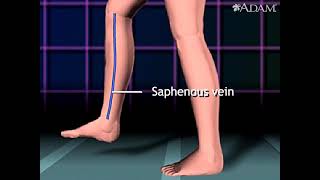
Heart bypass surgery begins with an incision in the chest, and the breastbone is cut exposing the heart. Next, a portion of the saphenous vein, which is very large, is harvested from the inside of the leg. Pieces of this large vein are used to bypass the blocked coronary arteries, which are arteries that supply blood to the heart. The venous graft is sewn to the aorta, the main artery of the body, and to the affected coronary artery, to bypass the blocked site.
The internal mammary artery from the chest may also be used to bypass a clogged artery.
Several arteries may be bypassed depending on the condition of the heart. After the graft is created, the breastbone and chest are closed.

https://bit.ly/3HIStRc #shorts
Coloscopy | Colon Polyp Resection | Polypectomy
Colonoscopies are essential for detecting colorectal abnormalities, including colon polyps. Polypectomy, the surgical removal of these growths, can prevent them from becoming cancerous. This article offers a brief overview of colonoscopies, colon polyps, and polypectomy procedures.
A colonoscopy is an endoscopic examination allowing healthcare providers to visualize the colon and rectum using a colonoscope. The colonoscope, a flexible tube with a camera and light source, helps detect abnormalities, including polyps or tumors.
Colon polyps are abnormal growths arising from the colon's inner lining. While most polyps are benign, some can become malignant. Adenomatous polyps have a higher potential to become cancerous, whereas hyperplastic and inflammatory polyps pose a lower risk.
Polypectomy involves removing colon polyps during a colonoscopy. Two primary techniques include snare polypectomy, using a wire loop to cut the polyp, and cold forceps polypectomy, which employs forceps to grasp and remove smaller polyps.
Following a polypectomy, patients may experience mild discomfort or bleeding. Regular surveillance is crucial to minimize colorectal cancer risk. The frequency of surveillance colonoscopies depends on the number, size, and type of polyps found, as well as the patient's overall risk factors.
Colonoscopies and polypectomies play vital roles in detecting and removing colon polyps, reducing the risk of colorectal cancer, and maintaining optimal colon health.
Do you want to learn more about colon polyps and colonoscopy? check our:
Article @ https://bit.ly/41w5Ooq
For more free resources, find us on Pinterest & Facebook pages:
https://www.pinterest.ca/medicalartsofficial/
https://www.facebook.com/Medicalartsofficial
https://www.youtube.com/@medic....alarts?sub_confirmat
https://www.instagram.com/medicalartsofficial/
https://www.tiktok.com/@medicalarts
#endoscopicsurgery #digestivesystem
coloscopy
polyp
colon polyp
polypectomy
colonoscopy
colon polyp animation
gi endoscopy
@MedicalArts , 2023.

Dr. Nick demonstrates how easy it is to have stitches taken out and that it is not painful!
#shorts #satisfying #reaction
 MAKE SURE TO SUBSCRIBE FOR ALL THE NEW SURGICAL AND EDUCATIONAL VIDEOS COMING!!
👉🏻For more information visit :
https://drnickcampi.com
👉🏻Follow me on TikTok!!
https://vm.tiktok.com/ZMeXLbc5F/
👉🏻Connect with me!!
https://www.instagram.com/drnickcampitelli
👉🏻Check out this video of how we remove an ingrown toenail!
https://youtu.be/JyZo8aZDYds
👉🏻Dr. Nick Campitelli Performs latest Minimally Invasive Bunion Surgery! Watch this video!
https://youtu.be/eRpABMsCbOU
Dr. Nick Campitelli is a podiatrist who specializes in foot and ankle surgery in the Akron and Cleveland Ohio area. He is the Residency Director of the Western Reserve Hospital / University Hospital Podiatric Medicine and Surgery Residency Program.
*** All content found on the this YouTube video including: text, images, audio, or other formats were created for informational purposes only. The Content is not intended to be a substitute for professional medical advice, diagnosis, or treatment. Always seek the advice of your physician or other qualified health provider with any questions you may have regarding a medical condition. Never disregard professional medical advice or delay in seeking it because of something you heard on this video. ***

Examination of Peripheral Vascular System - Clinical Skills OSCE Revision - Dr Gill
In this video, we demonstrate the peripheral vascular examination - a less common examination, but still vitally important, particularly amongst the older population
Starting with the examination of the hands looking for clinical signs of vascular compromise, we then check the pulses of the major arteries of the upper body - the radial, brachial and carotid arteries, before moving down to assess for an abdominal aortic aneurysm.
At this point, I feel it's a practical step to check the femoral pulses before doing the overview of the legs.
After visually assessing we must examine the major vascular areas of leg.- namely the popliteal pulses, before wrapping up around the ankle with the posterior tibial and dorsalis pedis pulses
For completeness, the cardiovascular examination is demonstrated here
https://www.youtube.com/watch?v=ECs9O5zl6XQ&t=2s
#PeripheralVascular #ClinicalSkills #DrGill

This video - produced by students at Oxford University Medical School - demonstrates how to perform an examination of the respiratory system. It also indicates common pathologies encountered. It is part of a series of videos covering basic clinical examinations and is linked to Oxford Medical Education (www.oxfordmedicaleducation.com).

Elbow Exam - Orthopaedic OSCE - Clinical Skills - Dr Gill
The elbow examination is a core skill - in this video, we demonstrate how to perform an elbow EXAM for an Orthopaedic Clinical Skills OSCE, which should be one of the more accessible examination stations for medical students.
For a passing grade in your Clinical Skills OSCE, an elbow assessment should follow the LOOK, FEEL, MOVE approach
Initially looking for erythema, scars, swelling and position
Palpating the elbow - specifically the olecranon, medial and lateral epicondyles, and radial head for heat, oedema and crepitus
Finally assess range of movement with flexion and extension at the elbow, before determining for tennis and golfers' elbows
Watch further orthopaedic examinations for your OSCE revision:
The Elbow - Deep Dive
https://youtu.be/SX5buhtCVDw
The Spine Examination:
https://youtu.be/pJxMHa6SCgU
The Knee examination
https://youtu.be/oyKH4EYfJDM
The Hip examination
https://youtu.be/JC9GKq5nSdQ
The GALS examination
https://youtu.be/5qJaf7gW-B0 - Gait, Arms, Legs, Spine - GALS screen
------------
Please note that there is no ABSOLUTE way to perform a clinical examination. Different institutions and even clinicians will have differing degrees of variations - the aim is the effectively identify medically relevant signs.
However during OSCE assessments. Different medical schools, nursing colleges and other health professional courses will have their own preferred approach to a clinical assessment - you should concentrate on THEIR marks schemes for your assessments.
The examination demonstrated here is derived from Macleods Clinical Examination - a recognised standard textbook for clinical skills.
Some people viewing this medical examination video may experience an ASMR effect
#clinicalskills #Elbow #DrGill

The most reliable clinical sign to detect ascites is checking for bilateral flank dullness. If a patient with ascites is lying supine, fluid accumulates in the flank regions, leading to dullness on percussion. At the same time, the air-filled bowel loops are forced upwards by the free fluid due to buoyancy, resulting in tympanitic percussion. To locate specifically where dullness shifts to tympany, or the air-fluid level, percussion should be performed from the sides towards the middle. To confirm that the dullness is caused by ascites, ask the patient to switch to a lateral decubitus position. If ascites is present, the air-filled bowel loops will shift accordingly and remain at the surface of the fluid. As a result, the air-fluid level will shift as well. This is known as shifting dullness.
Subscribe to AMBOSS YouTube for the latest clinical examination videos, medical student interviews, study tips and tricks, and live webinars!
Free 5 Day Trial: https://go.amboss.com/amboss-YT
Instagram: https://www.instagram.com/amboss_med/
Facebook: https://www.facebook.com/AMBOSS.Med/
Twitter: https://twitter.com/ambossmed
Blog: https://blog.amboss.com/us
#AMBOSSMed #ClinicalExamination
