
- Physical Examination
- Surgical Examination
- Ophthalmology
- Clinical Skills
- Orthopedics
- Surgery Videos
- Laparoscopy
- Pediatrics
- Funny Videos
- Cardiothoracic Surgery
- Nursing Videos
- Plastic Surgery
- Otorhinolaryngology
- Histology and Histopathology
- Neurosurgery
- Dermatology
- Pediatric Surgery
- Urology
- Dentistry
- Oncology and Cancers
- Anatomy Videos
- Health and Fitness
- Radiology
- Anaesthesia
- Physical Therapy
- Pharmacology
- Interventional Radiology
- Cardiology
- Endocrinology
- Gynecology
- Emergency Medicine
- Psychiatry and Psychology
- Childbirth Videos
- General Medical Videos
- Nephrology
- Physiology
- Diet and Food Health
- Diabetes Mellitus
- Neurology
- Women Health
- Osteoporosis
- Gastroenterology
- Pulmonology
- Hematology
- Rheumatology
- Toxicology
- Nuclear Medicine
- Infectious Diseases
- Vascular Disease
- Reproductive Health
- Burns and Wound Healing
- Kidney Zone Podcast
- Other





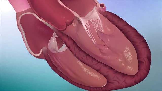





Review of heart sounds

Educational only: videos on MedicalVideos.com are for general information and are not medical advice. Always consult your own doctor about your health.
Heart sounds are reviewed in this cardiology listening exercise for clinical examination training. The recording works through the sounds heard with a stethoscope during assessment of the heart. It is a study resource for students learning cardiac auscultation basics and examination technique.
This video in 30 seconds
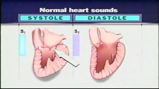
A short teaching review of normal heart sounds, recorded ahead of an auscultation exercise. The narration explains that valve closure produces the first and second heart sounds, S1 and S2, how systole and diastole fall between them, and where the sounds are heard across the aortic, pulmonic, tricuspid and mitral areas.
Key points
- The narration links S1 to mitral valve closure and S2 to aortic valve closure
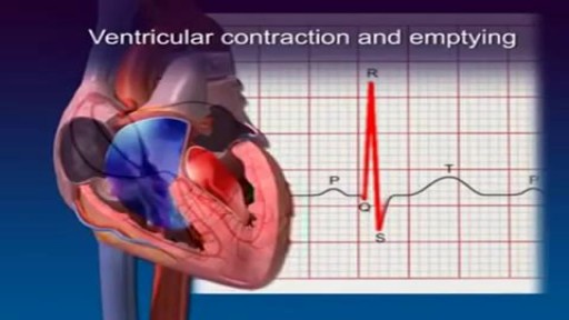
- Systole is placed between S1 and S2, and diastole between S2 and the next S1
- The longer length of diastole is given as a way to tell the two sounds apart
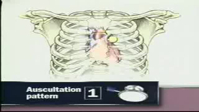
- Listening areas on the chest are named for the aortic, pulmonic, tricuspid and mitral positions
- The aortic and pulmonic areas together are called the base of the heart
Video Transcript
A short teaching review of the heart sounds, recorded ahead of an auscultation exercise. The narration explains how valve closure produces the first and second sounds, S1 and S2, how systole and diastole fall between them, and where each sound is heard — the aortic, pulmonic, tricuspid and mitral areas — noting that the aortic and pulmonic areas together are called the base of the heart.
Read the full transcript
Before auscultating the heart, let's review normal heart sounds. Closure of the heart valves creates a pair of audible heart sounds. The first sound, S1, accompanies mitral valve closure. The second sound, S2, accompanies aortic valve closure. Pulmonic and tricuspid valve closure may contribute to these sounds. Ventricular systole occurs between S1 and S2. Ventricular diastole occurs between S2 and the next S1. Because diastole usually lasts longer than systole, you can identify the two sounds. One-two, one-two, one-two. You can hear S1 and S2 in the aortic area in the right second interspace close to the sternum.
Pulmonic area in the left second interspace close to the sternum. Left third interspace. Tricuspid area in the left fourth and fifth interspaces. And mitral area at the apical impulse. The aortic and pulmonic areas together are sometimes called the base of the heart.
Transcript auto-generated from the video’s audio for accessibility and study. Educational content only — not medical advice.
Video Overview
From our Cardiology collection: ECG interpretation, echocardiograms, angioplasty, stents and heart conditions explained by cardiologists. Browse all Cardiology videos →
Medical Videos
Free medical and surgery videos for patients, students and professionals — trusted since 2007.
Founded by Dr. Mohamed Ibrahim, MD, PhD — physician-scientist.
Follow us on Facebook · Instagram
SORT BY-
Top Comments
-
Latest comments