- Physical Examination
- Surgical Examination
- Ophthalmology
- Clinical Skills
- Orthopedics
- Surgery Videos
- Laparoscopy
- Pediatrics
- Funny Videos
- Cardiothoracic Surgery
- Nursing Videos
- Plastic Surgery
- Otorhinolaryngology
- Histology and Histopathology
- Neurosurgery
- Dermatology
- Pediatric Surgery
- Urology
- Dentistry
- Oncology and Cancers
- Anatomy Videos
- Health and Fitness
- Radiology
- Anaesthesia
- Physical Therapy
- Pharmacology
- Interventional Radiology
- Cardiology
- Endocrinology
- Gynecology
- Emergency Medicine
- Psychiatry and Psychology
- Childbirth Videos
- General Medical Videos
- Nephrology
- Physiology
- Diet and Food Health
- Diabetes Mellitus
- Neurology
- Women Health
- Osteoporosis
- Gastroenterology
- Pulmonology
- Hematology
- Rheumatology
- Toxicology
- Nuclear Medicine
- Infectious Diseases
- Vascular Disease
- Reproductive Health
- Burns and Wound Healing
- Other
Top videos

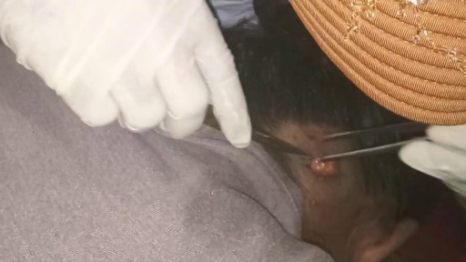
Surgical cutting and removal of a deep skin cyst Medical Videos
Pregnancy Tips : How Early Can You Take a Blood Test for Pregnancy?
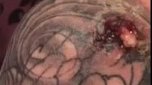
Infected Tattoo Abscess
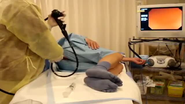
Colonoscopy is a test that allows your doctor to look at the inner lining of your large intestine (rectum and colon). He or she uses a thin, flexible tube called a colonoscope to look at the colon. A colonoscopy helps find ulcers, colon polyps, tumors, and areas of inflammation or bleeding.
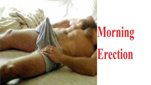
Morning erections have colloquially been termed as “morning wood” while scientifically it is called nocturnal penile tumescence. It is a normal and healthy physiological reaction and response that most men experience in their lives. Morning erections are really the ending of a series of erections that happen to men during the night. Healthy men can, on average, have anywhere between three to five erections in a full night of sleep, each of which lasts from 25-35 minutes.

http://vitiligo-home-remedies.plus101.com ---- white spots on skin, small white spots on face, what causes white spots on skin, vitiligo. The Link Between Vitiligo and Auto Immune Disorders. If you suffer with Vitiligo, a condition that strips your skin of its natural pigment or coloring, the odds are good that you are also battling some sort of autoimmune disorder. It took years for doctors to connect the two disorders, but recent research shows that at least 20 percent of Vitiligo suffers also get autoimmune thyroid disease, and that's juts the beginning. Many more suffer a multitude of other disorders. Until recently, the link between the Vitiligo and autoimmune problems were not clear. Doctors seemed to see a link, but nothing substantial could be proven. Until now. In march 2013, The National Institute's of Health's National Institute of Arthritis and Musculoskeletal and Skin Diseases (NIAMS) announced an amazing discovery: a connection between a specific gene named NALP1, Vitiligo and a host of autoimmune diseases including thyroid disease, pernicious anemia, rheumatoid arthritis, lupus and Addison's Disease. According to lead researcher, Richard Spritz M.D., the discovery of this gene may make newer, more effective Vitiligo treatments possible within the next few years. But that's not all. It will also be able to help treat certain auto immune disorders. By finding ways to block the inflammatory response of the NALP1 gene, doctors may some day be able to cure certain autoimmune disorders. A long-term solution for vitiligo should address the internal causes of vitiligo by tackling all vitiligo contributing factors. Only by controlling the nutritional, hormonal, psychological and environmental triggers of vitiligo, using a multidimensional and holistic approach to healing you can reverse the "internal vitiligo environment"- the only, safe, natural and effective way you could ever achieve lasting vitiligo freedom. More Info: http://vitiligo-home-remedies.plus101.com
A Pap smear (Papanicolau smear; also known as the Pap test) is a screening test for cervical cancer. The test itself involves collection of a sample of cells from a woman's cervix (the end of the uterus that extends into the vagina) during a routine pelvic exam
Educational video of male patient receiving an anoscopy.

The OrthoIllustrated® animation for total knee replacement is an educational tool to help patients better understand the diagnosis and treatment of arthritis.
- - - - -
Why Work Arthrex https://www.arthrex.com/job-seeker
Find an Arthrex Surgeon: https://doctorfinder.orthoillustrated.com
- - - - -
Join the Community:
LinkedIn: https://www.linkedin.com/company/arthrex
Facebook: https://www.facebook.com/Arthrex
Instagram: https://www.instagram.com/arthrex_inc/
Twitter: https://twitter.com/Arthrex
TikTok: https://www.tiktok.com/@arthrex
- - - - -
Arthrex Inc., headquartered in Naples, Florida, is a global leader in orthopedic surgical device design, research, manufacturing, and medical education. Arthrex develops and releases more than 1,000 new products and procedures every year to advance minimally invasive orthopedics worldwide.
For more information, visit https://www.arthrex.com
- - - - -
OrthoPedia is an innovative educational website that was created for anyone interested in learning about orthopedics from the first-year student to the experienced orthopedic surgeon.
Visit https://www.orthopedia.com to experience the future of Medical Education.

Watch that video of a Snake bite causes girl’s leg to rot away with necrosis
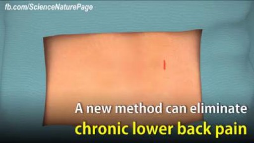
New method can eliminate lower back pain. watch to learn how.
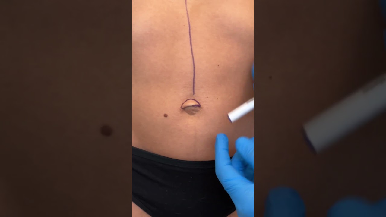
Check out @barrettplasticsurgery on TikTok!
Like and subscribe for more! #shorts #medical #plasticsurgery
More information:
www.drdanielbarrett.com
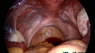
http://drraewynteirney.com.au/video/
http://drraewynteirney.com.au/....about-dr-raewyn-teir
Dr Raewyn Teirney - fertility specialist and Gynaecologist in Sydney shows a video recording of a laparoscopy for a woman with infertility and pelvic pain.
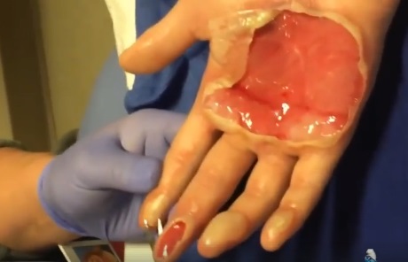
watch that video of Popping a Huge Hand Burn Blister

Patient Greg Grindley communicates with host Bryant Gumbel and his wife for the first time while undergoing deep brain stimulation surgery at University Hospital's Case Medical Center in Cleveland, Ohio.
➡ Subscribe: http://bit.ly/NatGeoSubscribe
About National Geographic:
National Geographic is the world's premium destination for science, exploration, and adventure. Through their world-class scientists, photographers, journalists, and filmmakers, Nat Geo gets you closer to the stories that matter and past the edge of what's possible.
Get More National Geographic:
Official Site: http://bit.ly/NatGeoOfficialSite
Facebook: http://bit.ly/FBNatGeo
Twitter: http://bit.ly/NatGeoTwitter
Instagram: http://bit.ly/NatGeoInsta
Greg's First In-Surgery Conversation | Brain Surgery Live
https://youtu.be/zvqV_2zncNU
National Geographic
https://www.youtube.com/natgeo
Full examination of the female from head to toe by Loyola Medical School, Chicago. Part 2

Acclaimed sexologist Hanny Lightfoot-Klein, author of several highly illuminating books on genital mutilation, discusses compromises in orgasm after male circumcision. Also commenting is cultural anthropologist James De Meo.From the groundbreaking documentary film, "Whose Body, Whose Rights?"

Visit our website to learn more about using Nucleus content for patient engagement and content marketing: http://www.nucleushealth.com/
#LaparoscopicColectomy #ColonSurgery #LargeIntestine
A colectomy is usually done to treat diseases that inflame your colon, a bowel obstruction, colon cancer, or a damaged or injured colon. The anatomy of the colon, and the laparoscopic procedure done to remove a portion of the colon, are depicted.
ANH18221
The gold standard treatment for bladder outlet obstruction.This is an endoscopic procedure in which a resectoscope is placed transurethrally and the obstructing lobes of the prostate are removed as chips of tissue. TURP results in improvement of flow rate, and symptom scores are superior to that of other minimally invasive therapies