- Physical Examination
- Surgical Examination
- Ophthalmology
- Clinical Skills
- Orthopedics
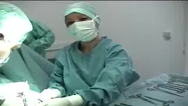
- Surgery Videos
- Laparoscopy
- Pediatrics
- Funny Videos
- Cardiothoracic Surgery
- Nursing Videos
- Plastic Surgery
- Otorhinolaryngology
- Histology and Histopathology
- Neurosurgery
- Dermatology
- Pediatric Surgery
- Urology
- Dentistry
- Oncology and Cancers
- Anatomy Videos
- Health and Fitness
- Radiology
- Anaesthesia
- Physical Therapy
- Pharmacology
- Interventional Radiology
- Cardiology
- Endocrinology
- Gynecology
- Emergency Medicine
- Psychiatry and Psychology
- Childbirth Videos
- General Medical Videos
- Nephrology
- Physiology
- Diet and Food Health
- Diabetes Mellitus
- Neurology
- Women Health
- Osteoporosis
- Gastroenterology
- Pulmonology
- Hematology
- Rheumatology
- Toxicology
- Nuclear Medicine
- Infectious Diseases
- Vascular Disease
- Reproductive Health
- Burns and Wound Healing
- Other
Top videos
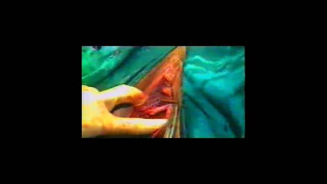
This video demonstrates the management of a large abscess in the emergency department. This abscess probably began as a sebaceous cyst that became infected.
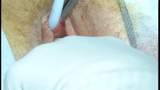
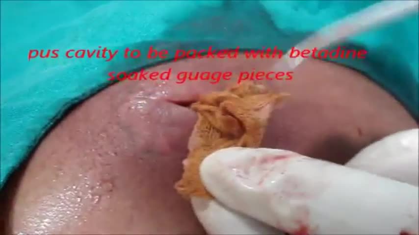
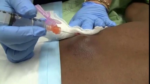
This poor old lady came with swelling in her left buttock for 10 days.She had history of injection in her buttocks two weeks back. She developed painful swelling and redness in her left gluteal region with difficulty in walking.It was diagnosed as injection abscess left gluteal region which needs incision and drainage under local anesthesia.Patient part painted and drapped.2% Lignocaine with adrenaline was infiltrated around the swelling for proper filed block.I use no-11 blade for stab incision over the swelling at the most fluctuating point of the abscess.You can watch how pus was flowing out from the cavity.The aim is to drain all pus from the abscess cavity.Finger exploration is essential to break all loculi inside the cavity, to know the depth and extend of the cavity and to fascilitate proper drainage of residual pus.after pus evacuation,, the cavity should be irrigated with normal saline and betadine solution.lastly the cavity to be packed with betadine soaked guage pieces.Proper dressing is essential.the dressing to be changed after 24 hours.daily dressing is essential with a good antibiotic coverage.the cavity usually obliterates within a period of seven to ten days.
![Female Foley Insertion (Urinary Catheter) [How to Insert Nursing Skills]](https://i.ytimg.com/vi/Mq4Yh0-iozY/maxresdefault.jpg)
Pass your tests and improve your grades with the below FREE resources:
1) A FREE 140 Must Know Meds book
Click here to get your FREE copy of the 140 Must Know Meds Book: https://bit.ly/41rxSt0
2) A FREE test-taking tips webinar
Join us for our free test-taking tips webinar to boost your exam scores: https://bit.ly/nursingtesttaking
You can now test your knowledge with a free lesson quiz on NURSING.com!
Click here to take a free quiz: https://bit.ly/3HwJr8t
FREE Nursing School Cheat Sheets at: http://www.NURSING.com
Get the full lesson on Female Foley Insertion here:
https://nursing.com/lesson/ski....lls-03-01-inserting-
Get the Male Foley Insertion lesson here:
https://nursing.com/lesson/ski....lls-03-02-inserting-
Get the Sterile glove application lesson here:
https://nursing.com/lesson/ski....lls-01-04-sterile-gl
Check out our new Nurse Care Plan Lessons here:
https://bit.ly/3BPRfPL
Get Access to Thousands of Lessons here:
https://nursing.com/courses/
Welcome to the NURSING Family, we call it the most supportive nursing cohort on the planet.
At NURSING.com, we want to help you remove the stress and overwhelm of nursing school so that you can focus on becoming an amazing nurse.
Check out our freebies and learn more at: (http://www.nursing.com)
Female Foley Insertion (Urinary Catheter)- Nursing Skills
In this video, we’re going to look at inserting a Foley catheter in a female. Of course make sure you’ve verified your order and told the patient what’s happening. You’ll also typically want to perform perineal care before you start. Then, you’ll want to assist the patient into the appropriate position. For females, that’s supine with their knees bent and feet close to their hips – allowing their knees to fall to the side. You may need a helper to help hold the patient in this position. We love you guys! Go out and be your best selves today! And, as always, happy nursing!
Bookmarks:
0.05 Female Foley insertion introduction
0.15 Patient positioning
0.27 Opening the sterile kit
1.41 Setting up the sterile field
2.25 Prepping the remaining Foley kit items
2.34 Catheter lubrication
3.00 Saline syringe attachment
3.10 Iodine, swabs and cleansing the area
3.52 Catheter insertion (into urethra)
4.06 Balloon inflation
4.25 Final catheter setting
4.31 Securing the catheter and bag
4.48 Discarding your supplies
5.00 Documentation
5.08 Foley insertion outro
Visit us at https://nursing.com/medical-disclaimer/ for disclaimer information.
NCLEX®, NCLEX-RN® are registered trademarks of the National Council of State Boards of Nursing, INC. and hold no affiliation with NURSING.com.
A video showing how to insert the Intra Uterine Device (IUD)
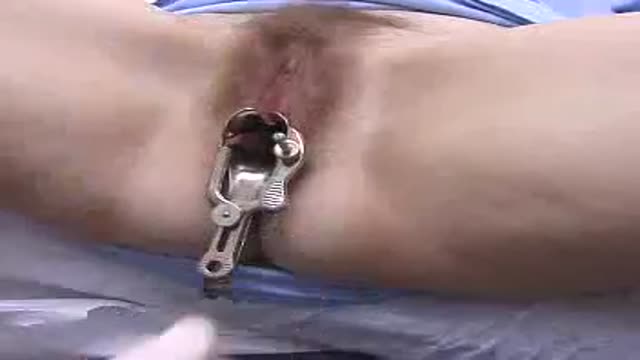
Adult circumcision video
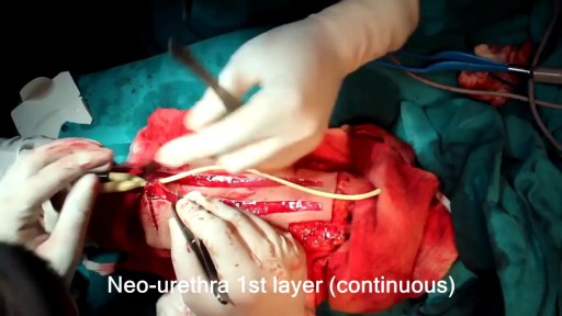
Sex reassignment surgery for male-to-female involves reshaping the male genitals into a form with the appearance of, and, as far as possible, the function of female genitalia. Prior to any surgeries, patients usually undergo hormone replacement therapy (HRT), and, depending on the age at which HRT begins, facial hair removal. There are associated surgeries patients may elect to, including facial feminization surgery, breast augmentation, and various other procedures
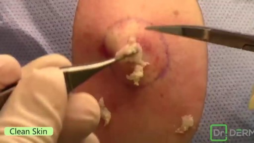
Popping Huge Cyst in the Back
Part 2. Full Obstetric examination and normal delivery by Egyptian doctor Hussein Sulayman and the video is in English showing: Obstetric Examination Episiotomy Obstetric Forceps Obstetric Instruments
Position the patient with her buttocks just at the edge or just over the edge of the exam table. If she is not down far enough, inserting the speculum can be more difficult for you and uncomfortable for her.
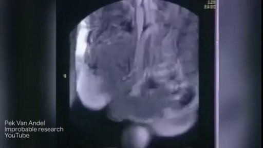
Watch that video of MRI Scans Human Body Internal Organs During Sex

http://www.vaginal-ultrasound.com A demonstration of a vaginal ultrasound.

Men and women have anatomical differences when it comes to genitals, but orgasms are fundamentally very similar. The female orgasm lasts longer than the male, ranging about 20 seconds compared to 3 to 10 seconds, but men do experience more orgasms.
Watch that Female to Male Gender Changing Surgery

Best Sex Position to Get Pregnant Fast

A digital rectal examination (DRE) is a simple procedure doctors use to examine the lower rectum and other internal organs. A DRE is done for a number of reasons. It's a quick, easy way to check the health of a man's prostate gland. It can detect conditions like an enlarged prostate
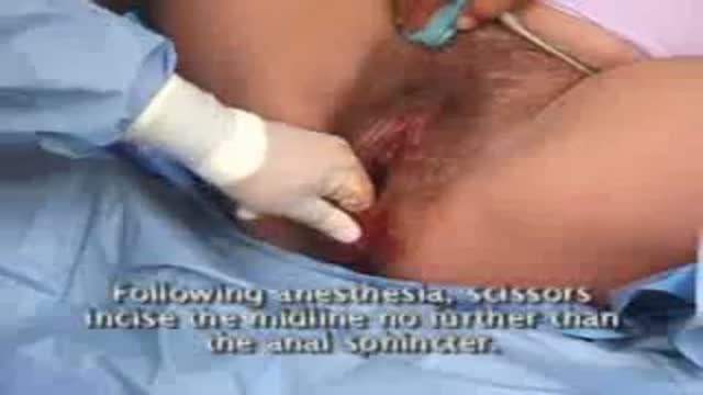
This video demonstrates the use of an episiotomy to facilitate vaginal delivery of a baby

An old video showing how to give an enema

An interesting documentary video from Discovery channel from the show "Human Files Night" explaining the anatomy and everything related to female genital tract in a very interesting professional way.

CORRECTION: After review of this video, it is clear that this video is of a baby who is near full term (40 weeks) based on the size. Late trimester "abortions" are defined only to viability of a baby (24 weeks) A 24 week baby is much smaller than this baby shown and by definition this is not a late "abortion" procedure. The proper labeling of this video should be management of a deceased breech baby with "head entrapment" as this was almost certainly a naturally occuring delivery and an OB nightmare (Reviewed by Dr. Frederick Bright)
