Top videoer

This baby was born with an adult sized tongue - and she just completed a surgery that will change her life.
Plasma cell dyscrasias are disorders of the plasma cells. Plasma cell dyscrasias are produced as a result of abnormal proliferation of a monoclonal population of plasma cells that may or may not secrete detectable levels of a monoclonal immunoglobulin or immunoglobulin fragment (paraprotein or M protein).
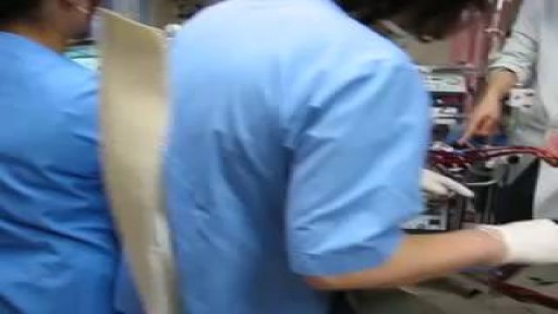
Start of CRRT circuit within ECMO
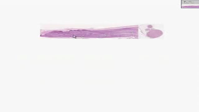
Histology of Peripheral Nerve
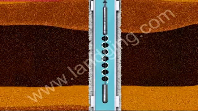
http://www.landging.com/through-tubing.html
This through-tubing perforation animation demonstrates new oil drilling technology.

Gynecology 3D Animation

At Nationwide Children’s, our Department of General Pediatric Surgery provides comprehensive surgical care for infants, children and adolescents with congenital and acquired conditions, including major congenital anomalies, traumatic and thermal injuries, and tumors. As the second largest pediatric treatment center in the United States our surgeons perform more than 4,000 operative procedures every year. We are dedicated to clinical excellence, generation of new knowledge through research and the training of the next generation of leaders in children’s surgery. Under the umbrella of a unified program, 11 surgical departments share a common mission, philosophy and approach to patient care.
Pediatric Surgery Program: https://bit.ly/3t4QZef
Pediatric Surgery Fellowship and Residency: https://bit.ly/3qWAWwd
Meet our Pediatric Surgery Team: https://bit.ly/3n39dJh
Fellowship Programs: https://bit.ly/3EX1JNX
Surgical Services: https://bit.ly/3eYDlB8
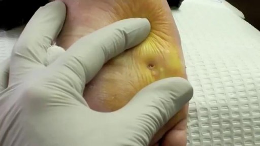
Curettage, electrosurgery, and laser surgery are more likely than cryotherapy to leave scars, so they are usually reserved for hard-to-remove or recurring warts. If you have a large area of warts, curettage may not be an effective treatment. Some surgical treatments may be too painful for some children.

Histology of Neurovascular Bundle
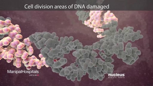
The drugs known as targeted therapy help stop cancer from growing and spreading. They work by targeting specific genes or proteins. These genes and proteins are found in cancer cells or in cells related to cancer growth, like blood vessel cells. Doctors often use targeted therapy with chemotherapy and other treatments.
complications from using a urinary catheter include: allergic reaction to the material used in the catheter, such as latex. bladder stones. blood in the urine. injury to the urethra. kidney damage (with long-term indwelling catheters) septicemia, or infection of the urinary tract, kidneys, or blood.
Foreign Body(FB) Airway (Whistle) was inhailed by a child causing intermitent stridor & respiratory distress.FForeign Body was removed successfully by rigid endoscopy under General Anesthesia (G/A).The relevant steps of procedure are shown

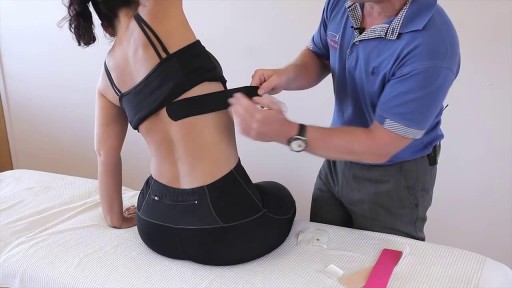
this video he is demonstrating how to apply Kinesiology Tape for a patient that presents with rib or intercostal pain
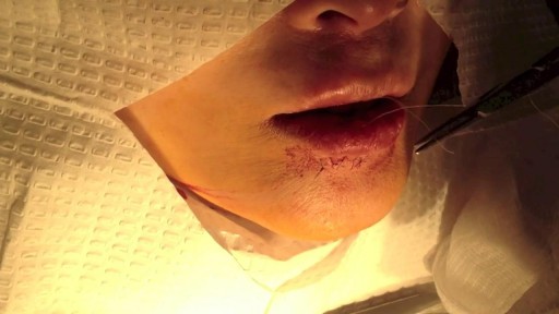
This video details the layered closure of a through-and-through facial laceration

UChicago Medicine organ transplant surgeon Dr. Rolf Barth explains a how the laparoscopic donor nephrectomy – also known as the single-port nephrectomy – procedure works to remove an organ donor’s kidney from their body to be transplanted into a recipient. This minimally invasive kidney donor transplant surgery allows living organ donors the get back to their lives more quickly than the traditional approach and leaves them with a nearly invisible scar in the belly button.
Learn more about living kidney donation: https://www.uchicagomedicine.o....rg/conditions-servic
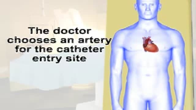
This video gives you an overview of how a cardiac catheterization is performed.
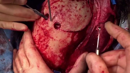
Mesenteric Vessel Ligation
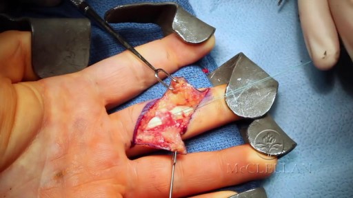
Flexor Digitorum Profundus (FDP) Finger Tendon Repair
