- Physical Examination
- Surgical Examination
- Ophthalmology
- Clinical Skills
- Orthopedics
- Surgery Videos
- Laparoscopy
- Pediatrics
- Funny Videos
- Cardiothoracic Surgery
- Nursing Videos
- Plastic Surgery
- Otorhinolaryngology
- Histology and Histopathology
- Neurosurgery
- Dermatology
- Pediatric Surgery
- Urology
- Dentistry
- Oncology and Cancers
- Anatomy Videos
- Health and Fitness
- Radiology
- Anaesthesia
- Physical Therapy
- Pharmacology
- Interventional Radiology
- Cardiology
- Endocrinology
- Gynecology
- Emergency Medicine
- Psychiatry and Psychology
- Childbirth Videos
- General Medical Videos
- Nephrology
- Physiology
- Diet and Food Health
- Diabetes Mellitus
- Neurology
- Women Health
- Osteoporosis
- Gastroenterology
- Pulmonology
- Hematology
- Rheumatology
- Toxicology
- Nuclear Medicine
- Infectious Diseases
- Vascular Disease
- Reproductive Health
- Burns and Wound Healing
- Other
Top videos
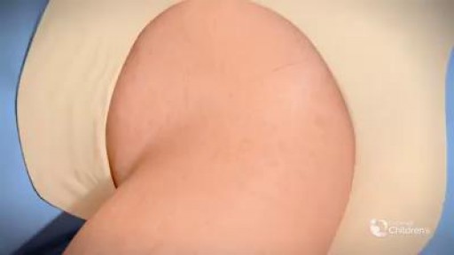
The definition of DDH is not universally agreed upon. Typically, the term DDH is used in referring to patients who are born with dislocation or instability of the hip, which may then result in hip dysplasia. More broadly, DDH may be defined simply as abnormal growth of the hip. Abnormal development of the hip includes the osseous structures, such as the acetabulum and the proximal femur, as well as the labrum, capsule, and other soft tissues. This condition may occur at any time, from conception to skeletal maturity. The author prefers to use the term hip dysplasia, considering it both simpler and more accurate. Internationally, this disorder is still referred to as congenital dislocation of the hip.
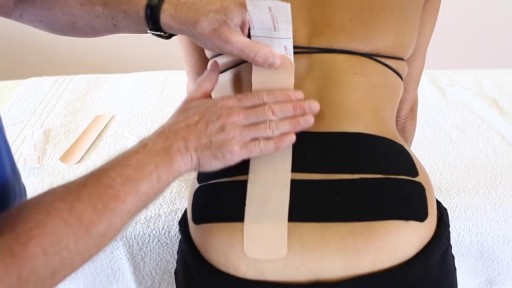
Dysfunction in the sacroiliac joint, also called the SI joint, can sometimes cause lower back and/or leg pain. Leg pain from sacroiliac joint dysfunction can be particularly difficult to differentiate from radiating leg pain caused by a lumbar disc herniation (sciatica) as they can feel quite similar.
Vaginal Yeast Infection
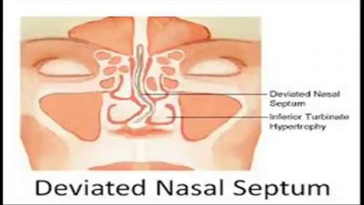
When a deviated septum is severe, it can block one side of your nose and reduce airflow, causing difficulty breathing. The additional exposure of a deviated septum to the drying effect of airflow through the nose may sometimes contribute to crusting or bleeding in certain individuals. Nasal obstruction can occur from a deviated nasal septum, from swelling of the tissues lining the nose, or from both. Treatment of nasal obstruction may include medications to reduce the swelling or nasal dilators that help open the nasal passages. To correct a deviated septum, surgery is necessar
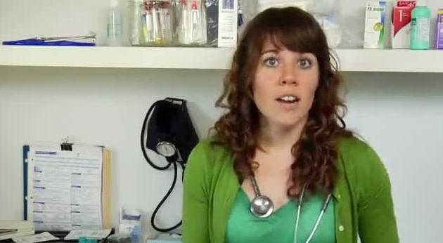
Possible causes include a combination of biological, psychological, and social sources of distress. Increasingly, research suggests these factors may cause changes in brain function, including altered activity of certain neural circuits in the brain. The persistent feeling of sadness or loss of interest that characterizes major depression can lead to a range of behavioral and physical symptoms. These may include changes in sleep, appetite, energy level, concentration, daily behavior, or self-esteem. Depression can also be associated with thoughts of suicide. The mainstay of treatment is usually medication, talk therapy, or a combination of the two. Increasingly, research suggests these treatments may normalize brain changes associated with depression.
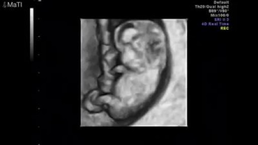
Pregnancy ultrasounds are performed mainly using transabdominal ultrasound. For many women, especially after 8 weeks gestation, sufficient information about the baby may be obtained with transabdominal ultrasound only. However, in the early pregnancy, the developing embryo is very small (at 6 weeks gestation, the baby is only 5-9mm long) and a transvaginal ultrasound may be required to get a better image of the baby. Transvaginal ultrasound is safe and commonly performed during all stages of pregnancy, including the first trimester. It will not harm you or your baby.
There many concerns and questions about how a breast augmentation procedure works. Dr. Linder a highly qualified plastic surgeon based in Beverly Hills, demystifies those worries. Dr. Stuart Linder is a Beverly Hills board certified plastic surgeon, specializing in body sculpting and reconstructive procedures including breast augmentation, reduction, lift, liposuction and tummy tuck. He is board-certified by the American Board of Plastic Surgery and is affiliated with the American College of Surgeons, the American Society of Plastic and Reconstructive Surgeons and the American Medical Association. Website: www.drlinder.com

#HerniaRepair #HerniaSurgery #LaparoscopicHerniaRepair #OpenHerniaRepair #InguinalHernia #UmbilicalHernia #VentralHernia #MeshRepair #HerniaRecovery #HerniaComplications
hernia operation
hernia treatment
hernia
hernia treatment at home
hernia operation and recovery
hernia surgery and recovery
terapi hernia
inguinal hernia treatment without surgery
harnia
hernia surgery
hernia surgery animation
harniya operation
hernia symptoms men
harniya
hernia ka ilaj
hernia laparoscopic surgery animation
abdominal hernia treatment without sur...
hernia symptoms
turun berok
abdominal hernia
hernia exercises without surgery
hernia operation in 3d animation
inguinal hernia surgery
umbilical hernia symptoms and treatment
harnia operation
harniya ka ilaj
hernia animation
hernia belt
hernias
how to treat hernia without surgery
inguinal hernia recovery after surgery
ngiri
open hernia surgery
skates
turun bero
3d surgery
after hernia surgery recovery
appam kaise banate hain
hernia belt for men
hernia belt how to use
hernia exam
hernia inguinal sintomas
hernia ka operation kaisa hota hai
hernia operation ke baad exercise
hernia operation video
hernia repair
hernia repair mesh complications
hernia repair surgery animation
hernia surgery recovery tips
hernie abdominale
herniya

The world's leading tubal reversal doctors explain tubal ligation reversal procedure and success rates
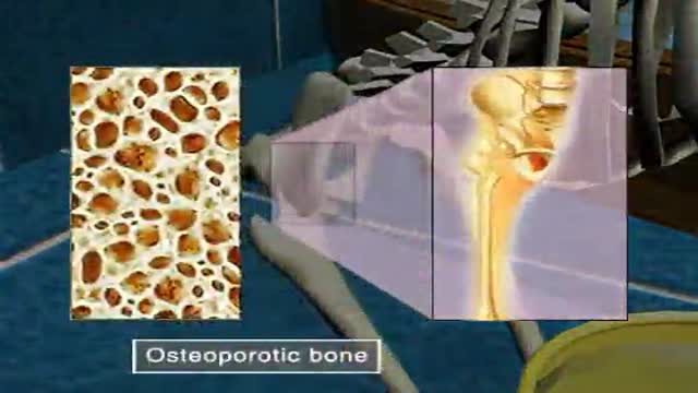
Osteoporosis
Podalic version is an obstetric procedure wherein the fetus is turned within the womb such that one or both feet present through the cervix during childbirth. It is used most often in cases where the fetus lies transversely or in another abnormal position in the womb.

Watch Dr. Robert Thomas, of Panorama Orthopedics & Spine Center, perform a Mako Knee replacement. He narrates each step of the process.

The OrthoIllustrated® animation for total knee replacement is an educational tool to help patients better understand the diagnosis and treatment of arthritis.
- - - - -
Why Work Arthrex https://www.arthrex.com/job-seeker
Find an Arthrex Surgeon: https://doctorfinder.orthoillustrated.com
- - - - -
Join the Community:
LinkedIn: https://www.linkedin.com/company/arthrex
Facebook: https://www.facebook.com/Arthrex
Instagram: https://www.instagram.com/arthrex_inc/
Twitter: https://twitter.com/Arthrex
TikTok: https://www.tiktok.com/@arthrex
- - - - -
Arthrex Inc., headquartered in Naples, Florida, is a global leader in orthopedic surgical device design, research, manufacturing, and medical education. Arthrex develops and releases more than 1,000 new products and procedures every year to advance minimally invasive orthopedics worldwide.
For more information, visit https://www.arthrex.com
- - - - -
OrthoPedia is an innovative educational website that was created for anyone interested in learning about orthopedics from the first-year student to the experienced orthopedic surgeon.
Visit https://www.orthopedia.com to experience the future of Medical Education.

Many mothers notice engorgement, or over-filled breasts, at some point or the other while they are breast-feeding their baby and it is especially common to experience when your baby is first born and you are just starting to make milk. So for the first couple of days you make colostrum and then 2-5 days later your milk comes in. And sometimes it comes in with a vengeance and all of the sudden you feel really full and it can be painful and very uncomfortable. Normally your milk supply will even out and start to work well with your babys demand, so it is kind of a supply and demand type of function, but until then, if you feel engorgement, there are a few things you can do to relieve it. If you are nursing your baby on demand this will usually help to self-regulate and most young babies want to eat every 2-3 hours and sometimes even every hour. So, basically, the more often your breasts are emptied the more relief you will feel. But on the same hand, the more you nurse the more milk your body will probably produce. This is why it is good to go off of your babys cues because then you will make what your baby needs and hopefully not much more. But if you are making more than your baby needs and you find that you are still full after feedings you will probably have to either manually express some milk or pump it off, so have a good pump available in case you need to, and if you don't, you can manually express the milk by gently massaging from the armpit down towards the nipple. And you can also try using heat prior to nursing your baby or pumping milk off and this will also help to relax things and help you to get the milk out. Take a warm shower and then feed your baby or use a warm compress.

Few facts and information are summed up in this short video related to Mental Health, Psychiatry and Depression.
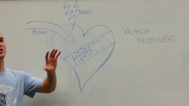
The maneuver is commonly used during some activities: Straining to have a bowel movement Blowing a stuffy nose Certain medical tests or exams As a pressure equalization technique by scuba divers, sky divers and airplane passengers The effect of the Valsalva Maneuver is a drastic increase in the pressure within the thoracic cavity.


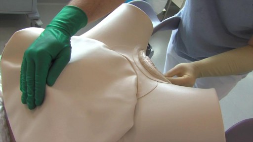
A cervical rib in humans is an extra rib which arises from the seventh cervical vertebra. Sometimes known as "neck ribs", their presence is a congenital abnormality located above the normal first rib. A cervical rib is estimated to occur in 0.2% (1 in 500 people) to 0.5% of the population.
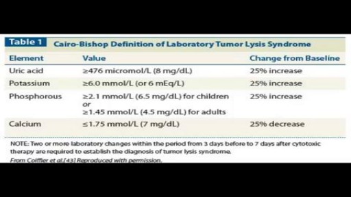
Presence of abdominal pain and distension. Presence of urinary symptoms - Such as dysuria, oliguria, flank pain, and hematuria. Occurrence of any symptoms of hypocalcemia - Such as anorexia, vomiting, cramps, seizures, spasms, altered mental status, and tetany. Symptoms of hyperkalemia - Such as weakness and paralysis.
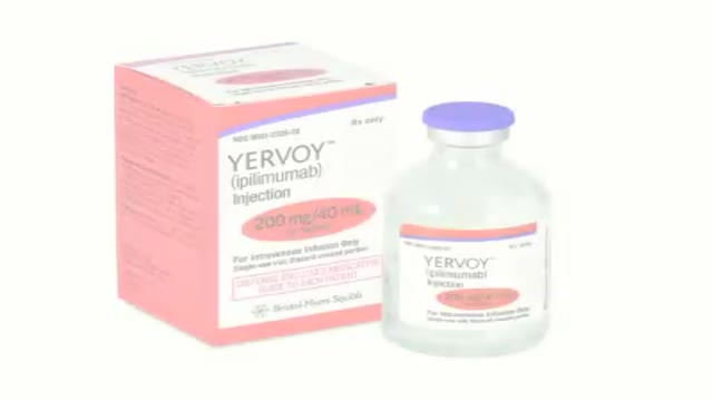
How revolutionizing advancements helps patients with metastatic melanoma kick start the body’s immune system to increase survival.
