- Physical Examination
- Surgical Examination
- Ophthalmology
- Clinical Skills
- Orthopedics
- Surgery Videos
- Laparoscopy
- Pediatrics
- Funny Videos
- Cardiothoracic Surgery
- Nursing Videos
- Plastic Surgery
- Otorhinolaryngology
- Histology and Histopathology
- Neurosurgery
- Dermatology
- Pediatric Surgery
- Urology
- Dentistry
- Oncology and Cancers
- Anatomy Videos
- Health and Fitness
- Radiology
- Anaesthesia
- Physical Therapy
- Pharmacology
- Interventional Radiology
- Cardiology
- Endocrinology
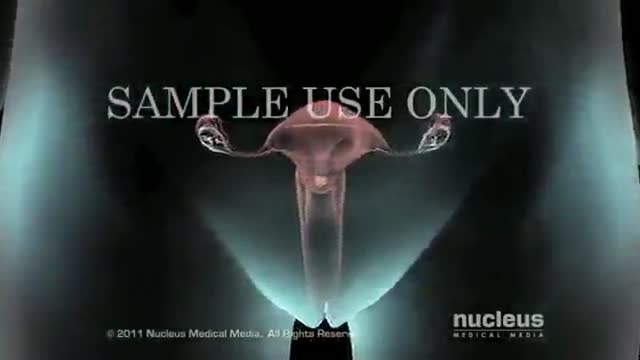
- Gynecology
- Emergency Medicine
- Psychiatry and Psychology
- Childbirth Videos
- General Medical Videos
- Nephrology
- Physiology
- Diet and Food Health
- Diabetes Mellitus
- Neurology
- Women Health
- Osteoporosis
- Gastroenterology
- Pulmonology
- Hematology
- Rheumatology
- Toxicology
- Nuclear Medicine
- Infectious Diseases
- Vascular Disease
- Reproductive Health
- Burns and Wound Healing
- Other
Top videos
The goal of COPD management is to improve a patient’s functional status and quality of life by preserving optimal lung function, improving symptoms, and preventing the recurrence of exacerbations. Currently, no treatments aside from lung transplantation have been shown to significantly improve lung function or decrease mortality; however, oxygen therapy (when appropriate) and smoking cessation may reduce mortality. Once the diagnosis of COPD is established, it is important to educate the patient about the disease and to encourage his or her active participation in therapy.
Kegel exercises strengthen the pelvic floor muscles, which support the uterus, bladder, small intestine and rectum. You can do Kegel exercises, also known as pelvic floor muscle training, just about anytime. Start by understanding what Kegel exercises can do for you — then follow step-by-step instructions for contracting and relaxing your pelvic floor muscles.
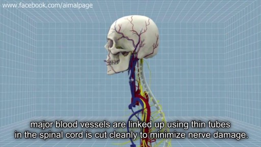
First Head Transplant Surgery
Catheter ablation is a minimally invasive procedure to treat atrial fibrillation. It can relieve symptoms and improve quality of life. During an ablation, the doctor destroys tiny areas in the heart that are firing off abnormal electrical impulses and causing atrial fibrillation. You will be given medicine to help you relax. A local anesthetic will numb the site where the catheter is inserted. Sometimes, general anesthesia is used. The procedure is done in a hospital where you can be watched carefully. Thin, flexible wires called catheters are inserted into a vein, typically in the groin or neck, and threaded up into the heart. There is an electrode at the tip of the wires. The electrode sends out radio waves that create heat. This heat destroys the heart tissue that causes atrial fibrillation or the heart tissue that keeps it happening. Another option is to use freezing cold to destroy the heart tissue. Sometimes, abnormal impulses come from inside a pulmonary vein and cause atrial fibrillation. (The pulmonary veins bring blood back from the lungs to the heart.) Catheter ablation in a pulmonary vein can block these impulses and keep atrial fibrillation from happening. View a slideshow of catheter ablation to see how the heart's electrical system works, how atrial fibrillation happens, and how ablation is done. Atrial Fibrillation: Should I Have Catheter Ablation? AV node ablation AV node ablation is a slightly different type of ablation procedure for atrial fibrillation. AV node ablation can control symptoms of atrial fibrillation in some people. It might be right for you if medicine has not worked, catheter ablation did not stop your atrial fibrillation, or you cannot have catheter ablation. With AV node ablation, the entire atrioventricular (AV) node is destroyed. After the AV node is destroyed, it can no longer send impulses to the lower chambers of the heart (ventricles). This controls atrial fibrillation symptoms. After AV node ablation, a permanent pacemaker is needed to regulate your heart rhythm. Nodal ablation can control your heart rate and reduce your symptoms, but it does not prevent or cure atrial fibrillation. AV node ablation helps about 9 out of 10 people.1 The procedure has a low risk of serious problems.2 View a slideshow of AV node ablation to see how the heart's electrical system works, how atrial fibrillation happens, and how AV node ablation is performed.
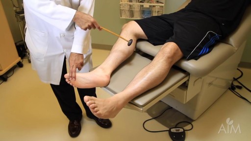
In a normal person, when a muscle tendon is tapped briskly, the muscle immediately contracts due to a two-neuron reflex arc involving the spinal or brainstem segment that innervates the muscle. The afferent neuron whose cell body lies in a dorsal root ganglion innervates the muscle or Golgi tendon organ associated with the muscles; the efferent neuron is an alpha motoneuron in the anterior horn of the cord. The cerebral cortex and a number of brainstem nuclei exert influence over the sensory input of the muscle spindles by means of the gamma motoneurons that are located in the anterior horn; these neurons supply a set of muscle fibers that control the length of the muscle spindle itself.
Vaginal Yeast Infection
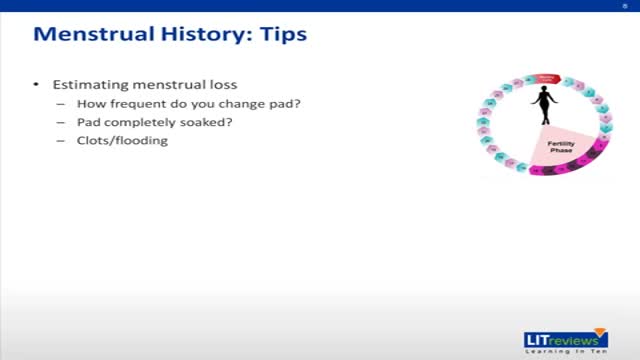
General Considerations Because a discussion of reproductive issues may be difficult for some women, it is important to obtain the history in a relaxed and private setting. The patient should be clothed, particularly if she is meeting the provider for the first time. Ordinarily, the patient should be interviewed alone. Exceptions may be made for children, adolescents, and mentally impaired women, or if the patient specifically requests the presence of a caretaker, friend, or family member. However, even in these circumstances, it is desirable for the patient to have some time to speak with the clinician privately. The manner of address should be formal using the title Mrs., Ms., Miss, or Dr. with the patient’s surname, unless the patient requests otherwise. In some settings, it may be appropriate for nursing staff to be involved with history taking. A nurse may be perceived as less threatening, and may be able to take the history in a less hurried manner.1 The provider can verify the history and focus on areas of concern. Alternatively, it may be helpful to ask the patient to complete a self-history form on paper or by computer prior to speaking with the provider. This allows the provider to devote time to addressing positive responses, and ensures that important questions are not missed. Hasley2 showed that responses to a computer-based questionnaire designed to update a patient’s gynecologic history were equivalent to those obtained during a personal interview. Several studies involving patients in non-gynecologic settings have shown that patients are more likely to provide sensitive information when responding to a computer-based questionnaire as opposed to a personal interview or even a paper questionnaire.3 In order to increase a patient’s level of comfort during the interview, questions should be asked in an open-ended and nonjudgmental way. Assumptions should not be made about aspects of the patient’s background such as sexual orientation. At the conclusion of the interview, patients should be asked whether there are concerns that they would like to discuss that were not addressed previously in the interview.

If you’re wondering ‘what’s the cause of my knee pain?’ or ‘what kind of knee pain do I have?’ the position of your knee pain can often tell you what type of knee pain you have.
You confirm this if you know the common symptoms an aggravations for each type of knee problem. So if you want to know ‘why my knee hurts’... here’s a quick look at the most common type of knee problems...
Patellofemoral Pain Syndrome (Or Runner’s Knee) (Old Name: Chondromalacia Patellae)
Infrapatellar Fat Pad Syndrome (Hoffa's Syndrome)
Patella Tendonitis (Jumper’s Knee)
Prepatellar Bursitis
Osgood-Schlatter Disease
Meniscus Tear
Medial Collateral Ligament Tear
Osteoarthritic Knee Pain
Pes Anserine Bursitis.
Iliotibial Band Syndrome
Quadriceps Tendinopathy
Popliteus Strain
Baker’s Cyst
ACL Or PCL Tear/Rupture
---------------------------------------
Check out my channel...
https://youtube.com/@BodyFixExercises
OTHER VIDEOS:
How To Fix Pain In The Front Of The Knee… (Runner's Knee) https://youtu.be/g0qmx_0enAA
Knee Strengthening Exercises To Prevent Knee Pain
https://youtu.be/Pk-ae_lyx7M
How To Treat Patellar Tendinopathy (Jumper’s Knee) & Quadriceps Tendinopathy
https://youtu.be/MkPwsb-rQwU
---------------------------------------
#bodyfixexercises #kneepainrelief #kneepain
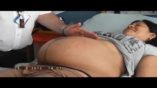
The obstetric examination is distinct from other examinations in that you, the clinician, are trying to assess the health of two individuals – the mother and the fetus – simultaneously. From the initial history, you should be able to judge the health of the pregnancy, any risk factors that need to be addressed, and any concerns from the parents. The history is an opportunity for you to find out how much the parents know about pregnancy, labour and delivery and if they have any preferences to which these events are carried out. A carefully taken history will also direct your attention to specific signs during the examination. As such, it is important that you develop a concise and systematic method of taking the history and carrying out the examination so that you do not miss any important information. This article focuses primarily on the examination. Pregnancy is a sensitive issue, especially for the primigravida’s. Therefore, extra care is needed when you approach a pregnant woman. Always obtain expressed informed consent before examining her and have a chaperone accompany you throughout the examination. A walk-through of what you will be doing is a good way of reassuring the patient and allows the examination to go on smoothly. It is also important to let your patient know that if the examination is too painful, she can stop at any time she wants. Finally, before you begin, you should always wash your hands, especially at an OSCE station.

From our beginnings in 1990 in primary healthcare, Healthway Medical has grown to become a respected medical group in Singapore. With over 100 clinics and medical centres, Healthway Medical has a wide network of medical centres and clinics in Singapore.
We offer comprehensive services including GP & family medicine clinics, health screening, adult specialists, baby & child specialists, dental services and allied healthcare services.
Intramedullary nailing of the tibia with suprapatellar entry and semi-extended positioning makes it technically easier to nail the proximal and distal fractures. The purpose of this article was to describe a simple method for suprapatellar nailing (SPN). A step-by-step run through of the surgical technique is described, including positioning of the patient. There are as yet only a few clinical studies that illustrate the complications with this method, and there has been no increased frequency of intraarticular damage. Within the body of the manuscript, information is included about intraarticular damage and comments with references about anterior knee pain.

https://bit.ly/3HIStRc #shorts
Tracheotomy and tracheostomy are surgical procedures that create an opening in the trachea (windpipe) to help patients breathe when they have difficulty doing so through the nose or mouth. Though they are similar in purpose, there are some key differences between them.
Tracheotomy is a temporary procedure that involves creating a small incision in the trachea to insert a breathing tube. The tube is typically removed once the patient no longer requires it, and the incision heals on its own. Tracheostomy, on the other hand, is a more permanent solution that involves creating a hole in the trachea and inserting a tracheostomy tube, which remains in place for an extended period.
Indications for these procedures include:
Airway obstruction due to trauma, tumors, or infection
Severe respiratory distress or failure
Prolonged mechanical ventilation
Inability to protect the airway due to neurological disorders or impaired consciousness
Steps for performing a tracheotomy and tracheostomy:
Preparation: The patient is positioned, and the neck area is cleaned and draped. Local anesthesia is often administered, although general anesthesia may be used in some cases.
Incision: A small incision is made in the neck, and the muscles and tissues are carefully separated to expose the trachea.
Tracheal opening: A small opening is made in the trachea, typically between the second and third tracheal rings.
Tube insertion: A tracheotomy tube is inserted through the incision and into the trachea for a tracheotomy, while a tracheostomy tube is inserted for a tracheostomy. Both tubes are secured in place.
Confirmation: Proper placement of the tube is confirmed by listening for breath sounds and checking for adequate ventilation.
Pre-operative care typically involves a thorough assessment of the patient's medical history, as well as any necessary imaging studies or lab tests to ensure the procedure is appropriate and safe. Informed consent should be obtained from the patient or their legal representative.
Post-operative care includes monitoring the patient's vital signs, ensuring the tube remains secure and patent, and managing any pain or discomfort. For tracheostomy patients, regular cleaning and maintenance of the stoma (the opening in the trachea) and the tracheostomy tube are essential to prevent infection and other complications. Long-term care may involve speech therapy, respiratory therapy, and support from a multidisciplinary team to address any ongoing needs.
It's crucial to remember that these procedures should only be performed by trained medical professionals in a clinical setting.
for additional information about this procedure check our article @ www.medicalartsshop.com
For more free resources, find us on Pinterest & Facebook pages:
https://www.pinterest.ca/medicalartsofficial/
https://www.facebook.com/Medicalartsofficial
https://www.youtube.com/@medic....alarts?sub_confirmat
https://www.instagram.com/medicalartsofficial/
https://www.tiktok.com/@medicalarts
This video and associated content are for entertainment and educational purposes only!!
A central venous catheter, also called a central line, is a long, thin, flexible tube used to give medicines, fluids, nutrients, or blood products over a long period of time, usually several weeks or more. A catheter is often inserted in the arm or chest through the skin into a large vein.

The world's leading tubal reversal doctors explain tubal ligation reversal procedure and success rates
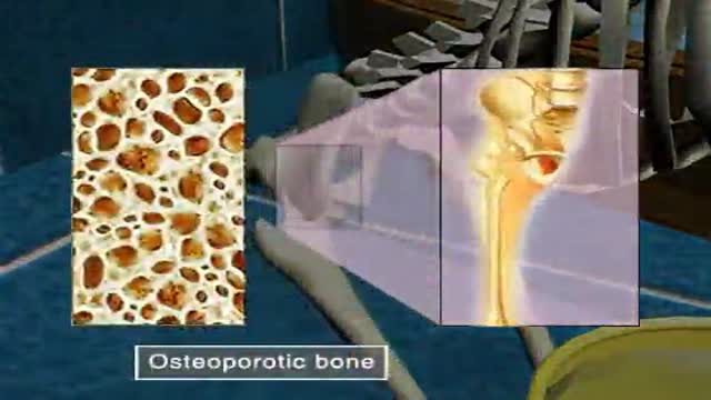
Osteoporosis
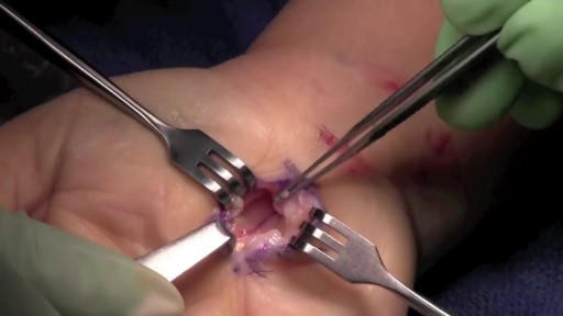
During open carpal tunnel release surgery, the transverse carpal ligament is cut, which releases pressure on the median nerve and relieves the symptoms of carpal tunnel syndrome. An incision is made at the base of the palm of the hand. This allows the doctor to see the transverse carpal ligament.
Dilation and Curettage D and C
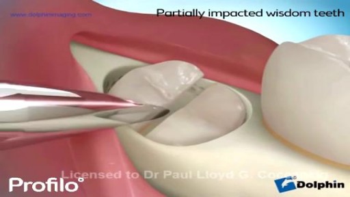
our dentist says it's time to remove your wisdom teeth. He may refer you to an oral surgeon, who will do the procedure in his office. It should only take a few days for you to heal and feel back to normal.
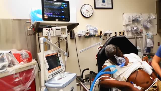
Pulmonary edema is almost always treated in the emergency room or hospital. You may need to be in an intensive care unit (ICU). Oxygen is given through a face mask or tiny plastic tubes are placed in the nose. A breathing tube may be placed into the windpipe (trachea) so you can be connected to a breathing machine (ventilator) if you cannot breathe well on your own. The cause of edema should be identified and treated quickly. For example, if a heart attack has caused the condition, it must be treated right away. Medicines that may be used include: Diuretics that remove excess fluid from the body Medicines that strengthen the heart muscle, control the heartbeat, or relieve pressure on the heart
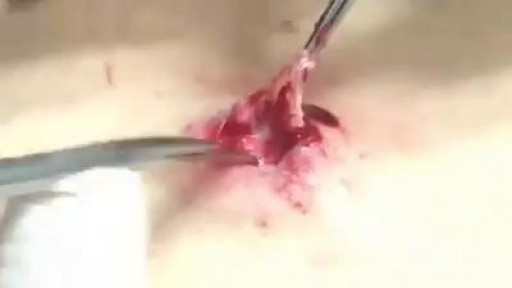
What is inside A Cyst? Watch it now
