Video teratas
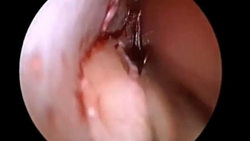
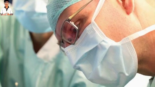
Repair Deviated Nasal Septum, Endoscopic Septoplasty, endoscopic surgery, Stapler repair of nasal septum, Dr B. Todd Schaeffer.
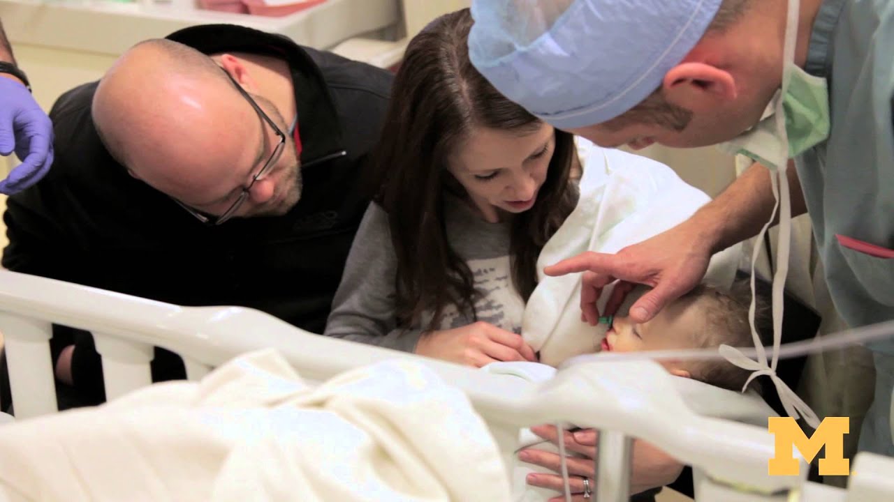
Cleft palate is among the most common birth defects affecting children in North America. The incomplete formation of the roof of the mouth can occur individually, or in addition to cleft lip. Cleft palate repair is a type of plastic surgery to correct this abnormal development both to restore function and a more normal appearance. This video explains what to expect for families scheduled for cleft palate surgery at the Craniofacial Anomalies Program at University of Michigan C.S. Mott Children's Hospital.
Learn more about our program at http://www.mottchildren.org/craniofacial
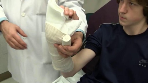
A boxer's fracture is a break through the bones of the hand that form the knuckles. Some doctors use the term "brawler's fracture" rather than "boxer's fracture" because a boxer is not likely to get this injury. The less well-trained brawlers have to learn how to punch without hurting themselves. The metacarpal bones in the hand connect the bones in the finger to the bones in the wrist. There are five metacarpal bones, one to connect each finger to the wrist. All of the metacarpal bones have the same anatomic structure. Each consists of the base, the shaft, the neck, and the head
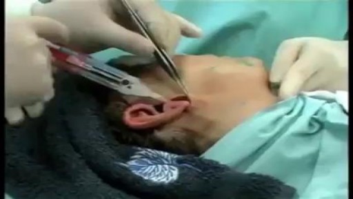
A bone-anchored hearing aid (BAHA) or bone-anchored hearing device,is a type of hearing aid based on bone conduction. It is primarily suited for people who have conductive hearing losses, unilateral hearing loss, single-sided deafness and people with mixed hearing losses who cannot otherwise wear 'in the ear' or 'behind the ear' hearing aids. They are more expensive than conventional hearing aids, and their placement involves invasive surgery which carries a risk of complications, although when complications do occur, they are usually minor. Two of the causes of hearing loss are lack of function in the inner ear(cochlea) and when the sound has problems in reaching the nerve cells of the inner ear. Example of the first include age-related hearing loss and hearing loss due to noise exposure. A patient born without external ear canals is an example of the latter for which a conventional hearing aid with a mould in the ear canal opening would not be effective. Some with this condition have normal inner ear function, as the external ear canal and the inner ear are developed at different stages during pregnancy. With normal inner anatomy, sound conducted by the skull bone improves hearing.
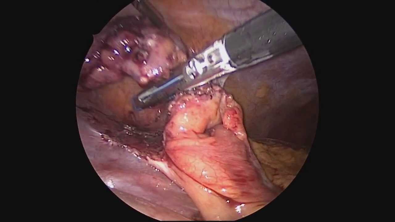
Dr. Celia Divino, Chief, Division of General Surgery at The Mount Sinai Hospital, performs a laparoscopic appendectomy. Visit the Division of General Surgery at http://bit.ly/18z944M. Click here to learn more about Dr. Celia Divino http://bit.ly/12RF0ee
Most people develop several moles (nevi) throughout adulthood. Moles can be found anywhere on the body, usually in sun-exposed areas, and are usually brown, smooth, and slightly raised. In most cases, a nevus is benign and doesn't require treatment. Rarely, they turn into melanoma or other skin cancers. A nevus that changes shape, grows bigger, or darkens should be evaluated for removal.
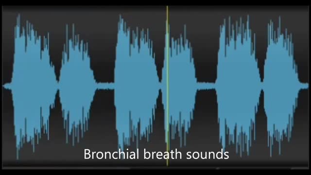
Physiological & pathological breath sounds
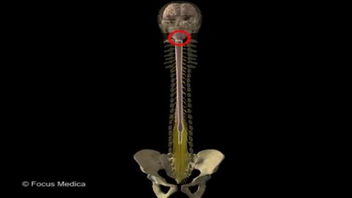
Back and Spinal cord Anatomy
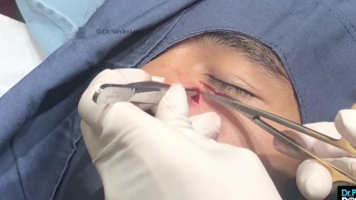
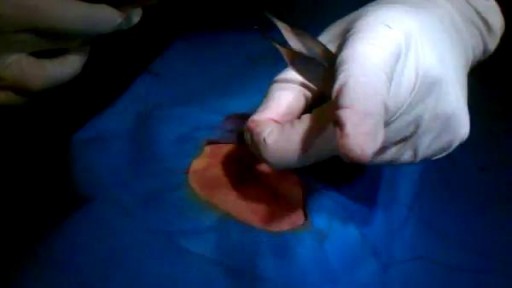
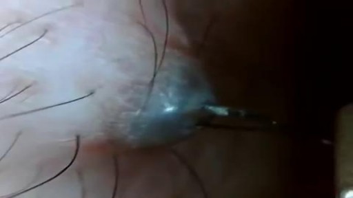
Nose Cyst Extraction

This video demonstrates why ears become clogged and why ear popping helps. The video also explains why ear popping may become difficult resulting in a persistent clogged or muffled ear especially after an ear infection.
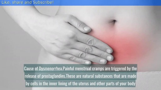
Can Birth Control Be a Dysmenorrhea Treatment? || Common gynaecological problems in women Dysmenorrhea is the medical term used for when you have painful menstrual cramps that occur immediately before or during your period. The pain can be so bad that it limits your daily activities. Dysmenorrhea is the most commonly reported menstrual disorder. It can affect up to 90 percent of young women. The Pill (as well as other hormonal contraceptives) can help in the treatment of dysmenorrhea.
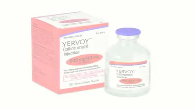
How revolutionizing advancements helps patients with metastatic melanoma kick start the body’s immune system to increase survival.

Immunomodulating effect of autohaemotherapy (a literature review). PMID 3534085 [PubMed indexed for MEDLINE]
J Hyg Epidemiol Microbiol Immunol. 1986;30(3):331-6.
Immunomodulating effect of autohaemotherapy (a literature review).
Klemparskaya NN, Shalnova GA, Ulanova AM, Kuzmina TD, Chuhrov AD.
Abstract
An analysis is presented of experimental and clinical data from different authors on the stimulating effect of autohaemotherapy with regard to the immunological reactivity of humans and animals as well as in vitro experiments with lymphocytes. Erythrolysate has been found to exert a more powerful effect than intact erythrocytes. The stimulating effect of autohaemotherapy on both irradiated and non-irradiated animals manifests itself in an increase in resistance to infection (increased LD50 in experimental infection), enhanced production of antibodies to microbial and tissue antigens and activated functioning of cell-mediated immune defence mechanisms. The favourable influences on radioresistance and the antitumour effect of authohaemotherapy are described. Induced desensitization plays an important part in the mechanism of action of autohaemotherapy. The administration of large doses of erythrocytes or of erythrolysate results in immunosuppression. Autohaemotherapy does not cause side effects and is feasible both on an in-and out-patient basis.
PMID: 3534085
[PubMed - indexed for MEDLINE]
http://www.ncbi.nlm.nih.gov/pubmed/3534085
Autohemotherapy: an immunization with our own blood
http://www.geocities.ws/autohemoterapiabr/
http://autohemoterapia.fortunecity.com/
http://www.geocities.ws/autohemoterapiabr/aht_english.htm
http://autohemoterapia.fortunecity.com/aht_english.htm
-
Auto-hemotherapy PDF files in GOOGLE sites:
https://sites.google.com/site/autohemotherapy/
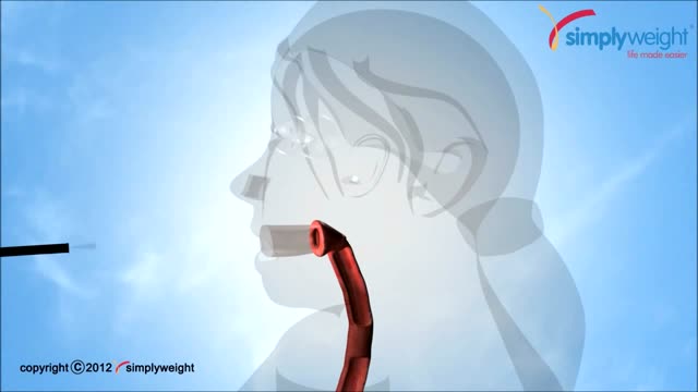
This animation shows how a balloon is placed inside the stomach with out an operation for weightloss. This is done through an endoscope which goes through the mouth.

What to expect during the day of a pediatric surgery at Sutter Children's Center Sacramento.
Plastic Surgeon in NY Doctor Michael Wolfeld of Wolfeld Plastic Surgery (http://www.drwolfeld.com) discusses case studies of of two patients who underwent a liposuction procedure.
This new surgical technique provide good stability for all type of fracture even severe comminution. Each fragment are reduced and several pin sleeves are inserted circumferentially and tighten by braded cable through the sleeve box. The final features of surgery seems blooming sunflower 'Himwari in Jananese'.
Cosmetic facial plastic surgery is surgery performed to enhance visual appearance of the facial structures and features. Common procedures include facelifts, eye lifts, rhinoplasty, chin and cheek implants, liposuction, and procedures to correct facial wrinkles.
