Mga nangungunang video
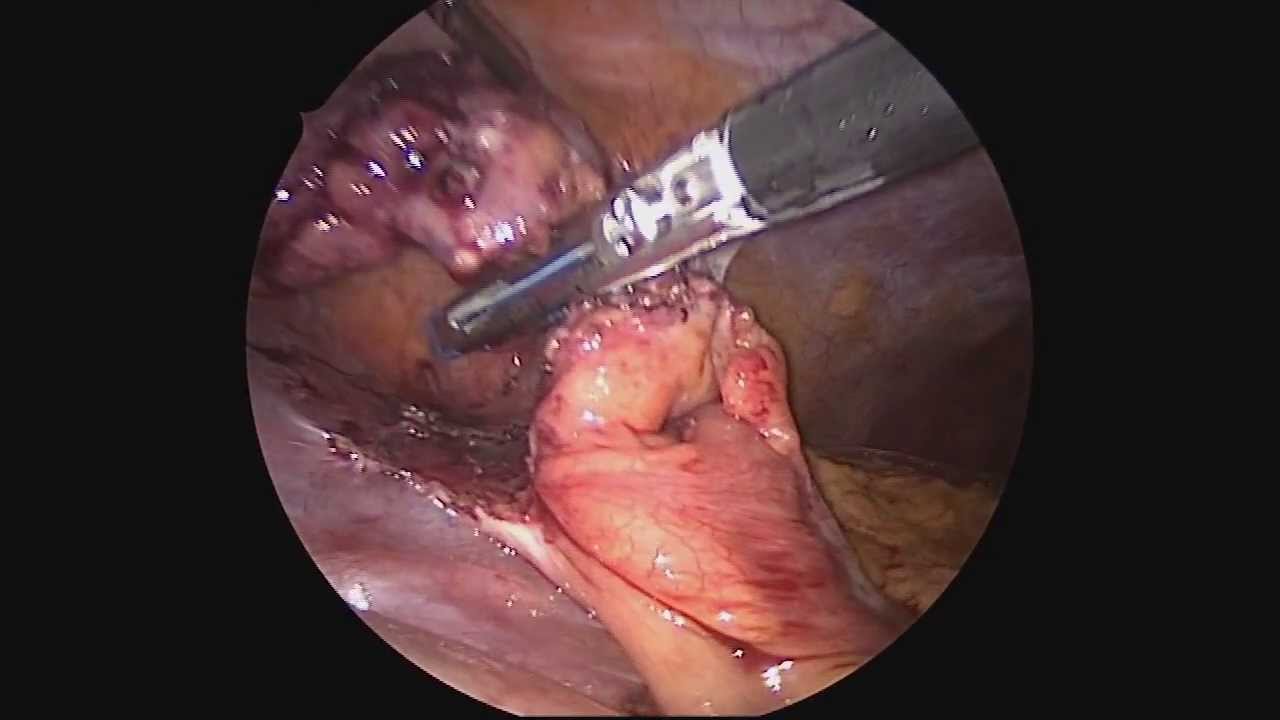
Dr. Celia Divino, Chief, Division of General Surgery at The Mount Sinai Hospital, performs a laparoscopic appendectomy. Visit the Division of General Surgery at http://bit.ly/18z944M. Click here to learn more about Dr. Celia Divino http://bit.ly/12RF0ee
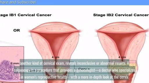
A cervical biopsy is a procedure that is sometimes done on women during an exam called a colposcopy to remove cervical tissue for examination. It is also called a punch biopsy. It is usually performed when a Pap smear result is either inconclusive or abnormal and a doctor wants to screen further for any cervical dysplasia or cervical cancer.
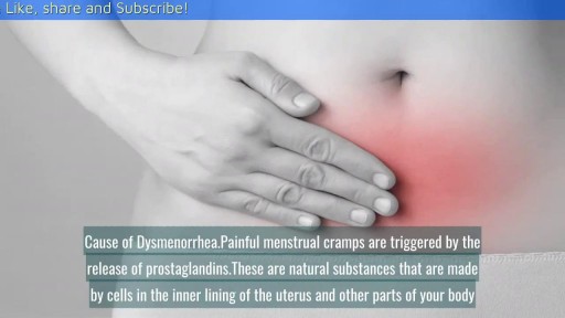
Can Birth Control Be a Dysmenorrhea Treatment? || Common gynaecological problems in women Dysmenorrhea is the medical term used for when you have painful menstrual cramps that occur immediately before or during your period. The pain can be so bad that it limits your daily activities. Dysmenorrhea is the most commonly reported menstrual disorder. It can affect up to 90 percent of young women. The Pill (as well as other hormonal contraceptives) can help in the treatment of dysmenorrhea.

Menorrhagia is the medical term for menstrual periods with abnormally heavy or prolonged bleeding. Although heavy menstrual bleeding is a common concern, most women don't experience blood loss severe enough to be defined as menorrhagia. With menorrhagia, you can't maintain your usual activities when you have your period because you have so much blood loss and cramping. If you dread your period because you have such heavy menstrual bleeding, talk with your doctor. There are many effective treatments for menorrhagia. Symptoms Signs and symptoms of menorrhagia may include: Soaking through one or more sanitary pads or tampons every hour for several consecutive hours Needing to use double sanitary protection to control your menstrual flow Needing to wake up to change sanitary protection during the night Bleeding for longer than a week Passing blood clots larger than a quarter Restricting daily activities due to heavy menstrual flow Symptoms of anemia, such as tiredness, fatigue or shortness of breath
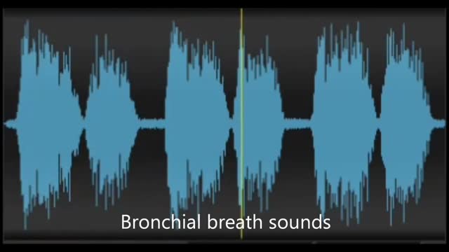
Physiological & pathological breath sounds

This video demonstrates why ears become clogged and why ear popping helps. The video also explains why ear popping may become difficult resulting in a persistent clogged or muffled ear especially after an ear infection.
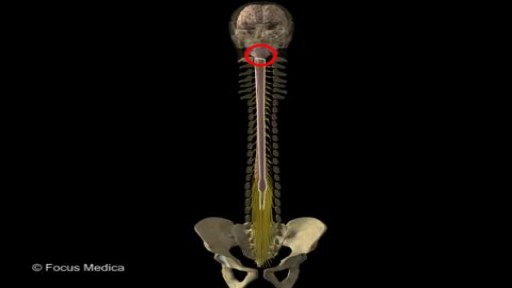
Back and Spinal cord Anatomy
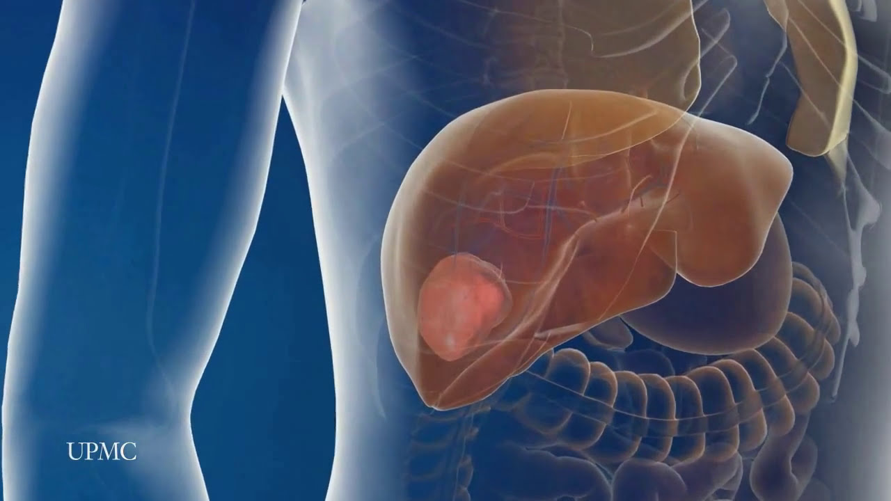
UPMC liver surgeons are among the most experienced in the world in performing minimally invasive liver surgery. Most patients benefit from less trauma and pain, minimal scarring, a shorter hospital stay, and faster recovery than from traditional surgery.
To learn more, please visit https://www.upmc.com/services/....liver-cancer/treatme
Repair Deviated Nasal Septum, Endoscopic Septoplasty, endoscopic surgery, Stapler repair of nasal septum, Dr B. Todd Schaeffer.
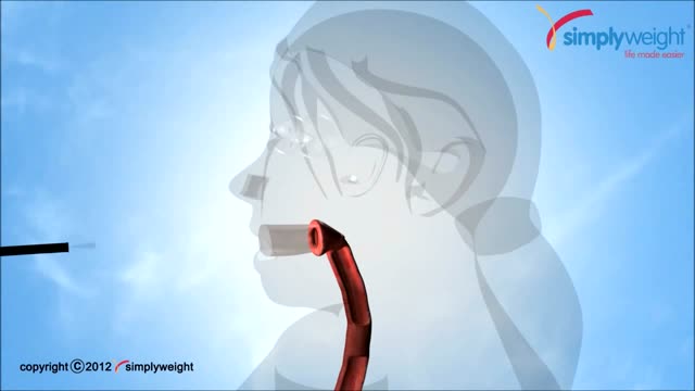
This animation shows how a balloon is placed inside the stomach with out an operation for weightloss. This is done through an endoscope which goes through the mouth.

Commentary:
0:24
He may not look like he’s in good condition but you can guesst that his somewhere in nirvana at this point
0:44
After the operation, this patient loses more than just color in his skin but apparently he loses his nipples as well
1:43
This sedated patient is equipped with his own hand-gun. No pun intended
2:17
His anesthesia dose came with the usual side effects of crazy talk with a dash of attitude and sarcasm
3:17
The only thing crazier than love is being sedated during an endometriosis surgery
4:36
This may come as a surprise to some but penguins don’t actually reside in Alaska. In case you didn’t know that well now you do
5:09
If the doctor advises you against something you can’t resist doing, how many of us would still listen to him?
6:35
When them meds start kicking in , it’s time to frame this experience as an excuse to divulge some of your secret fantasies
7:05
There’s a time and place dirty jokes but anesthesia told this guy any times the right time
7:24
Her 16 year old son talks about the last thing he remembers right after surgery and this is what he says
8:35
She’s definitely not in the mood at all. I wouldn’t wanna tick her off during this time if I were you
8:44
A feeling of relief after your operation may be followed by some emotional changes such as mood swings and over sensitivity
9:44
Even if you do say something you wouldn't normally say while you are under sedation, according to some doctors, “it's always kept within the operating room”
10:38
The beeping sounds of the medical equipments tip this patient over the edge. so she tries to drown out the noise with her own voice
11:08
Anyone who's received anesthesia can attest to feeling pretty loopy. Although many won't remember it's fairly common to say some wacky things after waking up
11:53
It's typical for people to feel sad or vulnerable after surgery. Kind of like how this girl is feeling right now
12:04
If she wasn’t under the influence in the hospital right now , it would be pretty hard to justify this type of behavior
12:17
Imagine working as an anesthesiologist. You might become numb to a lot of strange behaviors and everything unusual becomes the new norm for you
► Subscribe: https://bit.ly/3I4zXBT
Top Special Videos: https://bit.ly/3o64YOa
Acts Of Kindness: https://bit.ly/3E5FmXh
Try Not To Laugh Videos: https://bit.ly/3leRpdl
Social media:
► INSTAGRAM: https://www.instagram.com/topthings.tt/
► FACEBOOK: https://www.facebook.com/TopTh....ings-108385027422972
► TWITTER: https://twitter.com/TopThings10
► YOUTUBE: https://www.youtube.com/channe....l/UCArcrGQYzJhB_IfEl
#funnyvideos #anesthesia #anesthesiareactions
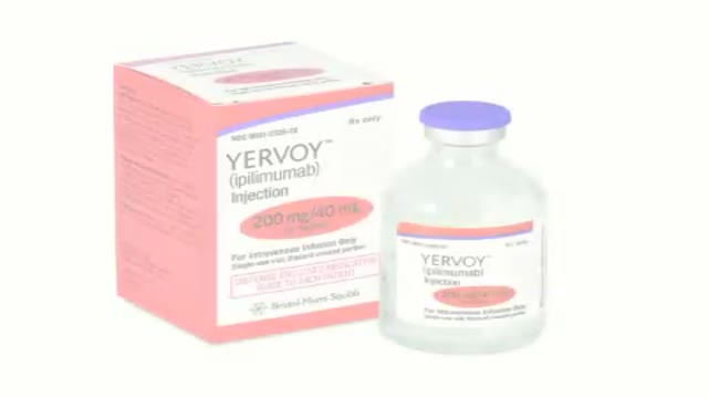
How revolutionizing advancements helps patients with metastatic melanoma kick start the body’s immune system to increase survival.
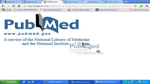
Immunomodulating effect of autohaemotherapy (a literature review). PMID 3534085 [PubMed indexed for MEDLINE]
J Hyg Epidemiol Microbiol Immunol. 1986;30(3):331-6.
Immunomodulating effect of autohaemotherapy (a literature review).
Klemparskaya NN, Shalnova GA, Ulanova AM, Kuzmina TD, Chuhrov AD.
Abstract
An analysis is presented of experimental and clinical data from different authors on the stimulating effect of autohaemotherapy with regard to the immunological reactivity of humans and animals as well as in vitro experiments with lymphocytes. Erythrolysate has been found to exert a more powerful effect than intact erythrocytes. The stimulating effect of autohaemotherapy on both irradiated and non-irradiated animals manifests itself in an increase in resistance to infection (increased LD50 in experimental infection), enhanced production of antibodies to microbial and tissue antigens and activated functioning of cell-mediated immune defence mechanisms. The favourable influences on radioresistance and the antitumour effect of authohaemotherapy are described. Induced desensitization plays an important part in the mechanism of action of autohaemotherapy. The administration of large doses of erythrocytes or of erythrolysate results in immunosuppression. Autohaemotherapy does not cause side effects and is feasible both on an in-and out-patient basis.
PMID: 3534085
[PubMed - indexed for MEDLINE]
http://www.ncbi.nlm.nih.gov/pubmed/3534085
Autohemotherapy: an immunization with our own blood
http://www.geocities.ws/autohemoterapiabr/
http://autohemoterapia.fortunecity.com/
http://www.geocities.ws/autohemoterapiabr/aht_english.htm
http://autohemoterapia.fortunecity.com/aht_english.htm
-
Auto-hemotherapy PDF files in GOOGLE sites:
https://sites.google.com/site/autohemotherapy/
Plastic Surgeon in NY Doctor Michael Wolfeld of Wolfeld Plastic Surgery (http://www.drwolfeld.com) discusses case studies of of two patients who underwent a liposuction procedure.
Before Dr. Benjamin Carson became the first person to successfully separate twins conjoined at the head, before he had a TV movie made about his life, before he became known for his "gifted hands" and before he became head of pediatric neurosurgery at Johns Hopkins, Ben Carson was headed down the wrong path in life.
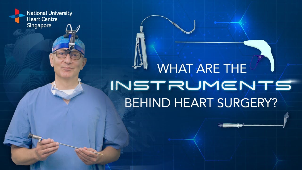
Instruments at work, innovation at play. 🔍
Watch on to discover the behind-the-scenes instruments utilised by our NUHCS cardiac surgery expert, A/Prof Theodoros Kofidis, Head of NUHCS' Department of Cardiac, Thoracic & Vascular Surgery (CTVS), for keyhole heart operations. 🔑
To find out more about Minimally Invasive Heart Surgery @ NUHCS, visit: https://[a]www.nuhcs.com.sg%2FOur-Services%2FSpecialties%2FPages%2FMinimally-Invasive-Cardiac-Surgery-Programme.aspx[/a]
Connect with us:
Instagram: @nuhcsofficial
Facebook: www.facebook.com/nuhcs
Website: www.nuhcs.com.sg
LinkedIn: www.linkedin.com/company/nuhcs
To make an appointment with the NUHCS Heart Clinic, email us at appointment@nuhs.edu.sg
#NUHCS #cardiacsurgery #heartsurgery #keyholesurgery #minimallyinvasive
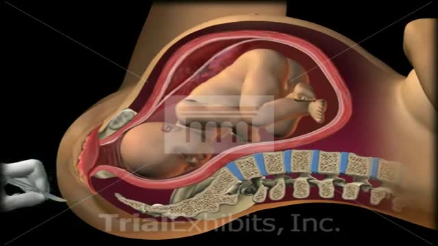
This medical 3D animation exhibit shows the left brachial plexus during birth and shoulder dystocia. Anatomy: symphysis pubis, uterus, sacrum, coccyx and fetus. "McRoberts Position". An episiotomy is cut. Brachial Plexus stretch injury. Retraction of head (turtle sign). Suprapubic pressure, gentle traction. To view our medical library of exhibits,
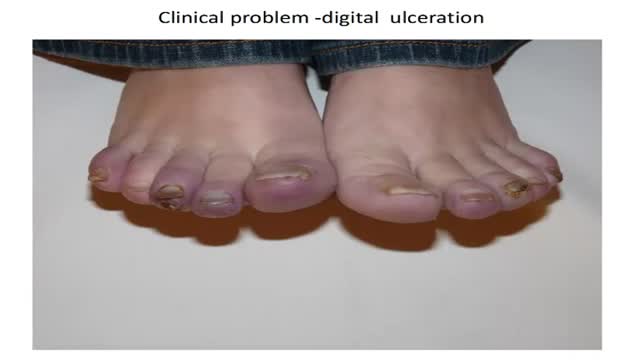
How to diagnose digital ulceration in out patient clinic.
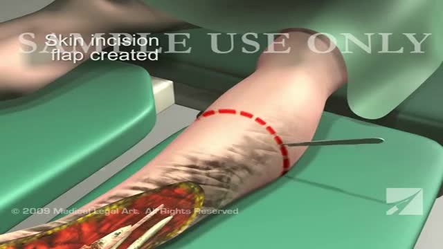
This 3d medical animation features a dramatic operative room overview of a left leg below the knee surgical amputation following severe trauma to the ankle and foot.
