
סרטונים מובילים


Gynecology 3D Animation
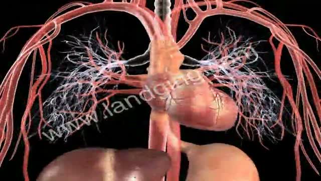
http://www.landging.com/circulatory-system-animation-cardiology.html
This circulatory system animation demonstrates cardiology mechanism of action and pharmacology in 3d.
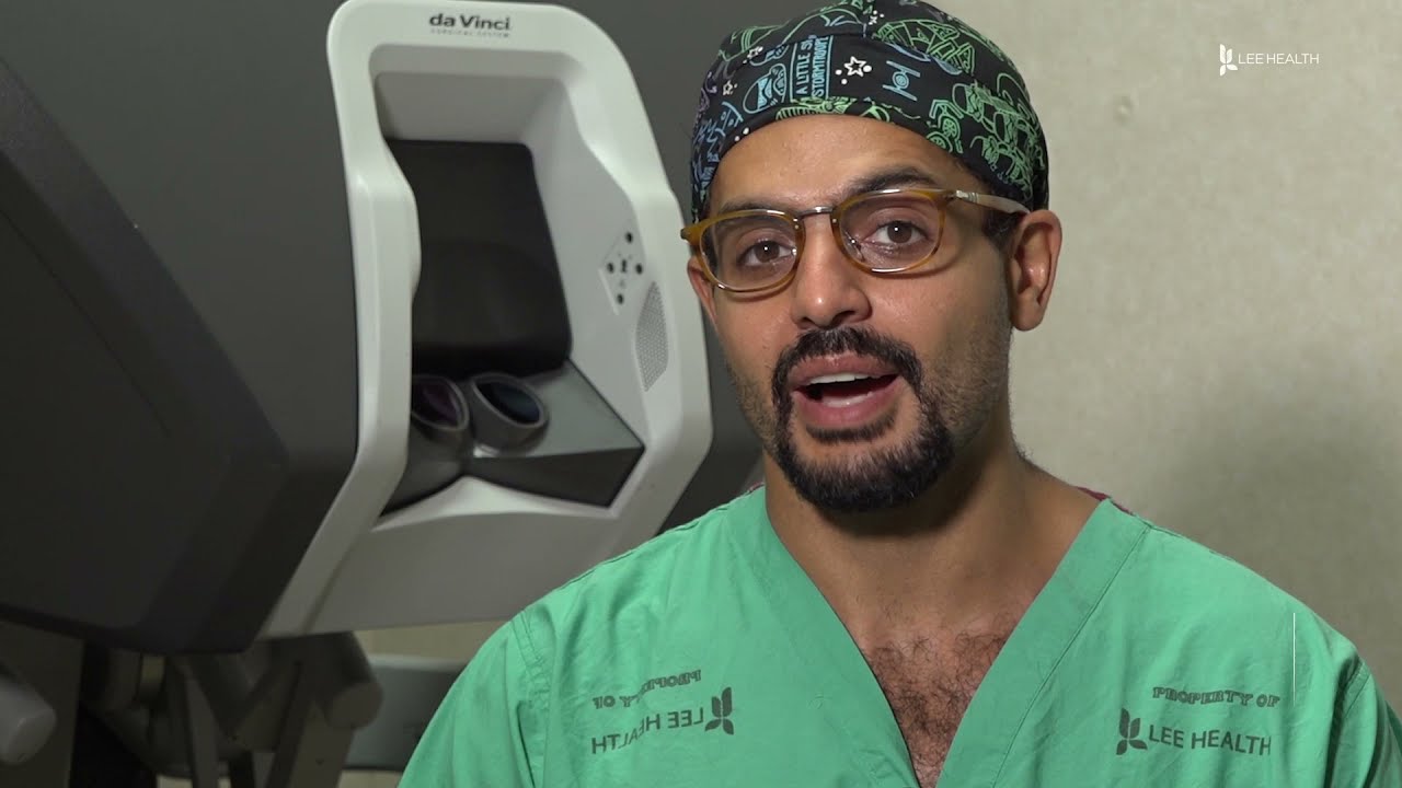
From across the room, using controls and pedals—pediatric surgeons at Golisano Children’s Hospital are now able to operate on patients without even touching them. “It allows performances of deep surgeries in the pelvis or abdomen through tiny, little incisions as opposed to a traditional, large incision to get access to the areas where urologists often operate,” explained Dr. Rahman Abd-El-Barr, a pediatric urologist with Golisano Children’s Hospital of Southwest Florida.
The DaVinci robot is a robotic platform that allows surgeons to do minimally invasive surgery, leaving patients with smaller incisions and a quicker recovery. “This is important because it allows us to minimize recovery time, pain, bleeding with surgery, and especially with kids, it helps them to get back on their feet right away,” he said.
So when high school athlete, Reagan Rebeor found out she needed to have kidney surgery, she decided to have it robotically. “Thankfully, I did that because if not, I would have had a long scar down my stomach instead of small holes, small incisions. I had pain for three days, three or four days. Then after that, I was fine,” she said.
While it’s not an option for all pediatric surgeries, doctors say it can be very beneficial for teenage and adult patients needing reconstructive surgery. An option that allows patients a quicker and easier recovery.
View More Health Matters video segments at LeeHealth.org/Healthmatters/
Lee Health in Fort Myers, FL is the largest network of health care facilities in Southwest Florida and is highly respected for its expertise, innovation and quality of care. For more than 100 years, we’ve been providing our community with personalized preventative health services and primary care to highly specialized care services and robotic assisted surgeries. Lee Health - Caring People. Inspiring Care.
Visit LeeHealth.org
Clinical examination of the anterior chest and lungs from the USMLE collection

Testosterone treatment in adolescent boys with constitutional delay
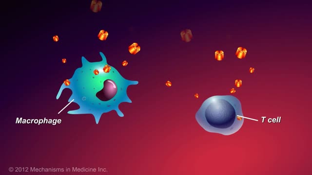
This animation describes what anti-TNF-alpha therapies are, how they work, and how patients with inflammatory bowel disease (IBD) can benefit.

Hysteroscopy adhesiolysis for treating Ashermann syndrome
Dilated cardiomyopathy (DCM) is a condition in which the heart's ability to pump blood is decreased because the heart's main pumping chamber, the left ventricle, is enlarged and weakened.
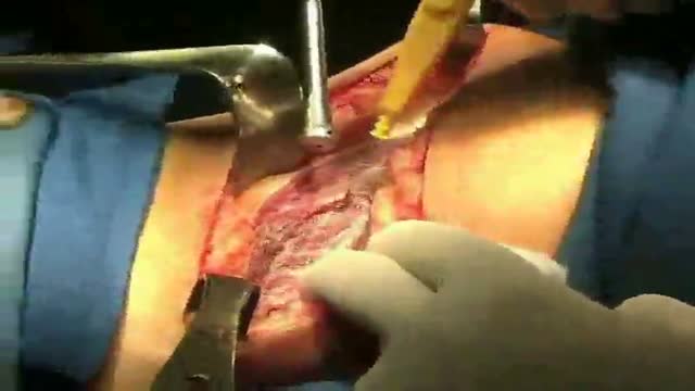
51 yr old female with right flank pain and recurrent UTI. IVP showed a UPJ calculus on the right.
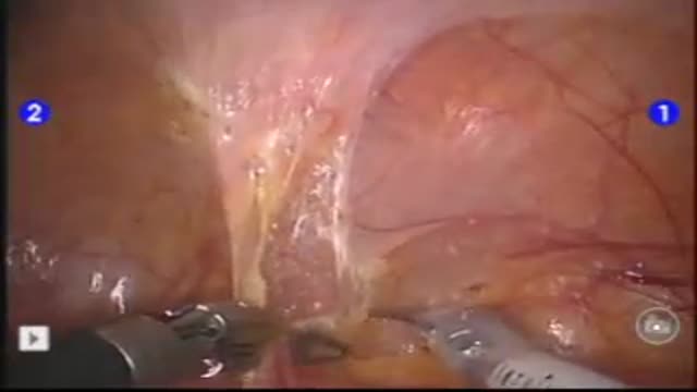
PURPOSE: Laparoscopic partial nephrectomy (LPN) is an alternative modality of treatment for small sized renal cell carcinoma. Robot assisted laparoscopic partial nephrectomy (RLPN) has also been performed with an advantage in repairing resected surface after tumor resection. We compare the periopera...
tive data of patients treated with laparoscopic partial nephrectomy with those of RLPN undertaken patients. MATERIAL AND METHOD: From September 2006 to April 2008, 22 patients were treated with LPN and 22 were RLPN. 3 arms were used for RLPN; camera was inserted through the 12mm, umbilical trocar port. The laparoscopic Bulldog clamp was used for the clamping of renal hilum. We retrospectively compared each group about tumor size, operation time, estimated blood loss, warm ischemic time and hospital stay. RESULT: Operation time of LPN was shorter than that of RLPN (p=0.033). Tumor size, estimated blood loss and hospital stay was not significant different in each group. No case had conversion to open surgery. 1 patient of RLPN group, however, had conversion to radical nephrectomy due to severe bleeding. CONCLUSION: RLPN was safe and feasible in small sized renal cell carcinoma. Warm ischemic time was reasonable and morbidity associated with RLPN was also low. RLPN LPN p-value Tumor Size (cm) 2.5 2.1 0.605 Op time (min) 169.3 140.8 0.033 EBL (ml) 243.2 213.2 0.878 Warm Ischemic Time (min) 29.2 26.4 0.237 Transfusion (%) 4.5 4.5 0.756 Hospital stay (day) 4.4 5.5 0.053

A 55-year-old man presented with recurrent epistaxis. After endoscopic sphenopalatine artery cauterization, the bleeding stopped. The patient was doing well at last follow up.
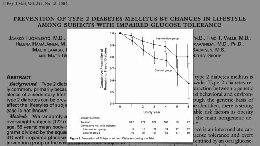
But here's the good news: it is possible to prevent prediabetes from developing into type 2 diabetes. Eating healthy food, losing weight and staying at a healthy weight, and being physically active can help you bring your blood glucose level back into the normal range.
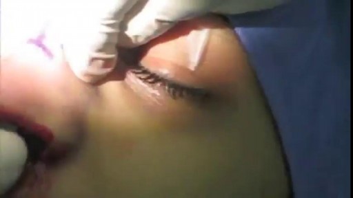
Nose Plastic Surgery: Open Rhinoplasty

Beta blockers, also known as beta-adrenergic blocking agents, are medications that reduce your blood pressure. Beta blockers work by blocking the effects of the hormone epinephrine, also known as adrenaline. When you take beta blockers, your heart beats more slowly and with less force, thereby reducing blood pressure. Beta blockers also help blood vessels open up to improve blood flow. Examples of beta blockers Some beta blockers mainly affect your heart, while others affect both your heart and your blood vessels. Which one is best for you depends on your health and the condition being treated. Examples of oral beta blockers include: Acebutolol (Sectral) Atenolol (Tenormin) Bisoprolol (Zebeta) Metoprolol (Lopressor, Toprol-XL) Nadolol (Corgard) Nebivolol (Bystolic) Propranolol (Inderal LA, InnoPran XL)
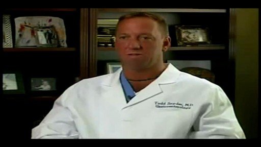
An esophageal stent is a flexible mesh tube, approximately 2cm (3/4 inch) wide, and is placed through the constricted area of your esophagus (food tube) to allow food and beverages to pass from your mouth to your stomach for digestion and absorption of nutrients.
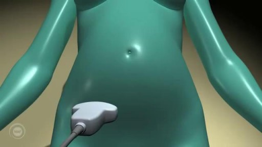
A prenatal ultrasound (also called a sonogram) is a noninvasive diagnostic test that uses sound waves to create a visual image of your baby, placenta, and uterus, as well as other pelvic organs. It allows your healthcare practitioner to gather valuable information about the progress of your pregnancy and your baby's health. During the test, an ultrasound technician (sonographer) transmits high-frequency sound waves through your uterus that bounce off your baby. A computer then translates the echoing sounds into video images that reveal your baby's shape, position, and movements. (Ultrasound waves are also used in the handheld instrument called a Doppler that your practitioner uses during your prenatal visits to listen to your baby's heartbeat.) You may have an early ultrasound at your practitioner's office at 6 to 10 weeks to confirm and date the pregnancy. Or you may not have one until the standard midpregnancy ultrasound between 16 and 20 weeks. That's when you may learn your baby's sex, if you like. (The technician will probably present you with a grainy printout of the sonogram as a keepsake.) You may also have a sonogram as part of a genetic test, such as the nuchal translucency test, chorionic villus sampling, or amniocentesis, or at any other time if there are signs of a problem with your baby. You'll have more frequent ultrasounds if you have diabetes, hypertension, or other medical complications.
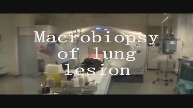
Spirotome macrobiopsy of a lung as a minimal invasive way to complete the diagnosis of lung lesions.
A detailed description of the Hepato-pulmonary syndrome including its definition, pathophysiology, diagnosis and treatment. The pathophysiology includes nitric oxide in the pulmonary vasculature which results in intrapulmonary vasodilatation. This causes the classical and unique symptom of platypnea and orthodeoxia.

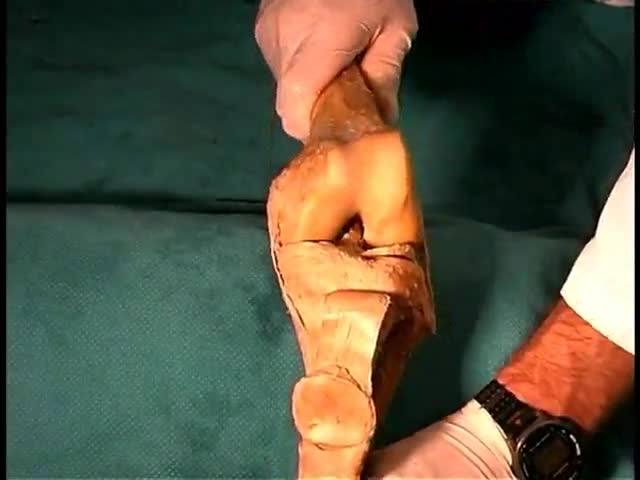
Anatomy of The Lower Limb Joints
