
- Physical Examination
- Surgical Examination
- Ophthalmology
- Clinical Skills
- Orthopedics
- Surgery Videos
- Laparoscopy
- Pediatrics
- Funny Videos
- Cardiothoracic Surgery
- Nursing Videos
- Plastic Surgery
- Otorhinolaryngology
- Histology and Histopathology
- Neurosurgery
- Dermatology
- Pediatric Surgery
- Urology
- Dentistry
- Oncology and Cancers
- Anatomy Videos
- Health and Fitness
- Radiology
- Anaesthesia
- Physical Therapy
- Pharmacology
- Interventional Radiology
- Cardiology
- Endocrinology
- Gynecology
- Emergency Medicine
- Psychiatry and Psychology
- Childbirth Videos
- General Medical Videos
- Nephrology
- Physiology
- Diet and Food Health
- Diabetes Mellitus
- Neurology
- Women Health
- Osteoporosis
- Gastroenterology
- Pulmonology
- Hematology
- Rheumatology
- Toxicology
- Nuclear Medicine
- Infectious Diseases
- Vascular Disease
- Reproductive Health
- Burns and Wound Healing
- Other
Top videos
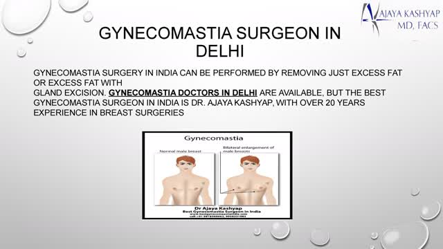

Best Gynecomastia surgeon in India is Dr. Ajaya Kashyap, with over 20 years experience in breast surgeries. He is the only active Indian member in American Society of Plastic Surgeons. Contact us to find out about your gynecomastia treatment in Delhi, India, maintaining high international standards, having a U.S. board certified surgeon, and latest technology and surgical techniques, our offices offer very economical costing. Contact us today inquire about gynecomastia surgery cost, in Delhi. You are sent query using WhatsApp and website.
For further information, are available visit our website: http://www.bestgynecomastiaindia.com/
Your Query for Chat and call +91-9818369662, 9958221983 (WhatsApp)

Soon enough, a robot will be doing surgeries on you!

Doctors save the life of an unborn baby who was injured along with her mother in a missile attack in the Syrian city of Aleppo. The video shows a team of emergency medical workers delivering the baby by Cesarean section and then treating the newborn for the shrapnel wounds covering her body and one very large gash in her head. “Medics can be seen frantically reviving the baby, after delivering her by emergency cesarean, as she lies motionless,” the article states. “Eventually the tiny newborn begins to cry and seemingly comes to life as she is given an oxygen mask and rubbed vigorously.” “According to Reuters, the woman also has three other children, all of whom were injured in the attack, but are reported by doctors in the hospital to be in a good condition,” the Daily Mirror article states. The article does not provide the gestational age of the baby before it was delivered. The article said the pregnant woman was hit by a barrel bomb – “crude explosives and shrapnel and dropped from helicopters used by [Syrian] President Bashir al-Assad’s regime." The article notes an estimated 7.6 million Syrians have been displaced by the ongoing civil war and that 320,000, including 11,000 children, have been killed in the conflict. The Daily Mirror also reports that the doctors suggested that the tiny girl be named Amal, which means hope in Arabic. UK Daily Mirror: Incredible footage shows Syrian doctors perform lifesaving caesarean after missile strike leaves shrapnel embedded in unborn baby's face
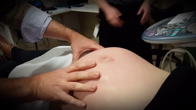
External cephalic version, or version, is a procedure used to turn a fetus from a breech position or side-lying (transverse) position into a head-down (vertex) position before labor begins. When successful, version makes it possible for you to try a vaginal birth.
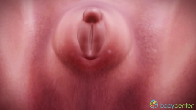
Your baby's sex is set at conception. At around 7 weeks, your baby's internal sex organs – such as ovaries and testes – begin to form in the abdomen. Male and female sex organs and genitalia look the same at this stage because they're derived from the same structures. At around 9 weeks, boys and girls begin to develop differently. In girls, a tiny bud emerges between the tissue of the legs. This bud will become the clitoris. The membrane that forms a groove below the bud separates to become the labia minora and the vaginal opening. By 22 weeks, the ovaries are completely formed and move from the abdomen to the pelvis. They already contain a lifetime supply of 6 million eggs. In boys, the bud develops into the penis and starts to elongate at around 12 weeks. The outer membrane grows into the scrotal sac that will later house the testicles. By 22 weeks, the testes have formed in the abdomen. They already contain immature sperm. Soon they'll begin their descent to the scrotum, but it's a long journey. They'll reach their destination late in pregnancy, or for some boys, after birth. If you're eager to find out whether you're having a girl or a boy, you'll have to wait until you're at least 17 weeks pregnant. That's when the genitals have developed enough to be seen on an ultrasound.
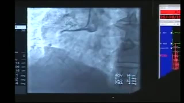
Cardiac catheterization (kath-uh-tur-ih-ZAY-shun) is a procedure used to diagnose and treat cardiovascular conditions. During cardiac catheterization, a long thin tube called a catheter is inserted in an artery or vein in your groin, neck or arm and threaded through your blood vessels to your heart. Using this catheter, doctors can then do diagnostic tests as part of a cardiac catheterization. Some heart disease treatments, such as coronary angioplasty, also are done using cardiac catheterization. Usually, you'll be awake during cardiac catheterization, but given medications to help you relax. Recovery time for a cardiac catheterization is quick, and there's a low risk of complications.
10 Animals Found Living Inside Humans
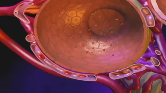
Bronchiectasis is an abnormal dilation of the proximal and medium-sized bronchi (>2 mm in diameter) caused by weakening or destruction of the muscular and elastic components of the bronchial walls. Affected areas may show a variety of changes, including transmural inflammation, edema, scarring, and ulceration, among other findings. Distal lung parenchyma may also be damaged secondary to persistent microbial infection and frequent postobstructive pneumonia. Bronchiectasis can be congenital but is most often acquired.[9] Congenital bronchiectasis usually affects infants and children. These cases result from developmental arrest of the bronchial tree. Acquired forms occur in adults and older children and require an infectious insult, impairment of drainage, airway obstruction, and/or a defect in host defense. The tissue is also damaged in part by the host response of neutrophilic proteases, inflammatory cytokines, nitric oxide, and oxygen radicals. This results in damage to the muscular and elastic components of the bronchial wall. Additionally, peribronchial alveolar tissue may be damaged, resulting in diffuse peribronchial fibrosis.[12] The result is abnormal bronchial dilatation with bronchial wall destruction and transmural inflammation. The most important functional finding of altered airway anatomy is severely impaired clearance of secretions from the bronchial tree. Impaired clearance of secretions causes colonization and infection with pathogenic organisms, contributing to the purulent expectoration commonly observed in patients with bronchiectasis. The result is further bronchial damage and a vicious cycle of bronchial damage, bronchial dilation, impaired clearance of secretions, recurrent infection, and more bronchial damage

Hyponatremia is a condition that occurs when the level of sodium in your blood is abnormally low. Sodium is an electrolyte, and it helps regulate the amount of water that's in and around your cells. In hyponatremia, one or more factors — ranging from an underlying medical condition to drinking too much water during endurance sports — causes the sodium in your body to become diluted. When this happens, your body's water levels rise, and your cells begin to swell. This swelling can cause many health problems, from mild to life-threatening. Hyponatremia treatment is aimed at resolving the underlying condition. Depending on the cause of hyponatremia, you may simply need to cut back on how much you drink. In other cases of hyponatremia, you may need intravenous fluids and medications
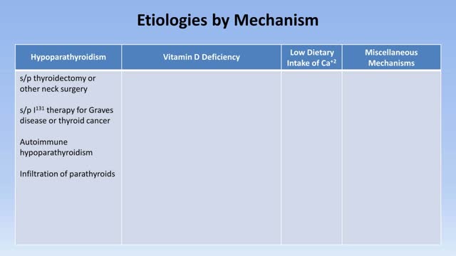
In neonates, hypocalcemia is more likely to occur in infants born of diabetic or preeclamptic mothers. Hypocalcemia also may occur in infants born to mothers with hyperparathyroidism. Clinically evident hypocalcemia generally presents in milder forms and is usually the result of a chronic disease state. In emergency department patients, chronic or subacute complaints secondary to mild or moderate hypocalcemia are more likely to be a chief complaint than severe symptomatic hypocalcemia. Once laboratory results demonstrate hypocalcemia, the first question is whether the hypocalcemia is true—that is, whether it is representative of a decrease in ionized calcium. The presence of chronic diarrhea or intestinal disease (eg, Crohn disease, sprue, chronic pancreatitis) suggests the possibility of hypocalcemia due to malabsorption of calcium and/or vitamin D. The patient's past medical history should be explored for pancreatitis, anxiety disorders, renal or liver failure, gastrointestinal disorders, and hyperthyroidism or hyperparathyroidism. Previous neck surgery suggests hypoparathyroidism; a history of seizures suggests hypocalcemia secondary to anticonvulsants. The patient may have a recent history of thyroid, parathyroid, or bowel surgeries or recent neck trauma. The length of time that a disorder is present is an important clue. Hypoparathyroidism and pseudohypoparathyroidism are lifelong disorders. Instead, acute transient hypocalcemia may be associated with acute gastrointestinal illness, nutritional deficiency, or acute or chronic renal failure. In an elderly patient, a nutritional deficiency may be associated with a low intake of vitamin D. A history of alcoholism can help diagnose hypocalcemia due to magnesium deficiency, malabsorption, or chronic pancreatitis. Inquire about recent use of drugs associated with hypocalcemia, including the following: Radiocontrast Estrogen Loop diuretics Bisphosphonates Calcium supplements Antibiotics Antiepileptic drugs Cinacalcet Other considerations in the history include the following: Family history of hypocalcemia Low-calcium diet Lack of sun exposure
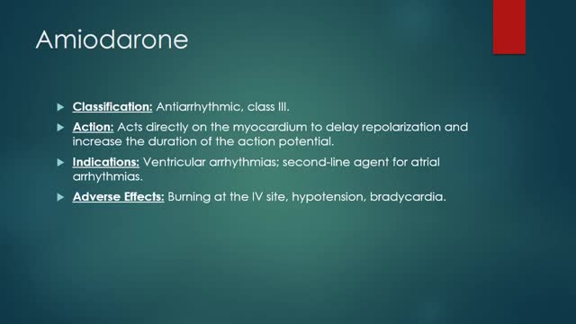
Why is this medication prescribed? Amiodarone is used to treat and prevent certain types of serious, life-threatening ventricular arrhythmias (a certain type of abnormal heart rhythm when other medications did not help or could not be tolerated. Amiodarone is in a class of medications called antiarrhythmics. It works by relaxing overactive heart muscles. How should this medicine be used? Amiodarone comes as a tablet to take by mouth. It is usually taken once or twice a day. You may take amiodarone either with or without food, but be sure to take it the same way each time.Follow the directions on your prescription label carefully, and ask your doctor or pharmacist to explain any part you do not understand. Take amiodarone exactly as directed. Do not take more or less of it or take it more often than prescribed by your doctor.

How to Insert a Tampon
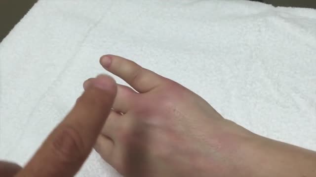
The dog bite victim needs to be taken to a safe place away from the assailant dog to prevent further attack and injury. Since dog bites can cause significant damage beneath the skin, a type of injury that cannot always easily be appreciated, medical care should be accessed by a health care practitioner. Wounds should be kept elevated and, if possible, washing the wound with tap water may be attempted. Information should be obtained from the dog's owner about the dog's rabies immunization status, but if this is not possible, hospital, animal control centers, or law enforcement personnel will help gather any required information.
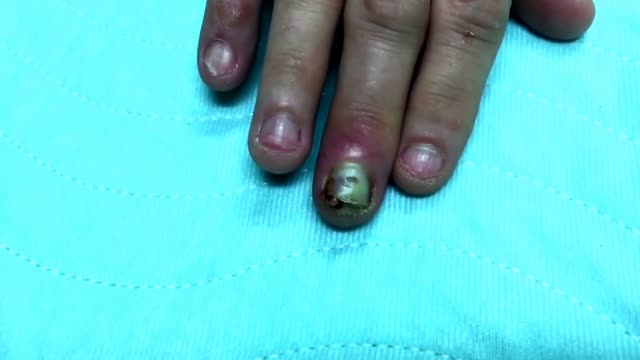
Finger infection Drainage
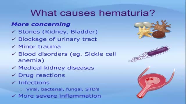
Seeing blood in your urine can cause anxiety. While in many instances there are benign causes, blood in urine (hematuria) can also indicate a serious disorder. Blood that you can see is called gross hematuria. Urinary blood that's visible only under a microscope is known as microscopic hematuria and is found when your doctor tests your urine. Either way, it's important to determine the reason for the bleeding. Treatment depends on the underlying cause.
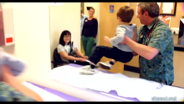
Chickenpox (varicella) is a viral infection that causes an itchy rash with small, fluid-filled blisters. Chickenpox is highly contagious to people who haven't had the disease or been vaccinated against it. Before routine chickenpox vaccination, virtually all people had been infected by the time they reached adulthood, sometimes with serious complications. Today, the number of cases and hospitalizations is down dramatically. For most people, chickenpox is a mild disease. Still, it's better to get vaccinated. The chickenpox vaccine is a safe, effective way to prevent chickenpox and its possible complications.

The key difference between monophasic and biphasic defibrillator is that the monophasic defibrillator is a type of defibrillation waveform where a shock is delivered to the heart from one vector as shown below. Whereas, in biphasic defibrillation, shock is delivered to the heart via two vectors.

The virus was first discovered in 1964 when Sir Michael Anthony Epstein and Ms. Yvonne Barr found it in a Burkitt lymphoma cell line. In 1968, the virus was linked to the disease infectious mononucleosis. Infection with Epstein-Barr virus (EBV) is common and usually occurs in childhood or early adulthood. EBV is the cause of infectious mononucleosis (also termed "mono"), an illness associated with fever, sore throat, swollen lymph nodes in the neck, and sometimes an enlarged spleen. It is also known as human herpes virus 4. Although EBV can cause mononucleosis, not everyone infected with the virus will get mononucleosis. Less commonly, EBV can cause more serious disease. Symptoms caused by EBV are usually mild and self-limited, but the virus persists in the body for life. It can be reactivated quietly without causing symptoms and may contaminate saliva. Thus, otherwise healthy people can spread the virus to uninfected people through kissing or sharing

An orgasm is a feeling of intense sexual pleasure that happens during sexual activity. It's sometimes called "coming" or "climaxing". Both men and women have orgasms.
Pulmonary embolism symptoms can vary greatly, depending on how much of your lung is involved, the size of the clots, and whether you have underlying lung or heart disease. Common signs and symptoms include: Shortness of breath. This symptom typically appears suddenly and always gets worse with exertion. Chest pain. You may feel like you're having a heart attack. The pain may become worse when you breathe deeply (pleurisy), cough, eat, bend or stoop. The pain will get worse with exertion but won't go away when you rest. Cough. The cough may produce bloody or blood-streaked sputum. Other signs and symptoms that can occur with pulmonary embolism include: Leg pain or swelling, or both, usually in the calf Clammy or discolored skin (cyanosis) Fever Excessive sweating Rapid or irregular heartbeat Lightheadedness or dizziness
