トップ動画
Certain high-surgical-risk patients with severe degenerative mitral regurgitation (DMR) now have a minimally invasive treatment option. MitraClip® therapy is a minimally-invasive transcatheter mitral valve repair (TMVr) procedure that has been established as a proven option with demonstrated safety and clinically important improvements. Used in more than 25,000 patients worldwide, MitraClip® is a well-established therapy. The MitraClip® device received CE Mark approval in Europe in 2008 and U.S. FDA approval in 2013, and has been approved for commercial use in 50 countries throughout the world.
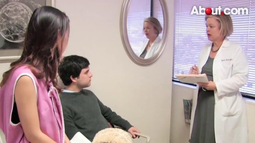
Start out with a visit to a doctor called a urologist. He'll give you a physical exam and ask you questions about your lifestyle and medical history, such as: Surgeries you've had Medications you take Your exercise habits Whether you smoke or take recreational drugs He may also have a frank discussion with you about your sex life, including any problems you've had or whether you have or ever had any STDs (sexually transmitted diseases). You'll probably be asked to give a sample of semen for analysis.
Disc Disease Videos Watch Disc Disease Videos There are several symptoms that are fairly consistent for people with lower back pain or neck pain from degenerative disc disease, including: Pain that is usually related to activity and will flare up at times but then return to a low-grade pain level, or the pain will go away entirely The amount of chronic pain—referred to as the patient's baseline level of pain—is quite variable between individuals and can range from almost no pain/just a nagging level of irritation, to severe and disabling pain Severe episodes of back or neck pain that will generally last from a few days to a few months before returning to the individual's baseline level of chronic pain Chronic pain that is completely disabling from degenerative disc disease does happen in some cases, but is relatively rare See Treating Chronic Pain and Depression from Degenerative Disc Disease
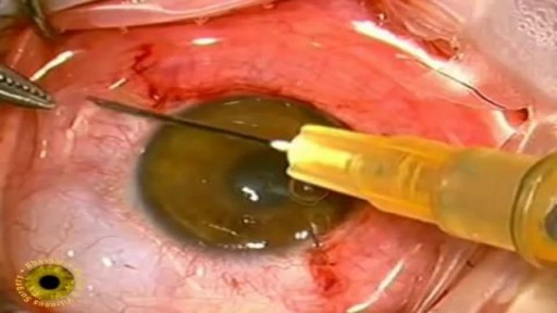
Eye cancers can be primary (starts within the eye) and metastatic cancer (spread to the eye from another organ). The two most common cancers that spread to the eye from another organ are breast cancer and lung cancer. Other less common sites of origin include the prostate, kidney, thyroid, skin, colon and blood or bone marrow. Melanomas (choroidal, ciliary body and uveal) -Early stages has no symptoms (the person does not know there is a tumor until an ophthalmology examination). As the tumor grows, symptoms can be blurred vision, decreased vision, double vision, eventual vision loss and if they continue to grow the tumor can break past the retina causing retinal detachment.

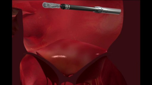
Amazing animation: General Dentistry in 3D

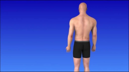
- with these 4 moves you can firm, lift and tone.
A small spontaneous pneumothorax may resolve without treatment; a pneumothorax arising as a result of lung disease or injury requires immediate treatment. Treatment may include insertion of a chest tube or aspiration of the free air in the chest cavity.Feb 19, 2016
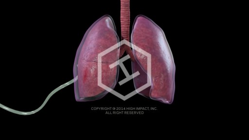
A small spontaneous pneumothorax may resolve without treatment; a pneumothorax arising as a result of lung disease or injury requires immediate treatment. Treatment may include insertion of a chest tube or aspiration of the free air in the chest cavity.
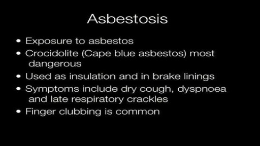
The most common symptoms of pneumoconiosis are cough and shortness of breath. The risk is generally higher when people have been exposed to mineral dusts in high concentrations and/or for long periods of time. Inadequate or inconsistent use of personal protective equipment (PPE) such as respirators (specially fitted protective masks) is another risk factor since preventing dusts from being inhaled will also prevent pneumoconiosis. Pneumoconiosis does not generally occur from environmental (non-workplace) exposures since dust levels in the environment are much lower.
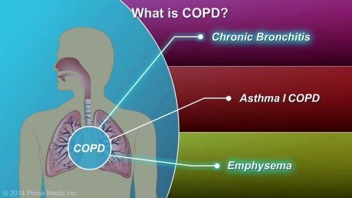
COPD, or chronic obstructive pulmonary disease, is a progressive disease that makes it hard to breathe. Progressive means the disease gets worse over time. COPD can cause coughing that produces large amounts of a slimy substance called mucus, wheezing, shortness of breath, chest tightness, and other symptoms. Cigarette smoking is the leading cause of COPD. Most people who have COPD smoke or used to smoke. However, up to 25 percent of people with COPD never smoked. Long-term exposure to other lung irritants—such as air pollution, chemical fumes, or dusts—also may contribute to COPD. A rare genetic condition called alpha-1 antitrypsin (AAT) deficiency can also cause the disease.
About Us Contact Disclaimer Get Published! Follow Us Epomedicine Medical Students Clinical Discussion Cases Emergencies Blog Medical Mnemonics Clinical Skills Search Subjects Clinical examination Gastrointestinal system Internal medicine Updated on January 31, 2017 Percussion of Spleen Traube’s semilunar space Borders: Superiorly: Left 6th rib superiorly Laterally: Left midaxillary line or Left anterior axillary line Inferiorly: Left costal margin Method: Patient’s position: supine with left arm slightly abducted. Percuss: from medial to lateral Interpretation: Resonance (Normal) and Dullness (Splenomegaly) Also: Pleural effusion or mass in stomach may cause dullness in Traube’s space.
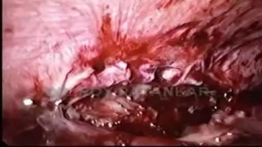
Bacterial abscess of the liver is relatively rare; however, it has been described since the time of Hippocrates (400 BCE), with the first published review by Bright appearing in 1936. In 1938, Ochsner's classic review heralded surgical drainage as the definitive therapy; however, despite the more aggressive approach to treatment, the mortality remained at 60-80%. [1] The development of new radiologic techniques, the improvement in microbiologic identification, and the advancement of drainage techniques, as well as improved supportive care, have reduced mortality to 5-30%; yet, the prevalence of liver abscess has remained relatively unchanged. Untreated, this infection remains uniformly fatal.
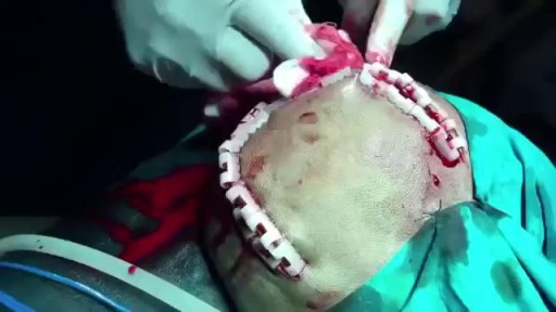
Epidural hematoma (EDH) is a traumatic accumulation of blood between the inner table of the skull and the stripped-off dural membrane. EDH results from traumatic head injury, usually with an associated skull fracture and arterial laceration.The inciting event often is a focused blow to the head, such as that produced by a hammer or baseball bat. In 85-95% of patients, this type of trauma results in an overlying fracture of the skull. Blood vessels in close proximity to the fracture are the sources of the hemorrhage in the formation of an epidural hematoma. Because the underlying brain has usually been minimally injured, prognosis is excellent if treated aggressively. Outcome from surgical decompression and repair is related directly to patient's preoperative neurologic condition. [1]
Tonsil stones are hard yellow or white formations that are located on or within the tonsils. It’s common for people with tonsil stones to not even realize they have them. Tonsil stones aren’t always easily visible and they can range from rice- to pea-sized. Tonsil stones rarely cause larger health complications. However, sometimes they can grow into larger tonsilloliths which can cause your tonsils to swell
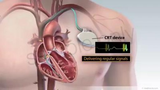
CRT is a clinically proven treatment option for some individuals with heart failure. A CRT device sends small electrical impulses to both lower chambers of the heart to help them beat together in a more synchronized pattern. This may improve the heart’s ability to pump blood and oxygen to your body. A CRT system is made up of two parts. The heart device, which is actually a tiny computer, plus a battery, contained in a small titanium metal case that is about the size of a pocket watch. Insulated wires, called leads, that are implanted to carry information signals from your heart to the heart device and to carry electrical impulses to your heart After the device system is implanted, an external computer, called a programmer, located at your doctor's office or clinic can be used to program the heart device and retrieve information from your heart device that will assist your doctor in your heart failure treatment. Your doctor will schedule periodic monitoring which may be done remotely if physician deems appropriate
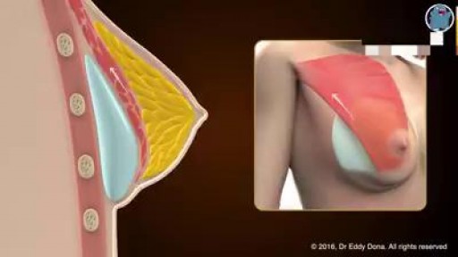
IMPLANT POCKETS - an educational animation explaining the different implant pockets
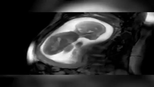
MRI scan of a 23-week-pregnancy

This device could prevent migraine headaches.
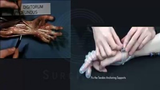
Exo-Glove Poly (Seoul National University). This wearable robot helps disabled patients regain control of their hands.

Always Love Your Mother Because You Will Never Get Another
