
- Physical Examination
- Surgical Examination
- Ophthalmology
- Clinical Skills
- Orthopedics
- Surgery Videos
- Laparoscopy
- Pediatrics
- Funny Videos
- Cardiothoracic Surgery
- Nursing Videos
- Plastic Surgery
- Otorhinolaryngology
- Histology and Histopathology
- Neurosurgery
- Dermatology
- Pediatric Surgery
- Urology
- Dentistry
- Oncology and Cancers
- Anatomy Videos
- Health and Fitness
- Radiology
- Anaesthesia
- Physical Therapy
- Pharmacology
- Interventional Radiology
- Cardiology
- Endocrinology
- Gynecology
- Emergency Medicine
- Psychiatry and Psychology
- Childbirth Videos
- General Medical Videos
- Nephrology
- Physiology
- Diet and Food Health
- Diabetes Mellitus
- Neurology
- Women Health
- Osteoporosis
- Gastroenterology
- Pulmonology
- Hematology
- Rheumatology
- Toxicology
- Nuclear Medicine
- Infectious Diseases
- Vascular Disease
- Reproductive Health
- Burns and Wound Healing
- Kidney Zone Podcast
- Other
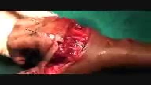
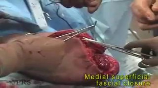
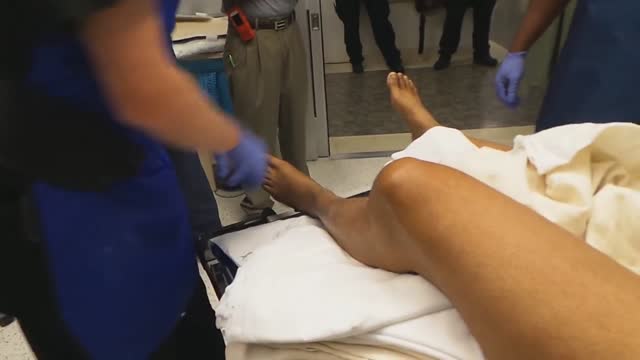
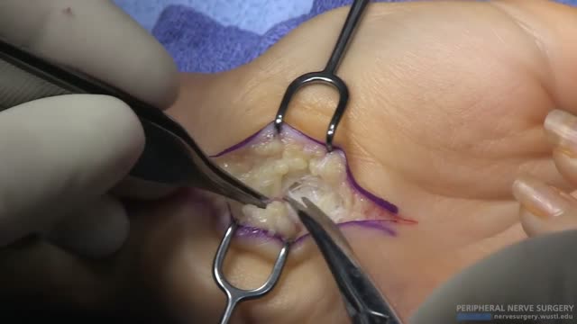
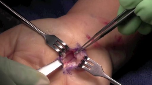
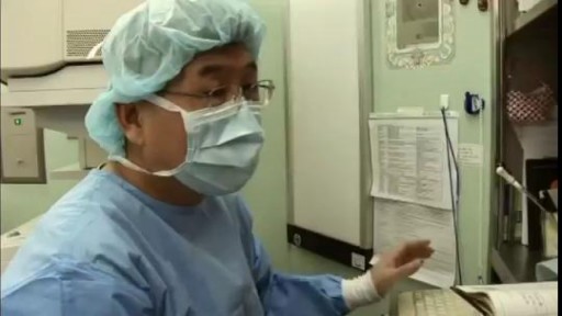
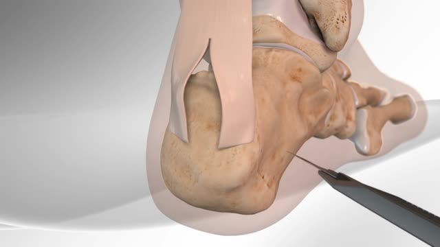
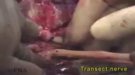

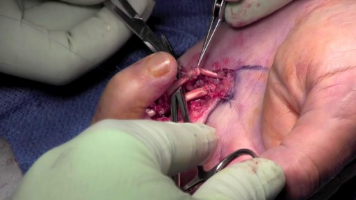

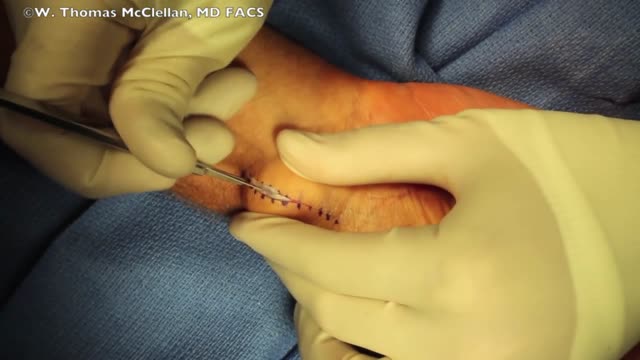
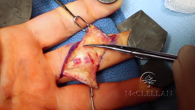
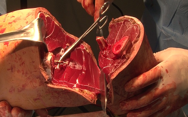
Amputated Hand Reattachment Surgery

Educational only: videos on MedicalVideos.com are for general information and are not medical advice. Always consult your own doctor about your health.
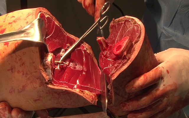
Microsurgical replantation — the reattachment of a severed hand — is documented in this surgery video, showing how blood vessels, nerves, and tissues are repaired to restore the limb. Replantation is a lengthy, specialized operation performed by trained microsurgeons. Presented for surgical education. Educational only — not medical advice.
This video in 30 seconds
A surgical team narrates the replantation of an amputated hand, with the amputation level through the metacarpals. The commentary follows bone fixation, tendon repair and vessel anastomoses until the hand is perfused, ending with checks of pulse and circulation.
Key points
- The narration identifies the amputation level through the metacarpals
- The team fixes the bones with K-wires across the metacarpals
- Flexor and extensor tendons are repaired
- Arterial and venous anastomoses are completed until the hand perfuses
- Final checks confirm pulse and circulation in the replanted hand
Chapters
- 0:28 — Amputated part and metacarpal level
- 1:41 — K-wire fixation of metacarpals
- 2:34 — Flexor tendon repair
- 2:47 — Arterial and venous anastomoses
- 6:42 — Final perfusion and pulse checks
Video Transcript
A surgical team's narration over replantation of an amputated hand, with the level through the metacarpals. The commentary follows K-wire fixation of the bones, repair of flexor and extensor tendons, arterial and venous anastomoses until the hand perfuses, and the final checks of pulse and circulation; the speech is heavily accented and parts of the automatic transcript are garbled, but the operative sequence is clear.
Read the full transcript
This was the amplitude part of the time. The level of transaction was just with the days of the summer. The time is to separate the master students that are not receiving the time from the end of the summer. They are only the best students and sub-summoner teachers. It's time to sit, sit and time to go to class. I am going to show you the cut side of the amplitude. If you clearly notice the metacarpal, as far as the thumb is concerned, what level is the amplitude? It's the metacarpal input.
It's the base of the functional value. So the whole force metacarpal is in the amplitude part. The fluxes and the metacarpals, they are seen in this part. The most important thing is the reasonably sharp part. This is the dorsal side, the dorsal view of the amplitude part. You know other ingredients in medicine, fingers, hands, nose, and other things. The amplitude part is detected partly and structures identified and ready for bone and shank session. Four files, K-wise, passing through all the four metacarpals and base of the thumb,
stabilize the hand. There was minimal shortening which was done. The external tendons are ready here. There are some veins which have been identified which could be anasthomas. We have moved on to the bowler side. On the bowler side, we have repaired all the FGP's, all four fingers. Inlex finger separately and mass repair of the lateral three fingers. The SPL of the thumb has been repaired. Now we are going for the arterial anasthomas. The arterial anasthomas is on the bowler side. Once the arterial starts to, the veins can be seen flowing on the dorsal.
We are now oscillating away. The arterial anasthomas is two arterial anasthomas. The second one was very good flow. The hand is perfused well. Now we are going on the back bone of the hand as we are doing this. This was reason for anasthomas to come out. If you take the time of the time of injury, you could pursue this kind of check-in on the finger, the SPL, and all the external tendons should be repaired. The nerves have not been repaired. We wanted to get some of the pain to some three and a half hours from here.
We wanted to work the teeth, the spine, the hand, the blood, the combination of internal and internal blood. And along with that work with slightly moving the hand. So Dr. Chahee is planning to give another booster and ores of the operation of the hand. So because at the end of the procedure, it was at 1.30 pm. The hand was perfused very nicely. There were two arteries which were anasthomas. One which was perfusing the thumb, one between the ring finger and the little finger. We notice the anasthomas artery here and the second anasthomas is here.
But the most important proof of this perfusion was... Lepido? Lendu, Hupan, Prasana, Sink. Fresh blood, nice, comfortable, ores. We have repaired all the flexor tendons, the STL, all four flexor tendons. On the dorsal side, we could do three venous anasthomas. One venous anasthomas is you notice here, the second is here, and the third is here. Three venous anasthomas as well, good hemostasis, all external tendons repaired. You hold the hand as well. Thank you. So this is the final replant over. Over. And you give me pulse of symmetry problem.
Good vascularity achieved, good flow here achieved. So then just do the footwork. All the important tendons are being repaired. The bone edification and stable, the hand is in a position of position. We'll be giving albous dressing for him. Now we are fixing the pulse proximity of the row and the indexing here. And we'll show you that on the one finger, very important, good pulse session and the procedure. This is the recording on the NCA4th v-plant. Very good pulse session, very good arterial signals heard over the radial side of the index finger.
Between the left finger and the ring finger, all the way to the left finger. All right, so the right finger, to the left finger, to the left finger.
Transcript auto-generated from the video’s audio for accessibility and study. Educational content only — not medical advice.
Video Overview
From our Surgery Videos collection: Surgery videos: watch real operations step by step — general, laparoscopic and specialist surgery performed in the OR, explained for students and surgeons. Browse all Surgery Videos videos →
Medical Videos
Free medical and surgery videos for patients, students and professionals — trusted since 2007.
Founded by Dr. Mohamed Ibrahim, MD, PhD — physician-scientist.
Follow us on Facebook · Instagram
SORT BY-
Top Comments
-
Latest comments