Лучшие видеоролики
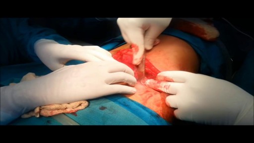
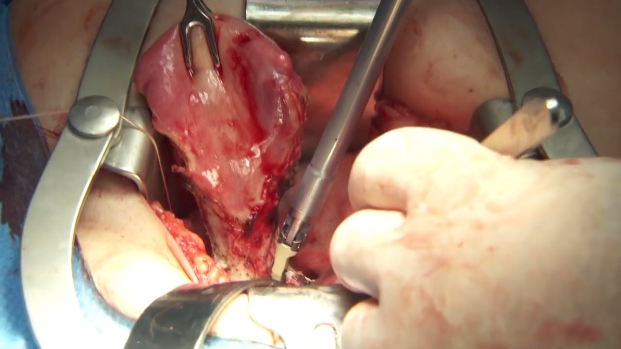
Majority of patients these days prefer PCNL ( Minimal Invasive Telescopic removal of kidney stones broken with lithoclast, removed through a button hole incision ). This patient with a big stone in the pelvis of the kidney wanted it open only so I did an open pyelolithotomy for this patient after a long time as I use to do it in routine in the past. Except for the long incision and scar as compared to PCNL the recovery time was the same and patient went home third day happily walking and eating.
Video showing normal vagina delivery and child birth

this video about identifying a hernia vs a cyst
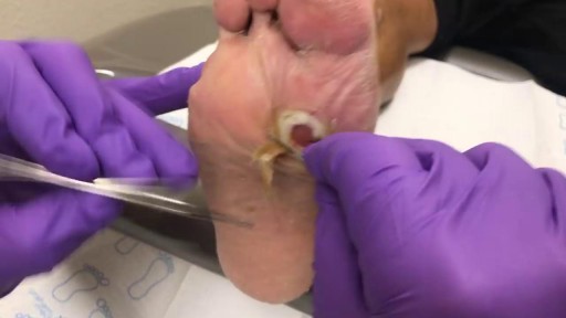
This is a diabetic foot ulcer. The patient reportedly went on vacation and noticed this ulcer upon their return. Debridement (removal of damaged tissue) to the level of healthy bleeding tissue is medically necessary as damaged tissue acts an impediment to wound healing. Due to their diabetic neuropathy, they did not feel any pain or indication that a wound was forming. This ulcer appeared to have penetrated to the level of subcutaneous tissue or even fascia, but turned out to be much deeper than that. These are serious wounds and are the beginnings of what lead to foot and leg amputations if they are not treated promptly by your healthcare provider, AKA Podiatrist.
The male orgasm is a common subject but usually misunderstood at the same time. Men are sometimes led to believe that ejaculating often is a bad thing, particularly if you masturbate. The truth is that ejaculation is important to every man due to a number of reasons. The main goal of this post is to shed some light on reasons why men need to ejaculate.
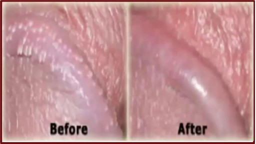
http://penilepapules.plus101.com/ ----- White Spots On Shaft, Pearly Penile Papules Treatment Cream, Single Red Bump On Shaft, Ppp Surgery. Common Home Made Remedies for Pearly Penile Papules. When it comes to treating pearly penile papules many people find it very difficult to reach one of the medical treatments. This is mainly because they are highly expensive and not many people can afford spending large amounts of money on surgery and recovery. In addition to that, these procedures have been reported as being quite risky, which make the men suffering from pearly penile papules think twice before going for one of the available surgeries. This is why, along the time, many homemade, natural treatments have been experienced, so that a cheaper and less risky way of curing pearly penile papules would be found. Some of the methods which have been tried proved to be very less effective, while some did not have any effect at all. Yet, there have also been methods which not only proved to be effective, but they were also considered to be much better than the medical treatment. Most of those who have tried the tea tree oil treatment reported significant diminish of the number of the papules from their penises. In addition to the clearing of the skin, they have also noticed that there were no side effects and the skin remained soft after the papules were removed. As the method was quite simple to put in practice (it requires the application of tea tree oil on the affected area with a cotton swab for three or four times per day), many men decided this was indeed a great solution to their problem.
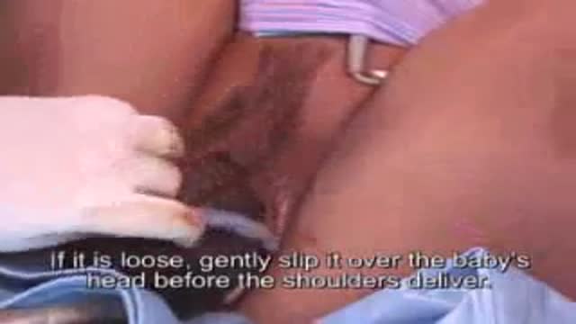
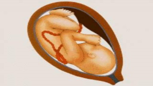
Umbilical Cord Around Fetal Neck During Delivery

In this video, Dr. Robert Rozbruch, chief of Limb Lengthening and Complex Reconstruction at Hospital for Special Surgery performs an osseointegration after a primary amputation. The patient, a 40 year old woman, had chronic nerve pain and compromised function of her residual limb.
For more information, visit: https://www.limblengthening.com/
https://www.hss.edu/limblengthening
https://www.hss.edu/LSARC
https://www.facebook.com/limblengtheningNYC
https://www.instagram.com/limblengthening
https://www.twitter.com/limblengthen
https://www.youtube.com/channe....l/UC-JL_X6ALjZXiXtcP
key words: Osseointegration, Amputee, Amputation, Limb Replacement, Tibia, Osseointegration
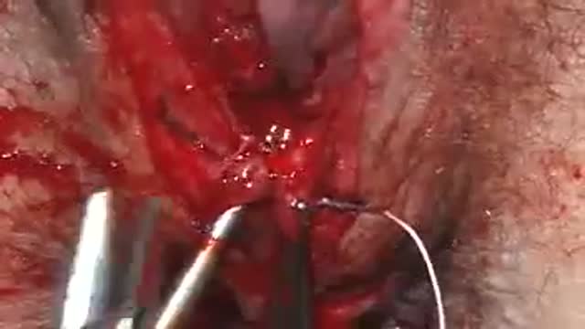
A video showing the repair of episiotomy
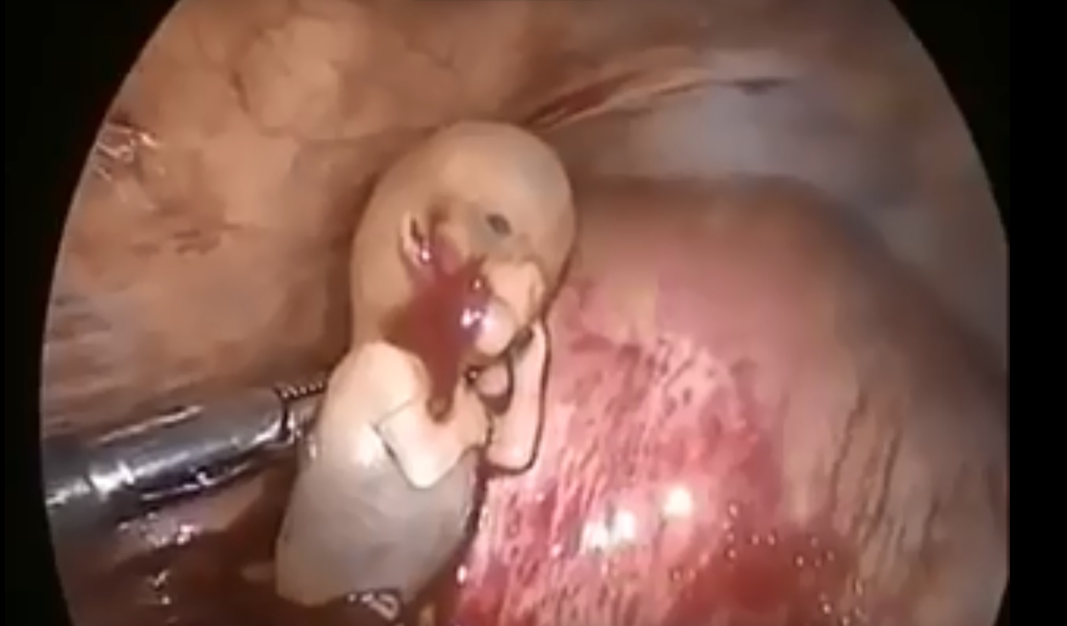
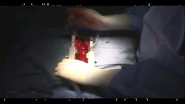
Watch that Ectopic Pregnancy Abortion Surgery
Watch that video to learn How To Insert Enema
Open heart (coronary artery bypass, or CABG) surgery is performed in order to reroute, or "bypass," blood around blocked arteries, thereby improving the supply of oxygen-rich blood to the heart. Surgeons usually use an artery from the chest wall to construct the "detour" around the blocked part of the artery. Veins from the legs are also used.
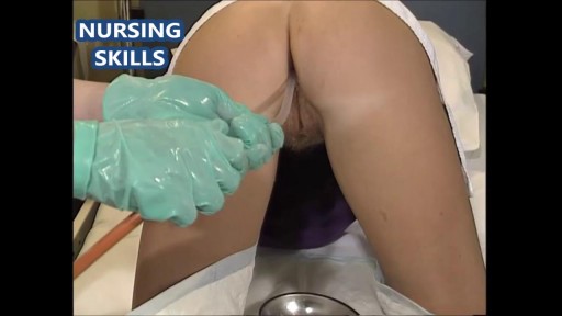
Routine Pap Smear and Pelvis Exam For Canadian Women
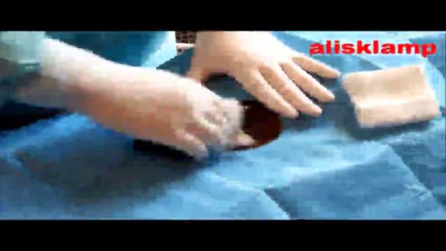
this video shows how the adult circumcision is easy by the alisklamp

Watch this clinical examination video to learn how to diagnose inguinal related groin pain.
This video clip is part of the FIFA Diploma in Football Medicine and the FIFA Medical Network. To enrol or to find our more click on the following link http://www.fifamedicalnetwork.com
The Diploma is a free online course designed to help clinicians learn how to diagnose and manage common football-related injuries and illnesses. There are a total of 42 modules created by football medicine experts. Visit a single page, complete individual modules or finish the entire course.
The network provides the opportunity for clinicians around the world to meet and share ideas relating to football medicine. Ask about an interesting case, debate current practice and discuss treatment strategies. Create a profile and log on to interact with other health professionals from around the globe.
This is not medical advice. The content is intended as educational content for health care professionals and students. If you are a patient, seek care of a health care professional.

Watch that video to know about the Anal Intercourse Medical Risks
Olympus has extended the value of its award-winning combined surgical energy device, THUNDERBEAT, to open surgical procedures. Watch Dr. Francois Blaudeau master use of THUNDERBEAT Open Extended Jaw (OEJ) in a total abdominal hysterectomy.
http://medical.olympusamerica.com/products/thunderbeat?utm_source=youtube&utm_campaign=Total%20Abdominal%20Hysterectomy%20Surgery%20-%20THUNDERBEAT&utm_medium=description&utm_term=energy&utm_content=surgical
Watch that video to know everything about male and female orgasm
Watch that Full Human Body Decay Process Video
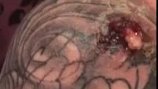
Infected Tattoo Abscess
