Top Videos

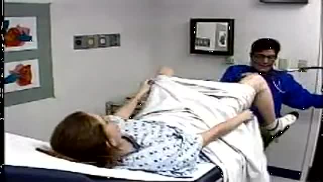
Proctoscope rectal examination

Squats are one of the essential exercises to do during pregnancy there are so many benefits from doing this functional type of exercise. Strengthening your glute muscles, that's your butt, helps to decrease lower back and pelvic pain.

The easy experimental answer to this question is 264 hours (about 11 days). In 1965, Randy Gardner, a 17-year-old high school student, set this apparent world-record for a science fair. Several other normal research subjects have remained awake for eight to 10 days in carefully monitored experiments. None of these individuals experienced serious medical, neurological, physiological or psychiatric problems. On the other hand, all of them showed progressive and significant deficits in concentration, motivation, perception and other higher mental processes as the duration of sleep deprivation increased. Nevertheless, all experimental subjects recovered to relative normality within one or two nights of recovery sleep. Other anecdotal reports describe soldiers staying awake for four days in battle, or unmedicated patients with mania going without sleep for three to four days.
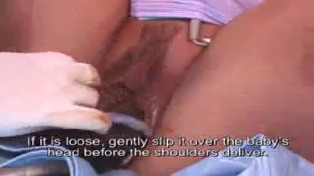
The umbilical cord is wrapped around the baby's neck in about 25% of deliveries. If loose, it usually has no impact on the delivery. If tight, it may need to be relieved before delivery of the baby can proceed safely
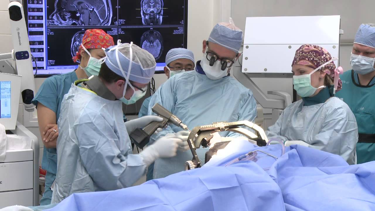
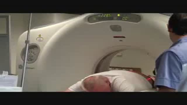
Dr. Jeffrey Ojemann, director of epilepsy surgery at Seattle Children's Hospital, explains a cutting-edge treatment for epilepsy: minimally invasive MRI-guided laser ablation surgery. Laser ablation surgery is much safer and more precise than other treatments, with fewer side effects.
A special thanks to patient Keoni Giauque.
For more information, visit: http://www.seattlechildrens.or....g/clinics-programs/n
"One Last Look" music rights via RoyaltyFreeMusic.com
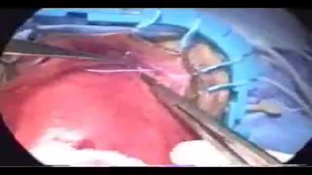
Microsurgical varicocelectomy is performed for patients with a varicocele and impaired semen parameters, testicular atrophy or pain due to the varicocele.
Rhabdomyolysis is a condition in which damaged skeletal muscle (Ancient Greek: rhabdomyo-) tissue breaks down rapidly (Greek –lysis). This damage may be caused by physical (e.g. crush injury), chemical, or biological factors. Breakdown products of damaged muscle cells are released into the bloodstream; some of these, such as the protein myoglobin, are harmful to the kidney and may lead to kidney dysfunction. The severity of the symptoms (which may include muscle pains, vomiting and confusion) depends on the extent of the muscle damage, and whether kidney failure develops. The mainstay of treatment is generous intravenous fluids, but could include dialysis or hemofiltration.
Rhabdomyolysis and its complications are significant problems for those injured in disasters such as earthquakes and bombing. Relief efforts in areas struck by earthquakes often include medical teams with skills and equipment for treatment of survivors with rhabdomyolysis. The disease and its mechanisms were first fully elucidated during the Blitz of London in 1941.

For patients looking to slim down their neck and achieve a more contoured and sculpted jaw line, then Tampa chin liposuction at the Artistic Lipo Sculpting Center is the answer! This one procedure can literally make patients look 10 years younger and 20 lbs lighter. Dr. Thomas Su is specialized in performing Liposuction procedures and consistently achieves stunning results for his Tampa patients. To find out more about Tampa neck lipo, visit http://www.artlipo.com/liposuction/liposuction-body-areas/lipo-chin---neck.html

A breech birth is the birth of a baby from a breech presentation. In the breech presentation the baby enters the birth canal with the buttocks or feet first as opposed to the normal head first presentation.
There are either three or four main categories of breech births, depending upon the source:
* Frank breech - the baby's bottom comes first, and his or her legs are flexed at the hip and extended at the knees (with feet near the ears). 65-70% of breech babies are in the frank breech position.
* Complete breech - the baby's hips and knees are flexed so that the baby is sitting crosslegged, with feet beside the bottom.
* Footling breech - one or both feet come first, with the bottom at a higher position. This is rare at term but relatively common with premature fetuses.
* Kneeling breech - the baby is in a kneeling position, with one or both legs extended at the hips and flexed at the knees. This is extremely rare, and is excluded from many classifications.
As in labour with a baby in a normal head-down position, uterine contractions typically occur at regular intervals and gradually cause the cervix to become thinner and to open. In the more common breech presentations, the baby’s bottom (rather than feet or knees) is what is first to descend through the maternal pelvis and emerge from the vagina.
At the beginning of labour, the baby is generally in an oblique position, facing either the right or left side of the mother's back. As the baby's bottom is the same size in the term baby as the baby's head. Descent is thus as for the presenting fetal head and delay in descent is a cardinal sign of possible problems with the delivery of the head.
In order to begin the birth, internal rotation needs to occur. This happens when the mother's pelvic floor muscles cause the baby to turn so that it can be born with one hip directly in front of the other. At this point the baby is facing one of the mother's inner thighs. Then, the shoulders follow the same path as the hips did. At this time the baby usually turns to face the mother's back. Next occurs external rotation, which is when the shoulders emerge as the baby’s head enters the maternal pelvis. The combination of maternal muscle tone and uterine contractions cause the baby’s head to flex, chin to chest. Then the back of the baby's head emerges and finally the face.
Due to the increased pressure during labour and birth, it is normal for the baby's leading hip to be bruised and genitalia to be swollen. Babies who assumed the frank breech position in utero may continue to hold their legs in this position for some days after birth.
Physical exam by a urologist including kidney, testicular and prostate exam.
Soft tissue biopsy from osteolytic lesions is a challenge for the interventionist. The Spirotome Bone is conceived for this intervention. The procedure is straigthforward and produces tissue specimens of high quality in sufficient amounts to allow quantitative molecular biology.

At one time, women who had delivered by cesarean section in the past would usually have another cesarean section for any future pregnancies. The rationale was that if allowed to labor, many of these women with a scar in their uterus would rupture the uterus along the weakness of the old scar. Over time, a number of observations have become apparent: Most women with a previous cesarean section can labor and deliver vaginally without rupturing their uterus. Some women who try this will, in fact, rupture their uterus. When the uterus ruptures, the rupture may have consequences ranging from near trivial to disastrous. It can be very difficult to diagnose a uterine rupture prior to observing fetal effects (eg, bradycardia). Once fetal effects are demonstrated, even a very fast reaction and nearly immediate delivery may not lead to a good outcome. The more cesarean sections the patient has, the greater the risk of subsequent rupture during labor. The greatest risk occurs following a “classical” cesarean section (in which the uterine incision extends up into the fundus.) The least risk of rupture is among women who had a low cervical transverse incision. Low vertical incisions probably increase the risk of rupture some, but usually not as much as a classical incision. Many studies have found the use of oxytocin to be associated with an increased risk of rupture, either because of the oxytocin itself, or perhaps because of the clinical circumstances under which it would be contemplated. Pain medication, including epidural anesthetic, has not resulted greater adverse outcome because of the theoretical risk of decreasing the attendant’s ability to detect rupture early. The greatest risk of rupture occurs during labor, but some of the ruptures occur prior to the onset of labor. This is particularly true of the classical incisions. Overall successful vaginal delivery rates following previous cesarean section are in the neighborhood of 70 This means that about 30of women undergoing a vaginal trial of labor will end up requiring a cesarean section. Those who undergo cesarean section (failed VBAC) after a lengthy labor will frequently have a longer recovery and greater risk of infection than had they undergone a scheduled cesarean section without labor. Women whose first cesarean was for failure to progress in labor are only somewhat less likely to be succesful in their quest for a VBAC than those with presumably non-recurring reasons for cesarean section. For these reasons, women with a prior cesarean section are counseled about their options for delivery with a subsequent pregnancy: Repeat Cesarean Section, or Vaginal Trial of Labor. They are usually advised of the approximate 70successful VBAC rate (modified for individual risk factors). They are counseled about the risk of uterine rupture (approximately 1in most series), and that while the majority of those ruptures do not lead to bad outcome, some of them do, including fetal brain damage and death, and maternal loss of future childbearing. They are advised of the usual surgical risks of infection, bleeding, anesthesia complications and surgical injury to adjacent structures. After counseling, many obstetricians leave the decision for a repeat cesarean or VBAC to the patient. Both approaches have risks and benefits, but they are different risks and different benefits. Fortunately, most repeat cesarean sections and most vaginal trials of labor go well, without any serious complications. For those choosing a trial of labor, close monitoring of mother and baby, with early detection of labor abnormalities and preparation for
Proctoscopy in Jackknife Position for examination of the rectum

Adenocarcinoma of the Transverse Colon taken by Dr. Julio Murra Saca This is the case of a 42 year-old male, with no significant past medical history presented with abdominal pain and no weight loss was reported. Adenocarcinoma of the colon is a primary cause of mortality and
morbidity in North America and Western Europe. Colonic cancers are the most common GI carcinomas and have the best prognosis. The 5-year survival rate is approximately 50%.
Survival rates may be improved by screening and removal of adenomatous polyps. Almost all colonic cancers are primary adenocarcinomas.
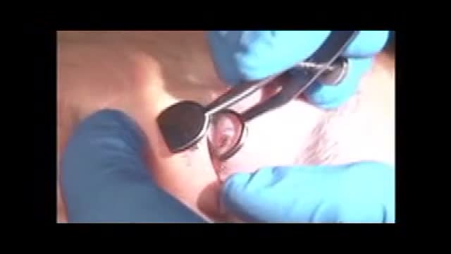
Surgical removal of a Chalazion from the eye lid
Pelvic Exam Video
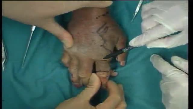
Kite flap, Guy Fouchier flap, 2nd finger to thumb. Cadaver dissection. Prof Steven Hovius demonstrates dissection technique and planning for a kite flap.
Perineal rectosigmoidectomy
Transurethral resection of the prostate (also known as TURP, plural TURPs and as a transurethral prostatic resection TUPR) is a urological operation. It is used to treat benign prostatic hyperplasia (BPH). As the name indicates, it is performed by visualising the prostate through the urethra and removing tissue by electrocautery or sharp dissection. This is considered the most effective treatment for BPH. This procedure is done with spinal or general anesthetic. A large triple lumen catheter is inserted through the urethra to irrigate and drain the bladder after the surgical procedure is complete. Outcome is considered excellent for 80-90% of BPH patients. Because of bleeding risks associated with the surgery, TURP is not considered safe for many patients with cardiac problems. As with all invasive procedures, the patient should first discuss medications they are taking with their doctor, most especially blood thinners or anticoagulants, such as warfarin (Coumadin), or aspirin. These may need to be discontinued prior to surgery. Postop complications include bleeding (most common), clotting and hyponatremia (due to bladder irrigation).
Additionally, transurethral resection of the prostate is associated with low but important morbidity and mortality.
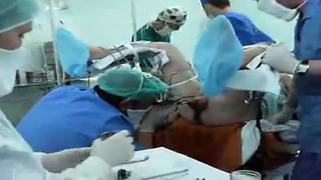
Circumcision by Dissection method
