
- Physical Examination
- Surgical Examination
- Ophthalmology
- Clinical Skills
- Orthopedics
- Surgery Videos
- Laparoscopy
- Pediatrics
- Funny Videos
- Cardiothoracic Surgery
- Nursing Videos
- Plastic Surgery
- Otorhinolaryngology
- Histology and Histopathology
- Neurosurgery
- Dermatology
- Pediatric Surgery
- Urology
- Dentistry
- Oncology and Cancers
- Anatomy Videos
- Health and Fitness
- Radiology
- Anaesthesia
- Physical Therapy
- Pharmacology
- Interventional Radiology
- Cardiology
- Endocrinology
- Gynecology
- Emergency Medicine
- Psychiatry and Psychology
- Childbirth Videos
- General Medical Videos
- Nephrology
- Physiology
- Diet and Food Health
- Diabetes Mellitus
- Neurology
- Women Health
- Osteoporosis
- Gastroenterology
- Pulmonology
- Hematology
- Rheumatology
- Toxicology
- Nuclear Medicine
- Infectious Diseases
- Vascular Disease
- Reproductive Health
- Burns and Wound Healing
- Kidney Zone Podcast
- Other





Female Pelvic Floor Part 1

The pelvic floor or pelvic diaphragm is composed of muscle fibers of the levator ani, the coccygeus, and associated connective tissue which span the area underneath the pelvis. The pelvic diaphragm is a muscular partition formed by the levatores ani and coccygei, with which may be included the parietal pelvic fascia on their upper and lower aspects. The pelvic floor separates the pelvic cavity above from the perineal region (including perineum) below.
The right and left levator ani lie almost horizontally in the floor of the pelvis, separated by a narrow gap that transmits the urethra, vagina, and anal canal. The levator ani is usually considered in three parts: pubococcygeus, puborectalis, and iliococcygeus. The pubococcygeus, the main part of the levator, runs backward from the body of the pubis toward the coccyx and may be damaged during parturition. Some fibers are inserted into the prostate, urethra, and vagina. The right and left puborectalis unite behind the anorectal junction to form a muscular sling . Some regard them as a part of the sphincter ani externus. The iliococcygeus, the most posterior part of the levator ani, is often poorly developed.
The coccygeus, situated behind the levator ani and frequently tendinous as much as muscular, extends from the ischial spine to the lateral margin of the sacrum and coccyx.
The pelvic cavity of the true pelvis has the pelvic floor as its inferior border (and the pelvic brim as its superior border.) The perineum has the pelvic floor as its superior border.
Some sources do not consider “pelvic floor” and “pelvic diaphragm” to be identical, with the “diaphragm” consisting of only the levator ani and coccygeus, while the “floor” also includes the perineal membrane and deep perineal pouch. However, other sources include the fascia as part of the diaphragm. In practice, the two terms are often used interchangeably.
Inferiorly, the pelvic floor extends into the anal triangle.
This video in 30 seconds
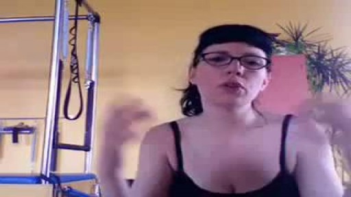
A Pilates instructor teaches the anatomy of the female pelvic floor using a pelvis model. She works through the pelvic bones and sacrum, explains how the sacrum moves during birth, connects the anatomy to post-birth back pain and rehabilitation, and then turns to the pelvic floor muscle layers.
Key points
- A pelvis model is used to show the hip bones and pelvic openings
- The ischial tuberosities, pubic bones and sacrum are identified
- Sacrum movement during birth is explained
- Links between the anatomy and post-birth back pain are discussed
- Pelvic floor muscle layers are introduced on the model
Chapters
- 1:02 — Hip bones and pelvis model
- 3:06 — Sacrum anatomy on the model
- 3:54 — Sacrum movement in birth
- 5:55 — Pelvic floor muscle layers
Video Transcript
Pilates instructor Mellie McCurray teaches the anatomy of the female pelvic floor using a pelvis model. She works through the bones — ilium, the ischial tuberosities you sit on, pubic bones, and sacrum — explains how the sacrum tips during birth, and connects the anatomy to post-birth back pain and rehabilitation before turning to the pelvic floor muscle layers.
Read the full transcript
Hi, I'm Mellie McCurray from SAMHSA Poll and Pilates, and Pilates Arts Tacoma. And this is a short educational video about the female pelvic floor. It's an upgrade from the one that's already posted on my SAMHSA YouTube site. This is the female pelvic floor. I love teaching the pelvic floor. When we see a pelvic floor like this, it's misleading in that it makes it look like this is truly the pelvic floor, but it includes so much more. But I'll point out here what is happening, and then I'll give you the kind of additional ideas of the anatomy that connects with the pelvic floor.
So these are often... I love my vagina. It's so long. These are the hip bones right here. We call them hip bones if you're just a layman, but they're the ilium, okay? And they create an upper and lower pelvis, really, right? You could see that top, bigger, down, smaller. So the ilium, these hip bones, really do a lot more than just our hip bones. Right here you could see this hole. That's where the head of the femur goes. This is called the acetabulum. It's the hip joint.
Most people think the hip joint's way out farther than it is, but if you have your finger right on the inside of the aces, that's the front here, the anterior superior iliac sign, then you're going to go right to the outside of it. So I'll show that on me right quick. Here's my hip bone, and if I went straight down, then actually my hip joint is here. That's the hip joint part. So then down here are your scissor bones. Those are the bones that you sit on.
They're called the ischial tuberosities from the bottom. They look like this. And if you're not familiar with where your scissor bones are, what you could do is pick up one side of your end, reach down, feel until you find kind of a round bump, and then notice things sit on it. In posture, you'd want to notice whether you sit behind it on top of it or in front of it. But this isn't really about posture here. It's about the pelvic bone. So you have your ischial tuberosities.
Then you have the pubic bone right here. You basically can see it's not just one solid unit, especially on a girl, it comes apart when we give birth. There's a right pubic bone and a left pubic bone. These are the obturators. I'll turn this around. Here's the sacrum. There's the tailbone. You can see it's several little tiny vertebra. The sacrum are fused, flat vertebra. If you hear clanging in the background, that's our boilers, by the way. Nothing to worry about. So you sacrum. And then here you can sort of see, let me see how to show this best.
This is the most bottom lumbar vertebra L5. And it sits right on top of the sacrum, which is often called S1 at the top. So because the sacrum are fused, flat, and vertebra, they have terms kind of like the rest of the spying vest. So the sacrum sits right in here between these ilium. And when a woman gives birth, the sacrum does a mutation, encounter a mutation. That means it tips back and forth. So when the baby begins to engage, then the sacrum, to make room for that baby coming through, tips backwards.
And then when the baby starts coming all the way through to allow the baby to come out the sacrum, tips forward so that the bottom of the sacrum tips out. What this often means for post-birth rehabilitation, whether you call it rehabilitation, or getting your body back together so it feels healthy, is that you're possibly going to be hypermobile in here, depending on how many babies you've had, too. And obviously, whether you had a vaginal birth and how that birth went, I mean, some women have smaller babies or easier or softer births,
and they don't have a lot of sacral issues. But in my practice, I find that the sacral issues after birth are pretty remarkable. But my back seems to have my baby. It just always aches right here. There's a lot of work that can be done to help that. Okay. So that's a bit about the bones that you see here. However, I do want to explain that the sacrum here, you can see it's tilted like this, so it bends backwards, right, behind the body. And the lower back comes forward like that.
It arcs forward. So they switch curves. And that vertebra at L5 can also create shear force, which when we're pregnant, you know, if the baby's really out that way, it can pull the lower back forward. And there's not a lot of support in here, and this can start to slide forward and mess with that disc, too. So lots of reasons why your back can hurt after you have a baby. And also, I mean, all these things can be an issue, even if you haven't had a baby, right?
So now let's talk more about the muscles. Muscles are the pelvic floor. So there's actually two layers. I can pull one layer off, isn't that fun? I'm going to put it in here. This is a pretty cool model. I like it a lot. I'm not going to give you all the names of the muscles because I don't want to fill your brain with too much, but I will explain kind of what they do and a general layman's idea of what they do. There are some really great books.
If you want the exact anatomy of what's happening, you can find that. So I'm going to try to get the light really good in here so we could really see what's going on. It's kind of hard. I have a little washout in here, and that's not going to work either. Let's see. Let me try to see. So washed out. Let's come back this way. Oh, that's not going to work. Okay, I'm going to try a different way. Okay, I got some darker. I got some shade behind me
to show the pelvic floor muscles better. Oh, here they are. I'm going to try to turn it and hold it really still. And I really wish there was a better closer, closer stop. Oh, that's great. I wish I could just balance it right here. Sorry for the Blair Witch Project vagina right now. Here's the clitoris. Here's what I make sure every person that ever studies with me knows. This is barely any of your clitoris. The clitoris comes up this way, the shaft, and it attaches to the suspensory ligament.
That, by the way, connects to the ovaries. Important to know. Okay, so then the clip has two legs. They come out this way, and they fork all the way down. Okay, so most of your clitoris is hidden. So that's where the clitoris is. And here's this little hole with red around it that's showing you the urethra and the urethral spongy material. And then of course, you got the introitis and the outer anal sphincter. Now, this is the sphincter layer of your pelvic floor. So partially, it's all about constricting to some degree or other,
just for different reasons. Obviously, the anal sphincter, it's a voluntary part of that sphincter and its job is to manage that area. Then up here, the introitis, it's surrounded by this part of the figurative system that creates when it gets engorged because there's spongy material in there. It makes a cuff, and that's why, once you're engorged and you're doing sexual play, it feels like the vagina is pulling together around whatever is inside there. And so that's the cuff system. There's also this whole system that takes care of the clitoris.
You have muscles that actually work with that, isn't that great? They kind of flank it. And they get engorged and put pressure on it and it swells and erects just like a man's penis. So when there's an erection of the clitoris, it actually retracts up and does this and this hardens in here.
Transcript auto-generated from the video’s audio for accessibility and study. Educational content only — not medical advice.
Video Overview
From our Gynecology collection: Pelvic exams, Pap smears, IUD insertion, hysteroscopy and women's health procedures explained step by step. Browse all Gynecology videos →
SORT BY-
Top Comments
-
Latest comments