
- Procedures & Tests
- Symptoms A-Z
- Symptom Checker
- Kidney Diet
- First Aid
- Anatomy Atlas
- Ask MedicalVideos
- Drug Guide
- Lab Values
- Vital Signs
- Muscles A–Z
- Clinical Signs
- Medical Abbreviations
- Medical Terminology
- Physical Examination
- Surgical Examination
- Ophthalmology
- Clinical Skills
- Orthopedics
- Surgery Videos
- Laparoscopy
- Pediatrics
- Funny Videos
- Cardiothoracic Surgery
- Nursing Videos
- Plastic Surgery
- Otorhinolaryngology
- Histology and Histopathology
- Neurosurgery
- Dermatology
- Pediatric Surgery
- Urology
- Dentistry
- Oncology and Cancers
- Anatomy Videos
- Health and Fitness
- Radiology
- Anaesthesia
- Physical Therapy
- Pharmacology
- Interventional Radiology
- Cardiology
- Endocrinology
- Gynecology
- Emergency Medicine
- Psychiatry and Psychology
- Childbirth Videos
- General Medical Videos
- Nephrology
- Physiology
- Diet and Food Health
- Diabetes Mellitus
- Neurology
- Women Health
- Osteoporosis
- Gastroenterology
- Pulmonology
- Hematology
- Rheumatology
- Toxicology
- Nuclear Medicine
- Infectious Diseases
- Vascular Disease
- Reproductive Health
- Burns and Wound Healing
- Kidney Zone Podcast
- Other







Hernia Exam

Educational only: videos on MedicalVideos.com are for general information and are not medical advice. Always consult your own doctor about your health.
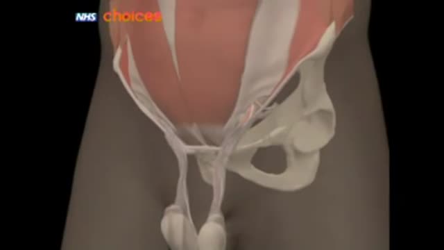
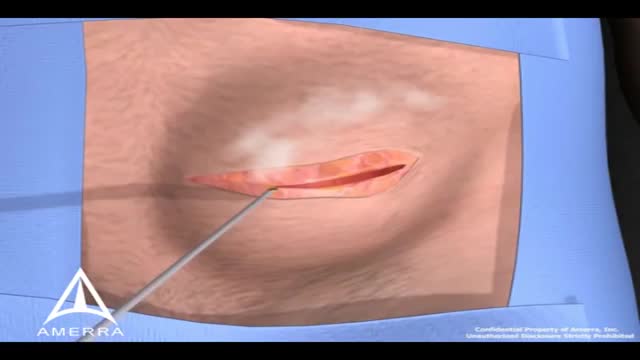
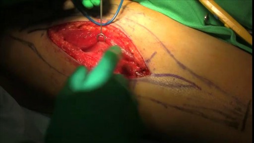
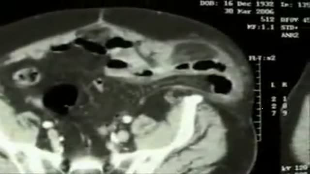
Inguinal hernia Diagram of an indirect, scrotal inguinal hernia ( median view from the left). Diagram of an indirect, scrotal inguinal hernia ( median view from the left). By far the most common hernias (up to 75% of all abdominal hernias) are the so-called inguinal hernias. For a thorough understanding of inguinal hernias, much insight is needed in the anatomy of the inguinal canal. Inguinal hernias are further divided into the more common indirect inguinal hernia (2/3, depicted here), in which the inguinal canal is entered via a congenital weakness at its entrance (the internal inguinal ring), and the direct inguinal hernia type (1/3), where the hernia contents push through a weak spot in the back wall of the inguinal canal. Inguinal hernias are more common in men than women while femoral hernias are more common in women.
This video in 30 seconds
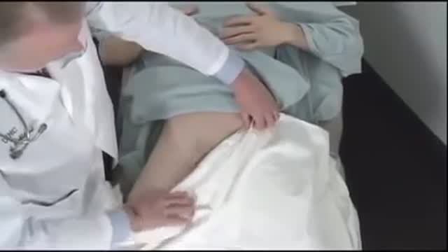
This narrated clinical-skills demonstration shows the hernia examination. The examiner inspects the inguinal and femoral regions while the patient bears down, then palpates for an inguinal hernia by following the spermatic cord to the external ring, and for a femoral hernia using the femoral pulse and inguinal ligament as landmarks. The recording also covers related genital inspection and palpation when the patient cannot stand.
Key points
- Inspection of the inguinal ring and femoral canal areas is shown while the patient bears down
- Palpation for an inguinal hernia follows the spermatic cord to the external ring
- The femoral pulse and inguinal ligament are used as landmarks for femoral hernia palpation
- Inspection and palpation are repeated on the opposite side
- Adaptations for a patient who cannot stand are described
Chapters
- 0:02 — Inspection for hernia bulges
- 0:16 — Palpating for inguinal hernia
- 0:41 — Palpating for femoral hernia
- 1:24 — Examination when patient cannot stand
Video Transcript
A narrated clinical-skills demonstration of the hernia examination. It works through inspection of the inguinal ring and femoral canal while the patient bears down, then palpation for inguinal hernia by following the spermatic cord to the external ring, and finally palpation for a femoral hernia using the femoral pulse and inguinal ligament as landmarks.
Read the full transcript
To assess for hernias, inspect the areas of the external inguinal ring and the femoral canal for bulges and continue to watch as the patient bears down. Please bear down. If none is seen, palpate for an inguinal hernia. Using your right index finger for the patient's right side, invaginate some loose scrotal skin and follow the spermatic cord up to the external inguinal ring, and if possible, through it to the inguinal canal. Ask the patient to bear down or cough and feel for a bulging mass that touches your finger.
Now palpate for a femoral hernia. Using the femoral pulse and inguinal ligament for orientation, place your fingers at the femoral canal and feel for swelling or tenderness as the patient again bears down or coughs. Please bear down. Repeat inspection and palpation on the left side using your left index finger to palpate the external inguinal ring. When a male patient cannot or should not stand up for this exam, perform it while he is lying down. This position also makes it easier to inspect all surfaces of the penis and scrotum,
cover the patient's legs and expose his genitalia. To assess sexual maturity, if indicated, inspect the patient's pubic hair and genitalia. Inspect the skin around the base of the penis for inflammation, excoriation, nits or lice. After retracting the foreskin, if necessary, inspect the glands and note the location of the urethral meatus. Compress the glands to open the meatus. If discharge is present, culture it. Then palpate the shaft of the penis. Continue by inspecting the scrotum, including its contours and all skin surfaces. Then palpate each testis and epididymis.
Feel each spermatic cord, including the vast efferents, and palpate it from the epididymis to the external inguinal ring.
Transcript auto-generated from the video’s audio for accessibility and study. Educational content only — not medical advice.
Medical Videos
Free medical and surgery videos for patients, students and professionals — trusted since 2007.
Founded by Dr. Mohamed Ibrahim, MD, PhD — physician-scientist.
Follow us on Facebook · Instagram
SORT BY-
Top Comments
-
Latest comments