- Physical Examination
- Surgical Examination
- Ophthalmology
- Clinical Skills
- Orthopedics
- Surgery Videos
- Laparoscopy
- Pediatrics
- Funny Videos
- Cardiothoracic Surgery
- Nursing Videos
- Plastic Surgery
- Otorhinolaryngology
- Histology and Histopathology
- Neurosurgery
- Dermatology
- Pediatric Surgery
- Urology
- Dentistry
- Oncology and Cancers
- Anatomy Videos
- Health and Fitness
- Radiology
- Anaesthesia
- Physical Therapy
- Pharmacology
- Interventional Radiology
- Cardiology
- Endocrinology
- Gynecology
- Emergency Medicine
- Psychiatry and Psychology
- Childbirth Videos
- General Medical Videos
- Nephrology
- Physiology
- Diet and Food Health
- Diabetes Mellitus
- Neurology
- Women Health
- Osteoporosis
- Gastroenterology
- Pulmonology
- Hematology
- Rheumatology
- Toxicology
- Nuclear Medicine
- Infectious Diseases
- Vascular Disease
- Reproductive Health
- Burns and Wound Healing
- Other
Top videos
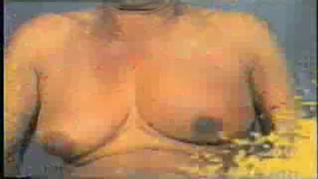
Breast masses are broadly classified as benign or malignant. Common causes of a benign breast mass include fibrocystic disease, fibroadenoma (see the image below), intraductal papilloma, and abscess.

Professional breast exam
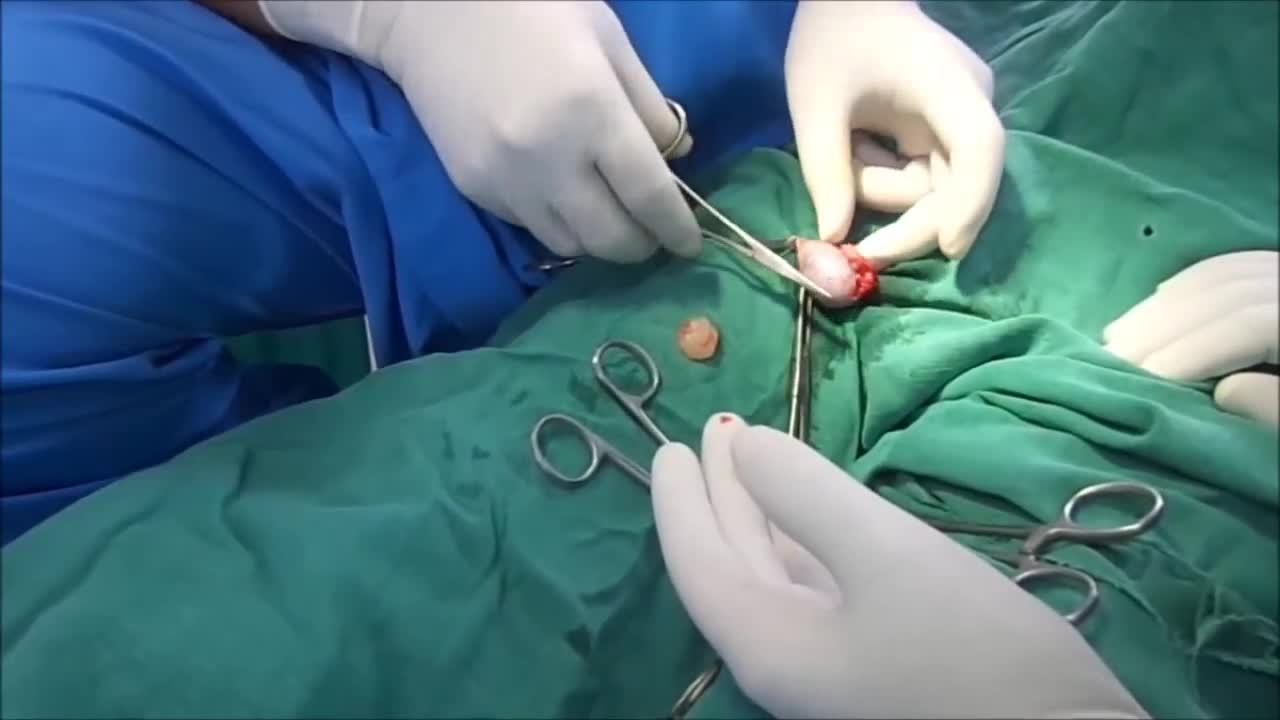
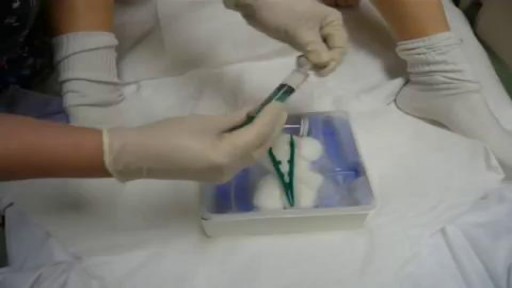
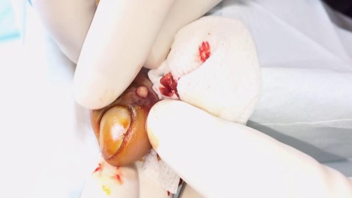
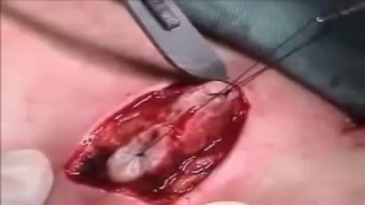
Surgery. A procedure to remove an epididymal cyst is carried out under general anaesthetic and involves removing the cysts through a small incision in your scrotum that is sealed with dissolvable stitches.
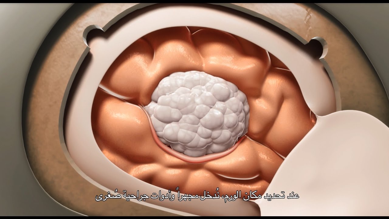
A craniotomy may be performed to treat brain tumors, blood clots, aneurysms, skull fractures, foreign objects, swelling of the brain, stroke or infection.
Our General Surgery team treats hernia patients on a daily basis. In fact, you could consider them to be hernia experts. We sat down with one of those experts, Dr. Heater Dunlap, to talk about the common signs and symptoms of hernias and to answer the question of when to see a doctor.

Watch that Female Foley Catheter Insertion Procedure
Catheterization of the Male and Female

This sqadia.com short video clip is a brief explanation of Epithelium.
Epithelium is one of the four basic tissues of the body and is derived from all three germ layers.
It is composed of very closely packed, contiguous cells, with very little or no extracellular material in the extracellular spaces.
----------------------------------------
Histology Lectures Collection -
https://www.sqadia.com/categor....ies/anatomy-histolog
----------------------------------------
Epithelial membranes can be: Simple squamous epithelium, Simple cuboidal epithelium, Simple columnar epithelium, and Pseudostratified epithelium.
----------------------------------------
5500+ Medical Videos
Try for FREE! - https://www.sqadia.com/categories/free
----------------------------------------
When there are two or more layers of cells epithelia is referred to as stratified, hence can be stratified squamous, stratified cuboidal and stratified columnar.
----------------------------------------
Facebook - https://www.facebook.com/sqadiacom
Instagram - https://www.instagram.com/sqadiacom
LinkedIN - https://www.linkedin.com/showcase/sqadia-com
Pinterest - https://www.pinterest.com/sqadiacom
TumblR - https://sqadiacom.tumblr.com
Twitter - https://twitter.com/sqadiacom
Vimeo - https://vimeo.com/sqadiacom
YouTube - https://www.youtube.com/sqadiacom
----------------------------------------

ThermiVa is a non-surgical vaginal tightening treatment for women who want to reclaim what childbirth or aging may have taken away. Using the same technology that’s used in ThermiTight and ThermiSmooth, radiofrequency energy is sent to the desired area (internally or externally), heating the tissue and stimulating the body’s own collagen. ThermiVa is performed in three treatments over the course of three months.

This is an educational medical video for Medical Students showing how to examine a hernia swelling

A patient at a British hospital played Mahler and Gershwin on the violin while surgeons removed a tumor from her brain, so doctors could preserve her ability to play music.
She left the hospital 3 days later and hopes to return to the symphony soon. https://abcn.ws/2SGY9mp
SUBSCRIBE to ABC NEWS: https://www.youtube.com/ABCNews/
Watch More on http://abcnews.go.com/
LIKE ABC News on FACEBOOK
https://www.facebook.com/abcnews
FOLLOW ABC News on TWITTER:
https://twitter.com/abc
GOOD MORNING AMERICA'S HOMEPAGE:
https://www.goodmorningamerica.com/
Paronychia Fingernail Abscess Infection Treatment
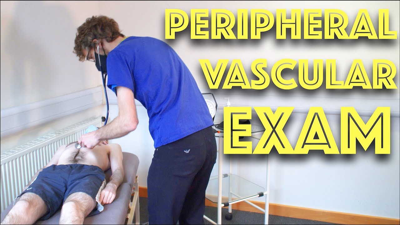
Peripheral Vascular Examination OSCE - Clinical Skills - Dr Gill
In the cardiovascular examination, particularly in the case of an OSCE station, we conclude the examination often by stating that the examiner would want to perform:
- An ECG
- Check full blood count
- and "do a peripheral vascular examination
In this video, we demonstrate that oft-talked about, but comparatively less common examination.
Starting off, with the examination of the hands, the radial, brachial and carotid pulses. before moving down to assess for a AAA, checking the femoral and popliteal pulses, before wrapping up around the ankle with the posterior tibial and dorsalis pedis pulses
For completeness, the cardiovascular examination is demonstrated here
https://www.youtube.com/watch?v=ECs9O5zl6XQ&t=2s
#PeripheralVascular #ClinicalSkills #DrGill
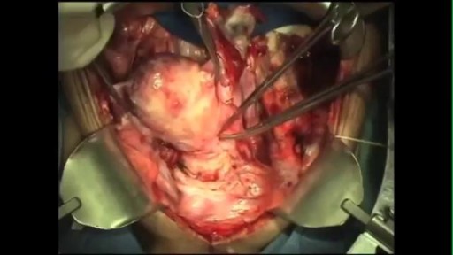
Care must be taken to prevent stenosis at the anastomotic site. If the diameter of the anastomosis is less than 2 cm, the anastomosis should be taken down and resected. A classic end-to-end anastomosis should be performed to ensure adequate diameter to the intestine. If the posterior wall of the colon has been preserved, care should be taken to close the colostomy prior to opening the peritoneal cavity. This will reduce intraperitoneal contamination from the stoma site. Copious irrigation of the wound should be made prior to primary closure. If gross contamination has occurred, delayed closure of the wound should be considered.

http://www.highimpact.com - This brain surgery animation was used to demonstrate a young girl's craniotomy, cranioplasty, and reconstructive skull surgery after her vehicle was struck by a tractor-trailer. The procedures included the evacuation of a large epidural hematoma, the draining of the epidural space, and the reassembly of bone fragments to repair the skull.
More Brain Surgery Animations: https://tinyurl.com/y6m4lkdf
WHAT HAPPENED
A teenage girl was riding home with her parents and boyfriend from a Wednesday night church service when a tractor-trailer struck the back driver’s side of their car as they were traveling through an intersection. The impact sent the car spinning into oncoming traffic where it struck another vehicle. When paramedics arrived, the 17-year-old was unresponsive with bleeding from her left ear and a laceration from behind her left ear.
She was rushed to the hospital where she underwent a series of CT scans that showed a severely comminuted open skull fracture with an underlying 1.1 cm subdural hematoma. She was taken to the operating room where an emergency craniotomy was performed to evacuate the hematoma and reassemble the skull fragments. The patient gradually began to wake up and was discharged six days later, after she showed she could maneuver up and down the hallway.
The biggest challenge in a traumatic brain injury case like this - where most of the damages are deeply underlying and undetectable on the surface - is that the only visual evidence is in the form of 2D black-and-white radiographic films. This can look ambiguous to the typical juror because it’s often difficult to discern where these snapshots are located inside the person’s skull. Tony Seaton, Esq., and Robert Bates, Esq., needed to reinforce this 2D radiographic evidence with maximum 3D context.
We equipped them with a custom Diagnostic Slice Chooser: an interactive presentation that presents radiographic slides within a three-dimensional model of the patient’s head. We also designed the model accurately to the patient’s likeness and colorized the films to highlight key areas of damage. The attorneys could show the complete depth and magnitude of his client’s injuries at every level both before and after the surgery. After establishing the full extent of damages, we also created an animation to walk viewers through the surgical experience the patient would undergo as a result of her injuries.
The visual presentation helped jurors understand the destructive impact this collision had on this young teenager’s life, and Mr. Seaton and Mr. Bates, Esq., were able to acquire a $4.5M settlement for his client.
Read the Full Case Study: https://tinyurl.com/yy4v2dyh

UPDATE 2/6/15: A new version of this animation is now available! https://www.youtube.com/watch?v=E1ljClS0DhM
This 3D medical animation depicts the surgical removal of the appendix (appendectomy) using laparoscopic instruments. The surgery animation begins by showing an inflamed appendix (appendicitis), followed by the placement of the laparoscope. Afterward, one can see the surgical device staple, cut and remove the inflamed appendix. Following the removal of the appendix the abdomen is flushed with a sterile saline solution to ensure all traces of infection have been removed.
ANCE00183
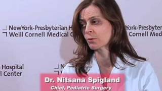
As a pediatric surgeon at NewYork-Presbyterian/Weill Cornell Medical Center, Dr. Nitsana Spigland treats newborns, children, teens, and young adults requiring surgical interventions. She specializes in antenatal counseling and newborn congenital malformations.
Learn more about Dr. Spigland at: https://www.nyp.org/physician/nspigland.
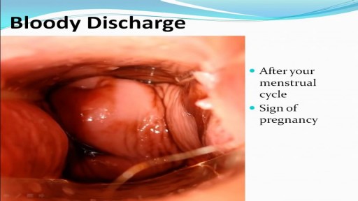
Watch that video to know the Types of Female Genital discharge
