- Physical Examination
- Surgical Examination
- Ophthalmology
- Clinical Skills
- Orthopedics
- Surgery Videos
- Laparoscopy
- Pediatrics
- Funny Videos
- Cardiothoracic Surgery
- Nursing Videos
- Plastic Surgery
- Otorhinolaryngology
- Histology and Histopathology
- Neurosurgery
- Dermatology
- Pediatric Surgery
- Urology
- Dentistry
- Oncology and Cancers
- Anatomy Videos
- Health and Fitness
- Radiology
- Anaesthesia
- Physical Therapy
- Pharmacology
- Interventional Radiology
- Cardiology
- Endocrinology
- Gynecology
- Emergency Medicine
- Psychiatry and Psychology
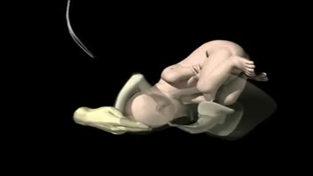
- Childbirth Videos
- General Medical Videos
- Nephrology
- Physiology
- Diet and Food Health
- Diabetes Mellitus
- Neurology
- Women Health
- Osteoporosis
- Gastroenterology
- Pulmonology
- Hematology
- Rheumatology
- Toxicology
- Nuclear Medicine
- Infectious Diseases
- Vascular Disease
- Reproductive Health
- Burns and Wound Healing
- Other
Top videos
a video showing the process of child birth or delivery using forceps

This minimally invasive technique allows surgeons to remove skull base tumors as large as softballs through the nose, with less trauma to the brain and critical nerves than with a traditional craniotomy.
To learn more, please visit https://www.upmc.com/
http://www.utexas.edu
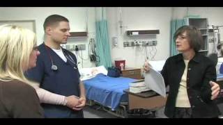
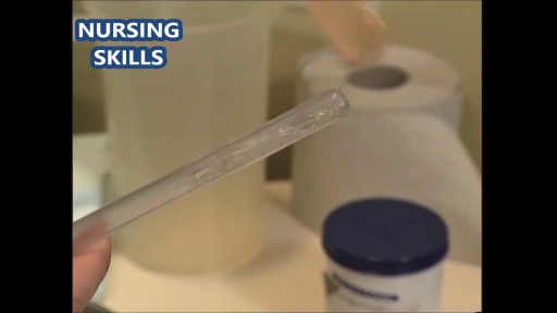
Nursing students practice their skills on mannequins and each other in the Nursing Skills Lab.
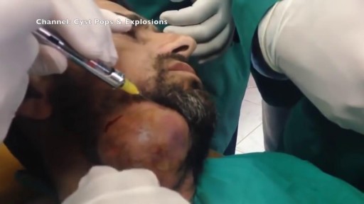
Watch that video of the Worlds largest Face Abscess Draining
exam
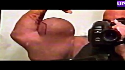
Bodybuilder Drains Synthol Hematoma From Bicep
Watch that video of Enema Medical Insertion Procedure
Delivery Video
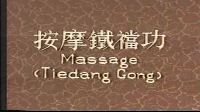
Mysterious massage from East Asia(CHINA).it can cure cure Erectile dysfunction,can let their life better.This video from mainland of China,so the language is Chinese mandarin.but you can see English show on the video too.Tiedang gong means kongfu of Iron penis&balls.
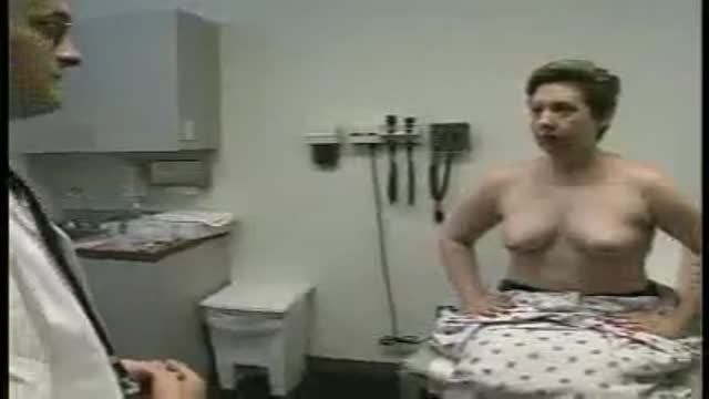
Full examination of the female from head to toe by Loyola Medical School, Chicago. Part 3

It’s called gamma knife surgery, but there’s no cutting involved.
It’s been used at Mayo Clinic for 30 years as an alternative to open brain surgery.
The patient’s head is held still during the procedure with a headframe, which also serves as a map for the radiation. Using 3D imaging — typically an MRI — as a guide, the gamma knife is targeted directly at the tumor.
And with no hospital stay and minimal side effects, it’s a procedure that is efficient and can be lifesaving.
More health and medical news on the Mayo Clinic News Network. https://newsnetwork.mayoclinic.org/
Journalists: Clean and nat sound versions of this pkg available for download at https://newsnetwork.mayoclinic.org/
Register (free) at https://newsnetwork.mayoclinic.org/request-account/
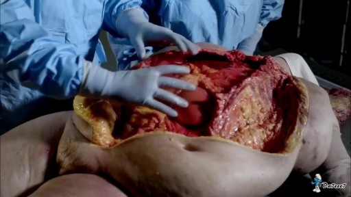
Watch that Cutting Inside Human Fat Body video
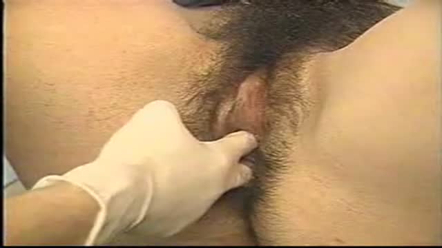
A video showing the examination of femoral hernia.

This video demonstrate Laparoscopic Cholecystectomy Full Length Skin to Skin Video with Infrared Cholangiography performed by Dr R K Mishra at World Laparoscopy Hospital. Infrared Cholegiography is performed by using Indocyanine Green during laparoscopic cholecystectomy surgery for gallbladder removal. Bile duct injury remains the most feared complication of laparoscopic cholecystectomy. Intraoperative cholangiography (IOC) is the current gold standard for biliary imaging and may reduce injury, but is not widely used because of the difficulties of doing it. Near-Infrared Fluorescence Cholangiography (NIRF-C) is a novel non-invasive method for real-time, radiation-free, intra-operative biliary mapping during laparoscopic cholecystectomy. We have experienced that NIRF-C is a safe and effective method for identifying biliary anatomy during laparoscopic cholecystectomy. Indocyanine green is a cyanine dye is very popular and used for many years in medical diagnostics. It is used for determining cardiac output, hepatic function, liver, and gastric blood flow, and for ophthalmic angiography. Now the use of this dye in lap chole has improved the safety of this surgery by NEAR INFRARED FLUORESCENT CHOLANGIOGRAPHY.
For more information please contact:
World Laparoscopy Hospital
Cyber City, Gurugram, NCR DELHI
INDIA 122002
Phone & WhatsApp: +919811416838, + 91 9999677788
Sex reassignment surgery for male-to-female involves reshaping the male genitals into a form with the appearance of, and, as far as possible, the function of female genitalia. Prior to any surgeries, patients usually undergo hormone replacement therapy (HRT), and, depending on the age at which HRT begins, facial hair removal. There are associated surgeries patients may elect to, including facial feminization surgery, breast augmentation, and various other procedures.

http://www.nucleushealth.com/ - This 3D medical animation depicts two operations, called craniotomy and craniectomy, in which the skull is opened to access the brain. The normal anatomy of the skull and tissues surrounding the brain are shown, including arteries and veins. The animation lists the common reasons for these procedures, and briefly introduces intracranial pressure.
Video ID: ANH13109
Transcript:
Your doctor may recommend a craniotomy or a craniectomy procedure to treat a number of different brain diseases, injuries, or conditions.
Your skull is made of bone and serves as a hard, protective covering for your brain. Just inside your skull, three layers of tissue, called meninges, surround your brain. The thick, outermost layer is the dura mater. The middle tissue layer is the arachnoid mater and the innermost layer is the pia mater. Between the arachnoid mater and the pia mater is the subarachnoid space, which contains blood vessels and a clear fluid called cerebrospinal fluid. Blood vessels, called bridging veins, connect the surface of your brain with the dura mater. Other blood vessels, called cerebral arteries, bring blood to your brain.
Inside your skull, normal brain function requires a delicate balance of pressure between the blood in your blood vessels, the cerebrospinal fluid that surrounds your brain, and your brain tissue. This is called normal intracranial pressure. Increased intracranial pressure may result from: brain tumors, head injuries, problems with your blood vessels, or infections in your brain or spinal cord. These conditions put pressure on your brain and may cause it to swell or change shape inside your skull, which can lead to serious brain injury.
Your doctor may recommend a craniotomy to remove: abnormal brain tissue, such as a brain tumor, a sample of tissue by biopsy, a blood clot, called a hematoma, excess cerebrospinal fluid, or pus from an infection, called an abscess.
A craniotomy may also be done to: relieve brain swelling,
stop bleeding, called a hemorrhage, repair abnormal blood vessels, repair skull fractures, or repair damaged meninges.
Finally, a craniotomy may also be done to: treat brain conditions, such as epilepsy, deliver medication to your brain, or implant a medical device, such as a deep brain stimulator.
The most common reason for a craniotomy is to remove a brain tumor.
#Craniotomy #Craniectomy #BrainSurgery

Funny Video from hospital waiting room

Children are special patients, and their medical needs are unique, including their surgical needs. At UNC Hospitals, an expert and experienced team of physicians treat children in a kid-friendly and family-centered environment. UNC Pediatric Surgeon Dr. Timothy Weiner explains
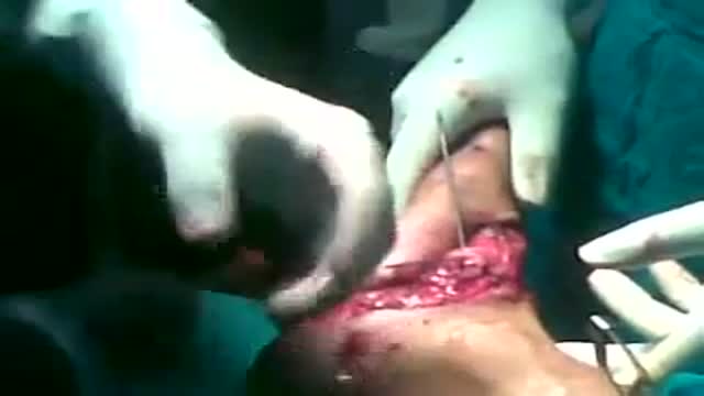
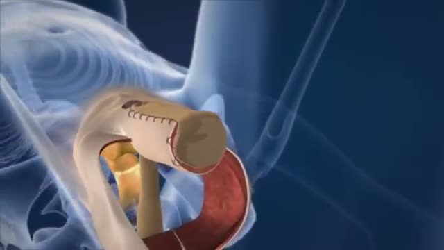
Kirschner wires or K-wires or pins are sterilized, sharpened, smooth stainless steel pins. Introduced in 1909 by Martin Kirschner, the wires are now widely used in orthopaedics and other types of medical and veterinary surgery. They come in different sizes and are used to hold bone fragments together (pin fixation) or to provide an anchor for skeletal traction. The pins are often driven into the bone through the skin (percutaneous pin fixation) using a power or hand drill. They also form part of the Ilizarov apparatus.
catheterization of the male urethra by a foley catheter
