- Physical Examination
- Surgical Examination
- Ophthalmology
- Clinical Skills
- Orthopedics
- Surgery Videos
- Laparoscopy
- Pediatrics
- Funny Videos
- Cardiothoracic Surgery
- Nursing Videos
- Plastic Surgery
- Otorhinolaryngology
- Histology and Histopathology
- Neurosurgery
- Dermatology
- Pediatric Surgery
- Urology
- Dentistry
- Oncology and Cancers
- Anatomy Videos
- Health and Fitness
- Radiology
- Anaesthesia
- Physical Therapy
- Pharmacology
- Interventional Radiology
- Cardiology
- Endocrinology
- Gynecology
- Emergency Medicine
- Psychiatry and Psychology
- Childbirth Videos
- General Medical Videos
- Nephrology
- Physiology
- Diet and Food Health
- Diabetes Mellitus
- Neurology
- Women Health
- Osteoporosis
- Gastroenterology
- Pulmonology
- Hematology
- Rheumatology
- Toxicology
- Nuclear Medicine
- Infectious Diseases
- Vascular Disease
- Reproductive Health
- Burns and Wound Healing
- Other
Top videos
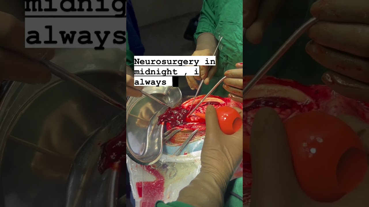
In this video, Dr Dhaval Patel, the best brain & spine surgeon in Surat South Gujarat, is performing Brain Hemorrhage Surgery. The Brain Hemorrhage Surgery was successfully done by the best neurosurgeon Dr Dhaval Patel in the midnight in Surat, South Gujarat.
Dr Dhaval Patel is the best and experienced brain & spine surgeon in Adajan, Vesu, Parvat Patiya, Surat, South Gujarat. Dr Dhaval is the expert of treatments and surgery for brain problems and spine problems.
.
Brain Hemorrhage Surgery, Best Brain & Spine Surgeon, Neurosurgeon, Brain Tumor Surgery, Brain Treatment Expert, Brain Expert, Brain & Spine Surgery, Neurosurgery in Surat, South Gujarat, Ahmedabad, Rajkot, Anand, Porbandar, patan, kutch, jamnagar, bhavnagar, junagadh, mehsana, nadiad, amreli, morbi, gandhinagar, verval, palanpur,godhra, gandhidham, botad, jetpur, kundal, kalol, disha, gondal, himatnagar, bhuj, modasa, lonavala, mandavi, kheda, khambhaliya, khambhat, dwarka, chhota udaipur, ambaji, dhoraji, idar, vallabhipur, una, dhandhuka, bhachau, mundra.
Dr. Dhaval Patel is an excellent neurosurgeon in Surat, South Gujarat. He is a Brain and Spine Surgeon; he is a reputable Neurosurgeon in Surat, South Gujarat. He has been practicing for the past five years. Till now, he has done 2500+ minor and major surgeries.
NEUROSURGEON DR. DHAVAL PATEL
Specialist in Brain & Spine Surgery
M.S.DNB (Neurosurgery - New Delhi)
Consultant Neurosurgeon
Surat Neuro Clinic Majura Gate, Ring Road, Surat.
Unity Hospital Parvat Patiya, Surat
United Green Hospital Adajan, Surat.
For more info. : +91-9687866766
#brainhemorrhage #brainsurgery #brainhemorrhagesurgery #brainstroke #heartdisease #brainconditions #brainsurgery #drdhavalpatel #spine #spinesurgery #unitedgreenhospital #surat_neuro_clinic #unity_hospital #drdhavalpatel #hormones #health #neuro #neurologiest #brain #surgery #recovery #patientreview #neurosurgeon #minimally_invasive #surgery #neurosurgery #stroke #heartattack #i3corporation
Breast abscesses are often linked to mastitis – a condition that causes breast pain and swelling (inflammation), and usually affects women who are breastfeeding. Infections can occur during breastfeeding if bacteria enter your breast tissue, or if the milk ducts (tiny tubes) become blocked. This can cause mastitis which, if not treated, can result in an abscess forming. Women who aren't breastfeeding can also develop mastitis if bacteria enter the milk ducts through a sore or cracked nipple, or a nipple piercing. White blood cells are sent to attack the infection, which causes tissue at the site of the infection to die. This creates a small, hollow area that fills with pus (an abscess).
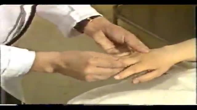
The examination room should be quiet, warm and well lit. After you have finished interviewing the patient, provide them with a gown (a.k.a. "Johnny") and leave the room (or draw a separating curtain) while they change. Instruct them to remove all of their clothing (except for briefs) and put on the gown so that the opening is in the rear. Occasionally, patient's will end up using them as ponchos, capes or in other creative ways. While this may make for a more attractive ensemble it will also, unfortunately, interfere with your ability to perform an examination! Prior to measuring vital signs, the patient should have had the opportunity to sit for approximately five minutes so that the values are not affected by the exertion required to walk to the exam room. All measurements are made while the patient is seated. Observation: Before diving in, take a minute or so to look at the patient in their entirety, making your observations, if possible, from an out-of-the way perch. Does the patient seem anxious, in pain, upset? What about their dress and hygiene? Remember, the exam begins as soon as you lay eyes on the patient. Temperature: This is generally obtained using an oral thermometer that provides a digital reading when the sensor is placed under the patient's tongue. As most exam rooms do not have thermometers, it is not necessary to repeat this measurement unless, of course, the recorded value seems discordant with the patient's clinical condition (e.g. they feel hot but reportedly have no fever or vice versa). Depending on the bias of a particular institution, temperature is measured in either Celcius or Farenheit, with a fever defined as greater than 38-38.5 C or 101-101.5 F. Rectal temperatures, which most closely reflect internal or core values, are approximately 1 degree F higher than those obtained orally. Respiratory Rate: Respirations are recorded as breaths per minute. They should be counted for at least 30 seconds as the total number of breaths in a 15 second period is rather small and any miscounting can result in rather large errors when multiplied by 4. Try to do this as surreptitiously as possible so that the patient does not consciously alter their rate of breathing. This can be done by observing the rise and fall of the patient's hospital gown while you appear to be taking their pulse. Normal is between 12 and 20. In general, this measurement offers no relevant information for the routine examination. However, particularly in the setting of cardio-pulmonary illness, it can be a very reliable marker of disease activity. Pulse: This can be measured at any place where there is a large artery (e.g. carotid, femoral, or simply by listening over the heart), though for the sake of convenience it is generally done by palpating the radial impulse. You may find it helpful to feel both radial arteries simultaneously, doubling the sensory input and helping to insure the accuracy of your measurements. Place the tips of your index and middle fingers just proximal to the patients wrist on the thumb side, orienting them so that they are both over the length of the vessel.
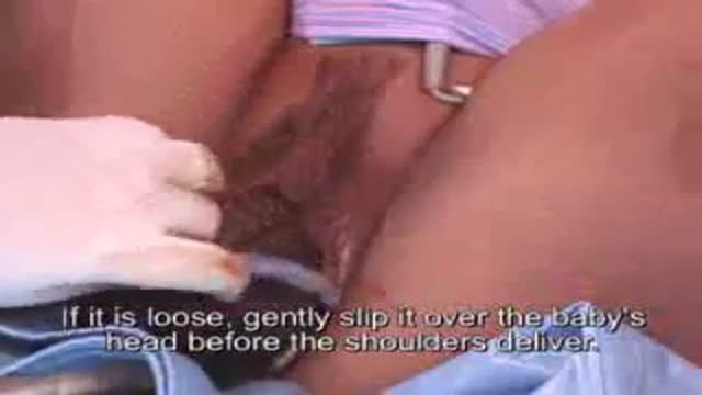
The umbilical cord is wrapped around the baby's neck in about 25% of deliveries. If loose, it usually has no impact on the delivery. If tight, it may need to be relieved before delivery of the baby can proceed safely
Open heart (coronary artery bypass, or CABG) surgery is performed in order to reroute, or "bypass," blood around blocked arteries, thereby improving the supply of oxygen-rich blood to the heart. Surgeons usually use an artery from the chest wall to construct the "detour" around the blocked part of the artery. Veins from the legs are also used.
Chinese Complete Physical Clinical Exam
A breech birth is the birth of a baby from a breech presentation. In the breech presentation the baby enters the birth canal with the buttocks or feet first as opposed to the normal head first presentation.
There are either three or four main categories of breech births, depending upon the source:
* Frank breech - the baby's bottom comes first, and his or her legs are flexed at the hip and extended at the knees (with feet near the ears). 65-70% of breech babies are in the frank breech position.
* Complete breech - the baby's hips and knees are flexed so that the baby is sitting crosslegged, with feet beside the bottom.
* Footling breech - one or both feet come first, with the bottom at a higher position. This is rare at term but relatively common with premature fetuses.
* Kneeling breech - the baby is in a kneeling position, with one or both legs extended at the hips and flexed at the knees. This is extremely rare, and is excluded from many classifications.
As in labour with a baby in a normal head-down position, uterine contractions typically occur at regular intervals and gradually cause the cervix to become thinner and to open. In the more common breech presentations, the baby’s bottom (rather than feet or knees) is what is first to descend through the maternal pelvis and emerge from the vagina.
At the beginning of labour, the baby is generally in an oblique position, facing either the right or left side of the mother's back. As the baby's bottom is the same size in the term baby as the baby's head. Descent is thus as for the presenting fetal head and delay in descent is a cardinal sign of possible problems with the delivery of the head.
In order to begin the birth, internal rotation needs to occur. This happens when the mother's pelvic floor muscles cause the baby to turn so that it can be born with one hip directly in front of the other. At this point the baby is facing one of the mother's inner thighs. Then, the shoulders follow the same path as the hips did. At this time the baby usually turns to face the mother's back. Next occurs external rotation, which is when the shoulders emerge as the baby’s head enters the maternal pelvis. The combination of maternal muscle tone and uterine contractions cause the baby’s head to flex, chin to chest. Then the back of the baby's head emerges and finally the face.
Due to the increased pressure during labour and birth, it is normal for the baby's leading hip to be bruised and genitalia to be swollen. Babies who assumed the frank breech position in utero may continue to hold their legs in this position for some days after birth.
exam
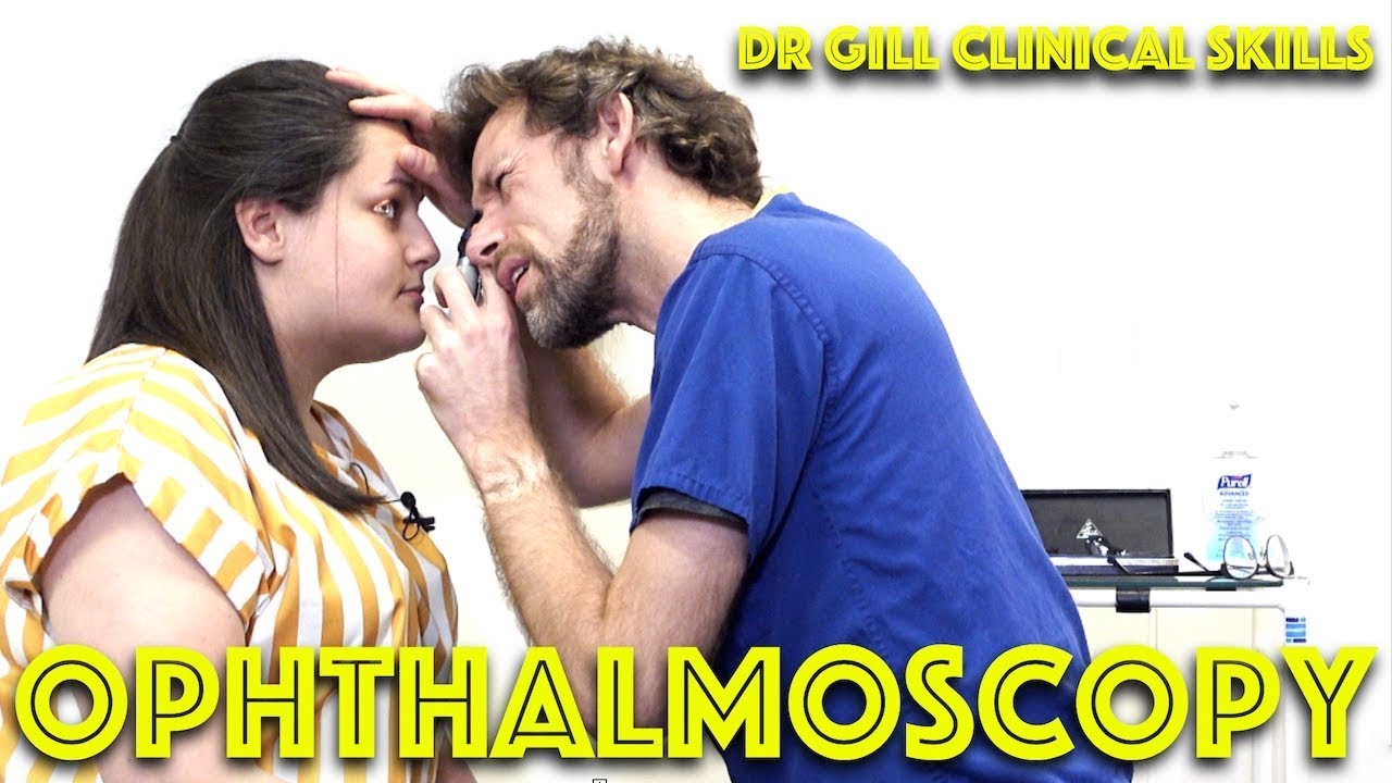
Ophthalmoscopy - Eye Clinical Examination - OSCE - Dr Gill
Direct Ophthalmoscopy use of the eyes is a very challenging clinical skill, incorporating both the examiner's knowledge of the retina, but also understanding the use of the ophthalmoscope
In this clinical skills tutorial, we look at the use of the direct ophthalmoscope as part of an ophthalmic examination
it should be noted that in the ideal circumstances, the room lights will be dimmed during the examination, and dilating eye drops used to improve the visualisation of the fundus
Some people may notice an ASMR effect from this clinical examination
#DrGill #Ophthalmoscopy #ClinicalSkills #EyeExam
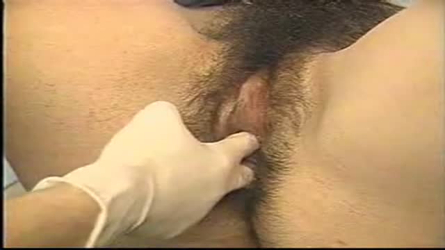
A vaginoplasty is a surgical procedure that tightens the vagina. This is done by removing excess vaginal lining and tightening the surrounding soft tissues and muscles. During delivery of a baby the vagina and surrounding tissues and muscles become stretched. After delivery the vagina may return to a more “normal” size, but it often fails to return to its’ pre pregnancy diameter. Generally, the more vaginal deliveries, the worse the condition gets. Many women will complain of decreased sensation and sexual satisfaction during intercourse. Commonly this is due to a lack of friction. Often their partner may notice a change although he may say nothing. Kegel exercises are often recommended but rarely succeed in restoring vaginal tightness.

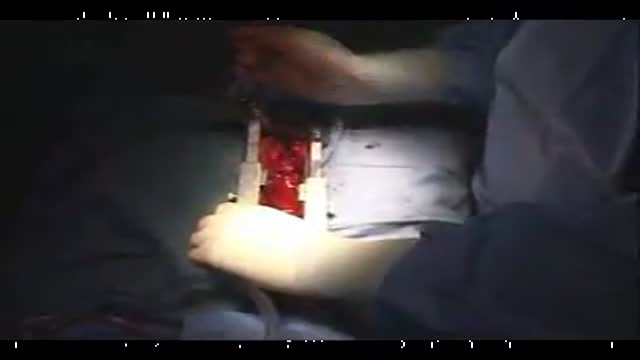
This 3D animation of brain surgery, shows how a ventriculostomy is performed, which is a neurosurgical procedure of creating a hole within a cerebral ventricle for drainage. It is most commonly performed on those with hydrocephalus, an abnormal buildup of fluid in the ventricles (cavities) deep within the brain. It's done by surgically penetrating the skull, dura mater, and brain such that the ventricular system ventricle of the brain is accessed.
When catheter drainage is temporary, it is commonly referred to as an external ventricular drain (EVD). When catheter drainage is permanent, it is usually referred to as a shunt.
There are many catheter-based ventricular shunts that are named for where they terminate, for example, a ventriculi-peritoneal shunt terminates in the peritoneal cavity, a ventriculoarterial shunt terminates within the atrium of the heart, etc. The most common entry point on the skull is called Kocher's point. An EVD ventriculostomy is done primarily to monitor the intracranial pressure as well as to drain cerebrospinal fluid (CSF), primarily, or blood to relieve pressure from the central nervous system (CNS).
For more information about custom medical animation, please visit https://www.amerra.com/.
Watch additional medical animations:
Craniectomy brain surgery - 3D animation: https://youtu.be/1RkseDeYS9g
Accessing an implantable port training - 3D animation: https://youtu.be/xSTpxjyv4O4
Open Suctioning with a Tracheostomy Tube - 3D animation: https://youtu.be/wamB7jpWCiQ
Suctioning the endotracheal tube - medical animation: https://youtu.be/pN6-EYoeh3g
Functional endoscopic sinus surgery (FESS) - 3D animation: https://youtu.be/qKTRyowwaLA
How to insert a nasogastric tube for NG intubation - 3d animation: https://youtu.be/Abf3Gd6AaZQ
Oral airway insertion - oropharyngeal airway technique - 3D animation: https://youtu.be/caxUdNwjt34
Nasotracheal suctioning (NTS) - 3D animation: https://youtu.be/979jWMsF62c
Learn about hemorrhoids with #3d #animation: https://youtu.be/R6NqlMpsiiY
LASIK eye surgery - 3D animation: https://youtu.be/Bb8bnjnEM00
CPR cardiopulmonary resuscitation - 3D animation: https://youtu.be/G87knTZnhks
What are warts (HPV)? - 3D animation: https://youtu.be/guJ1J7rRs1w
How Macular Degeneration Affects Your Vision - 3D animation: https://youtu.be/ozZQIZ_52YY
NeoGraft hair transplant procedure – animation: https://youtu.be/C-eTdH2UPXI
Watch that Ectopic Pregnancy Medical Abortion Procedure

The dilatation and Curettage procedure that is commonly performed (D and C)

Pediatric surgeons at Texas Children’s Hospital West Campus perform general surgical procedures such as circumcisions, removal of foreign objects, hernia repair, and suturing of minor lacerations. While more complex surgeries take place at the Texas Children’s Main Campus, pre-operative and follow-up outpatient care for those procedures is available at the West Campus.
Everything about Texas Children’s Hospital West Campus is dedicated to the health and wellness of children. As greater Houston's first suburban hospital designed exclusively for children, we offer the expert care you've come to trust from Texas Children's Hospital coupled with a location that's convenient and accessible for area families. Our facility is located just off the westbound feeder road of the Katy Freeway (at I-10 and Barker Cypress).
For more information about Texas Children's Hospital West Campus, visit http://www.texaschildrens.org/....Locate/In-the-Commun
Meet Dr. Allen Milewicz, chief of community surgery at Texas Children's West Campus
https://www.youtube.com/watch?v=uMoCdipuKfA&index=16&list=PLiN68C9rloPBD-E9ChWhVy73h7V3SEMlm
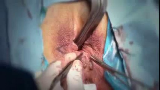
Huge Sebaceous Cyst Removal Medical Video procedure
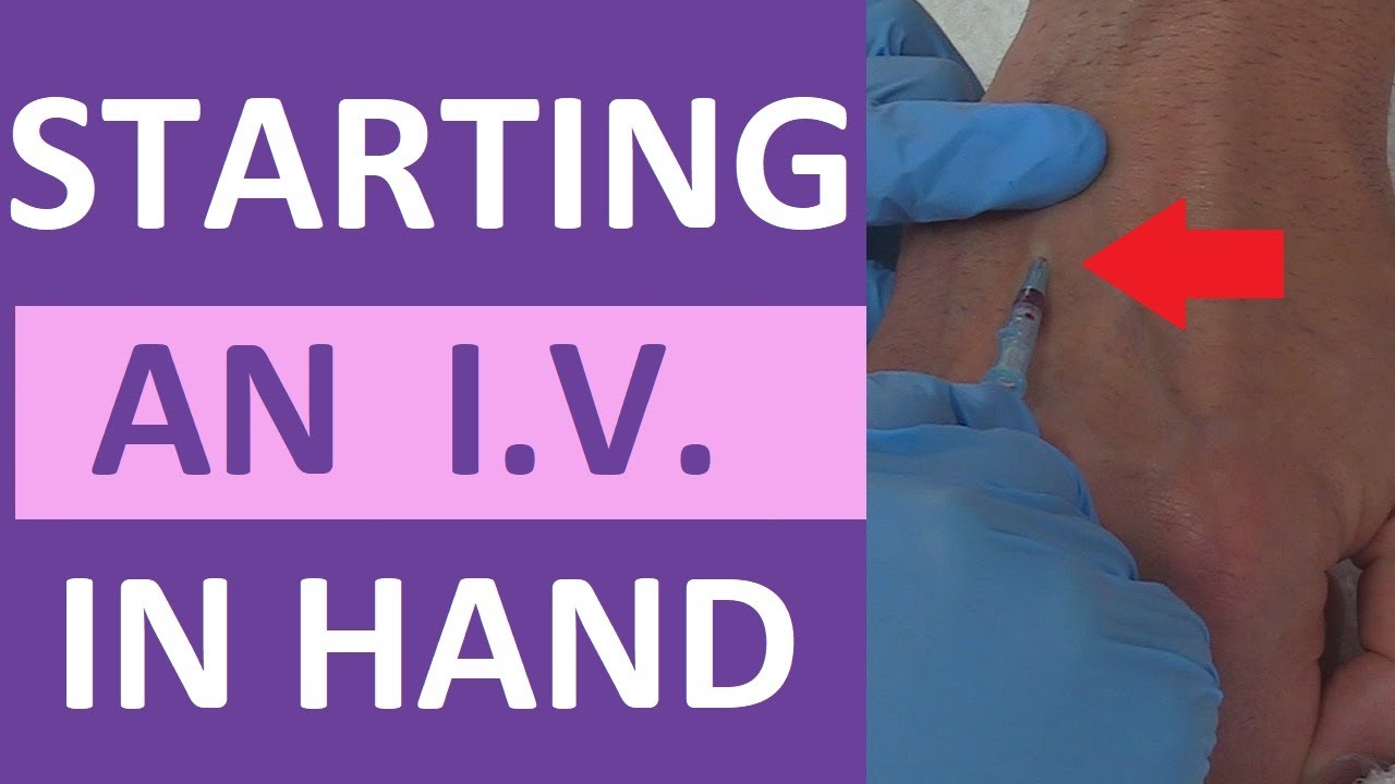
How to start a peripheral IV in the dorsum of the hand: clinical nursing skill technique.
Starting an IV (intravenous catheter) can be an intimidating experience for nurses, especially nursing students and new nurses. However, nurses will perform IV insertions often, so this is an important nursing skill to learn.
Before starting an IV, always follow the protocols of your facility, as well as manufacturer's instructions for any supplies used.
In this video, Nurse Sarah demonstrates how to start a peripheral IV in the dorsum of the hand. Prior to inserting the IV, you'll want to do the following:
-Gather supplies
-Perform hand hygiene
-Prepare supplies (including priming the saline flush, removing air from extension tubing, opening packages, completing labels, and any other steps required by your facility.
-Locate a suitable vein
-Perform hand hygiene
-Don gloves
If the patient has a lot of hair, you might want to use clippers to trim the hairs prior to starting the IV. You may also apply a tourniquet to help veins move near the surface of the skin.
Next, you'll want to clean the site using the cleaner that came in the IV start kit, such as ChloraPrep.
Once the site has dried completely, you can insert the IV. Stabilize the vein with your non-dominant hand, and insert the IV's needle into the vein, watching carefully for blood return (or a blood flash) in the chamber. Advance the IV around 2mm more to ensure the plastic cannula is in the vein, then thread the cannula into the vein and press the needle safety button.
Notes: https://www.registerednursern.....com/how-to-start-an-
IV Video Series: https://www.youtube.com/watch?v=MbG_1-_mnoo&list=PLQrdx7rRsKfXr6kruqEpIovf66sxo0gxh
This video also demonstrates how to flush the IV using the push-pause method, how to secure the IV using the Tegaderm dressing that came with the IV start kit, considerations of the different cap types and the clamp sequence, and more.
For more information, watch the complete tutorial.
#nurse #nursing #iv #startiv #ivtherapy
Website: https://www.registerednursern.com/
More Videos: https://www.youtube.com/watch?v=R2XMro13dD0&list=UUPyMN8DzkFl2__xnTEiGZ1w
Nursing Gear: https://teespring.com/stores/registerednursern
Instagram: https://www.instagram.com/registerednursern_com/
Facebook: https://www.facebook.com/RegisteredNurseRNs
Twitter: https://twitter.com/NursesRN
Popular Playlists:
NCLEX Reviews: https://www.youtube.com/playli....st?list=PLQrdx7rRsKf
Fluid & Electrolytes: https://www.youtube.com/playli....st?list=PLQrdx7rRsKf
Nursing Skills: https://www.youtube.com/playli....st?list=PLQrdx7rRsKf
Loyola Full Male Exam Part 2 A video from Loyola medical school, Chicago showing the full examination of the male
Tonsillectomy (ton-sih-LEK-tuh-me) is the surgical removal of the tonsils, two oval-shaped pads of tissue at the back of the throat — one tonsil on each side. A tonsillectomy was once a common procedure to treat infection and inflammation of the tonsils (tonsillitis). Today, a tonsillectomy is usually performed for sleep-disordered breathing but may still be a treatment when tonsillitis occurs frequently or doesn't respond to other treatments. A tonsillectomy may also be necessary to treat breathing and other problems related to enlarged tonsils and to treat rare diseases of the tonsils.

Otto Placik MD. a board certified Chicago based plastic surgeon presents Vulvar Vaginal Genital anatomy lesson reviewing the Vulva, Mons Pubis, clitoral hood, prepuce, frenulum, labia minora & majora, vagina, urethra and fourchette with surgical implications and techniques. Photos pictures and video of anatomic models are reviewed in detail on different models. Great for patients thinking about or planning before labiaplasty or vaginal cosmetic surgery
A c-section, or cesarean section, is the delivery of a baby through a surgical incision in the mother's abdomen and uterus. In some circumstances, a c-section is scheduled in advance. In others, the surgery is needed due to an unforeseen complication. If you or your baby is in imminent danger, you'll have an emergency c-section. Otherwise, it's called an unplanned section. According to the U.S. Centers for Disease Control, about 32 percent of American women who gave birth in 2015 had a cesarean delivery.
