- Physical Examination
- Surgical Examination
- Ophthalmology
- Clinical Skills
- Orthopedics
- Surgery Videos
- Laparoscopy
- Pediatrics
- Funny Videos
- Cardiothoracic Surgery
- Nursing Videos
- Plastic Surgery
- Otorhinolaryngology
- Histology and Histopathology
- Neurosurgery
- Dermatology
- Pediatric Surgery
- Urology
- Dentistry
- Oncology and Cancers
- Anatomy Videos
- Health and Fitness
- Radiology
- Anaesthesia
- Physical Therapy
- Pharmacology
- Interventional Radiology
- Cardiology
- Endocrinology
- Gynecology
- Emergency Medicine
- Psychiatry and Psychology
- Childbirth Videos
- General Medical Videos
- Nephrology
- Physiology
- Diet and Food Health
- Diabetes Mellitus
- Neurology
- Women Health
- Osteoporosis
- Gastroenterology
- Pulmonology
- Hematology
- Rheumatology
- Toxicology
- Nuclear Medicine
- Infectious Diseases
- Vascular Disease
- Reproductive Health
- Burns and Wound Healing
- Other
Top videos

The pelvic diaphragm is composed of muscle fibers of the levator ani, the coccygeus, and associated connective tissue which span the area underneath the pelvis. The pelvic diaphragm is a muscular partition formed by the levatores ani and coccygei, with which may be included the parietal pelvic fascia on their upper and lower aspects. The pelvic floor separates the pelvic cavity above from the perineal region (including perineum) below.
The right and left levator ani lie almost horizontally in the floor of the pelvis, separated by a narrow gap that transmits the urethra, vagina, and anal canal. The levator ani is usually considered in three parts: pubococcygeus, puborectalis, and iliococcygeus. The pubococcygeus, the main part of the levator, runs backward from the body of the pubis toward the coccyx and may be damaged during parturition. Some fibers are inserted into the prostate, urethra, and vagina. The right and left puborectalis unite behind the anorectal junction to form a muscular sling . Some regard them as a part of the sphincter ani externus. The iliococcygeus, the most posterior part of the levator ani, is often poorly developed.
The coccygeus, situated behind the levator ani and frequently tendinous as much as muscular, extends from the ischial spine to the lateral margin of the sacrum and coccyx.
The pelvic cavity of the true pelvis has the pelvic floor as its inferior border (and the pelvic brim as its superior border.) The perineum has the pelvic floor as its superior border.
Some sources do not consider “pelvic floor” and “pelvic diaphragm” to be identical, with the “diaphragm” consisting of only the levator ani and coccygeus, while the “floor” also includes the perineal membrane and deep perineal pouch.
Breast cancer is a malignant tumor that develops from the cells of
the breast. It is the most common type of cancer among women in
the United States. It is most often curable when found early. The
normal breast consists of three main components: the lobules
(milk-producing glands), the ducts (thin tubes that connect the
lobules to the nipple) and the stroma (fatty tissue and ligaments
surrounding the ducts and lobules, blood vessels, and lymphatic
vessels). About 80% of breast cancers start in the ducts.

Brought to you by http://nursing-resource.com
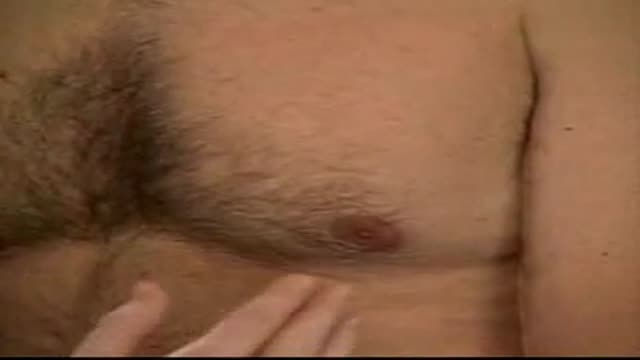
full pediatric examination of lymph nodes

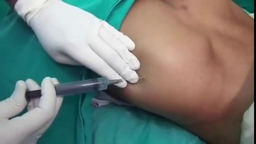
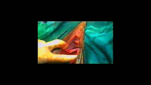
A video showing abscess incision and drainage
Incision and Drainage of a Huge Gluteal Abscess
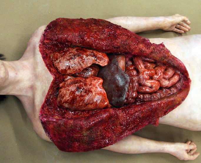
Watch that Full Human Body Medical Autopsy

Popping Cyst in the Ear Lobe
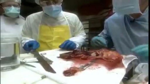
Watch that Poisoned Human Body Medical Dissect

Dr. Daniel Del Vecchio, Harvard trained plastic surgeon, performs his breast lift technique, filling the upper portion of the breast for added volume
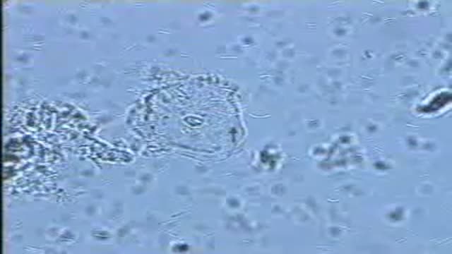
A normal vaginal epithelial cell is clear, with recognizable contents, and sharp, distinct cell borders.

A usage instruction on how to use a female condom (also know as a Femidom). Female Condom Application and Removal.

Watch that video to know how to treat premature ejaculation naturally
A great video showing Total Abdominal Hysterectomy

Vaginal Speculum and Bimanual Exam
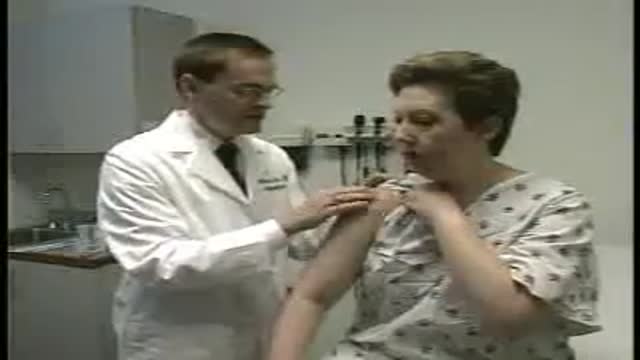
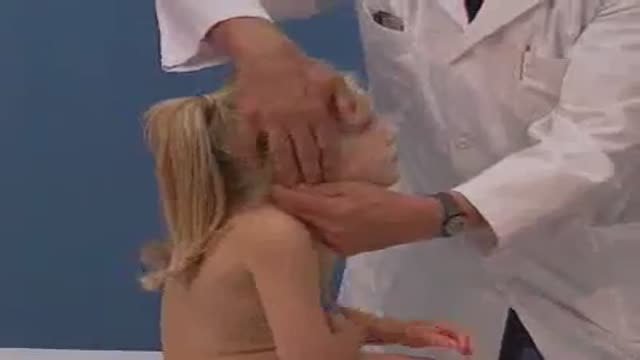
Full examination of the female from head to toe by Loyola Medical School, Chicago. Part 2
Part 2. Full Obstetric examination and normal delivery by Egyptian doctor Hussein Sulayman and the video is in English showing: Obstetric Examination Episiotomy Obstetric Forceps Obstetric Instruments
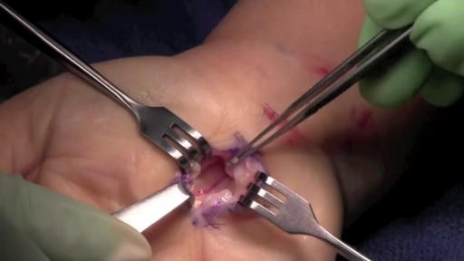
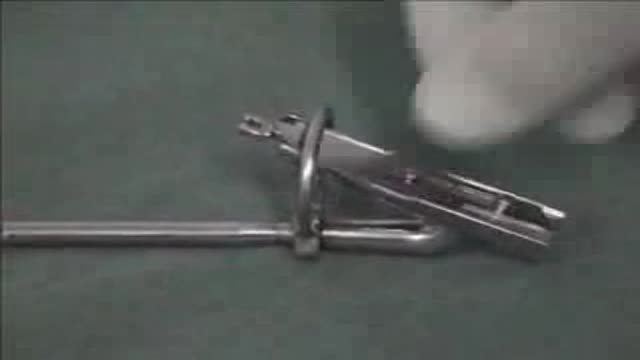
During open carpal tunnel release surgery, the transverse carpal ligament is cut, which releases pressure on the median nerve and relieves the symptoms of carpal tunnel syndrome. An incision is made at the base of the palm of the hand. This allows the doctor to see the transverse carpal ligament.

How to make your loved ones feel good and happy
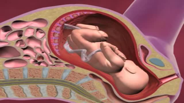
If you have placenta previa, it means that your placenta is lying unusually low in your uterus, next to or covering your cervix. The placenta is the pancake-shaped organ – normally located near the top of the uterus – that supplies your baby with nutrients through the umbilical cord.
