
- Physical Examination
- Surgical Examination
- Ophthalmology
- Clinical Skills
- Orthopedics
- Surgery Videos
- Laparoscopy
- Pediatrics
- Funny Videos
- Cardiothoracic Surgery
- Nursing Videos
- Plastic Surgery
- Otorhinolaryngology
- Histology and Histopathology
- Neurosurgery
- Dermatology
- Pediatric Surgery
- Urology
- Dentistry
- Oncology and Cancers
- Anatomy Videos
- Health and Fitness
- Radiology
- Anaesthesia
- Physical Therapy
- Pharmacology
- Interventional Radiology
- Cardiology
- Endocrinology
- Gynecology
- Emergency Medicine
- Psychiatry and Psychology
- Childbirth Videos
- General Medical Videos
- Nephrology
- Physiology
- Diet and Food Health
- Diabetes Mellitus
- Neurology
- Women Health
- Osteoporosis
- Gastroenterology
- Pulmonology
- Hematology
- Rheumatology
- Toxicology
- Nuclear Medicine
- Infectious Diseases
- Vascular Disease
- Reproductive Health
- Burns and Wound Healing
- Kidney Zone Podcast
- Other
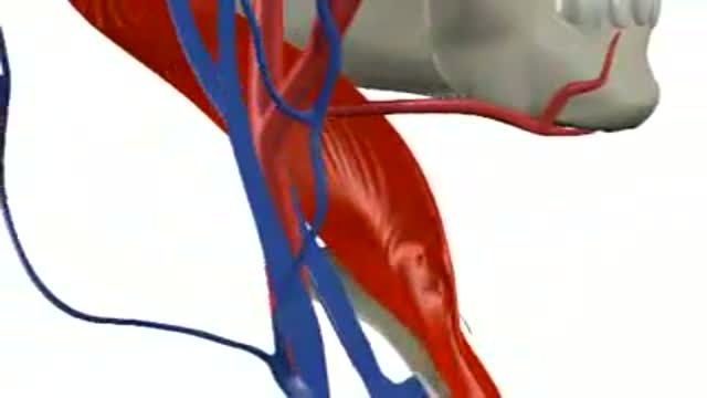
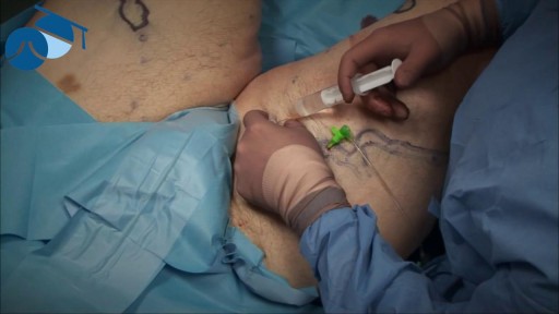
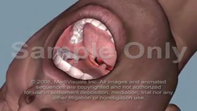
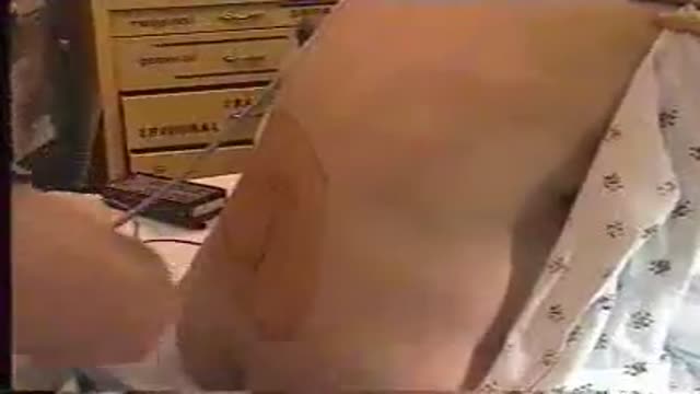
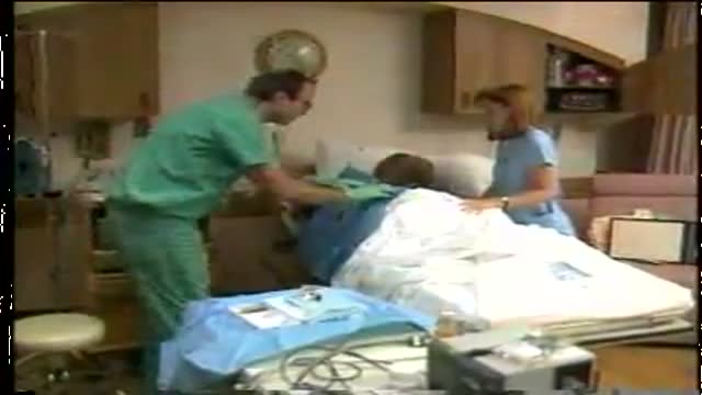
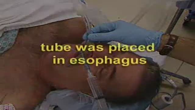
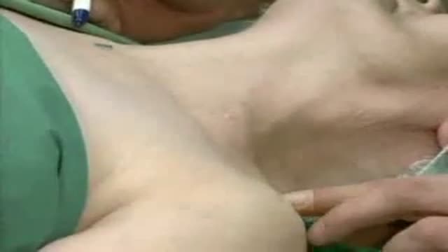
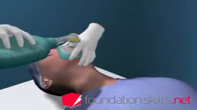
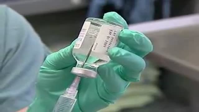
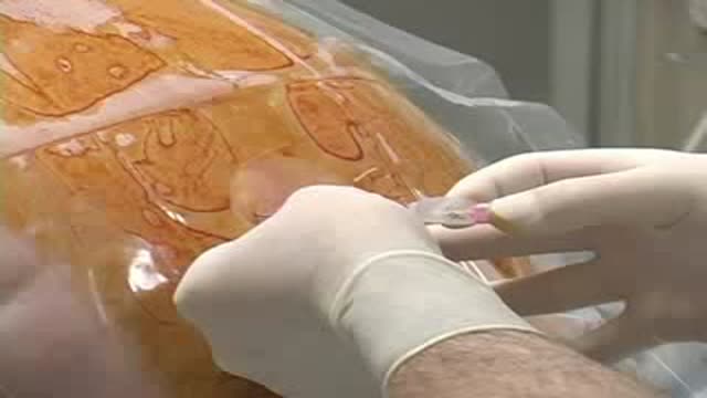
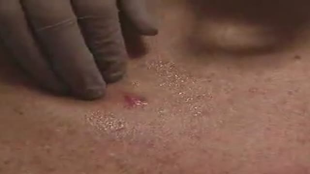
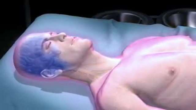
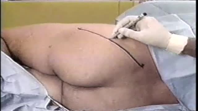
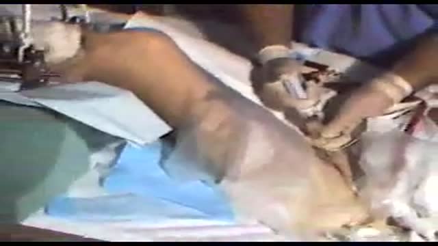
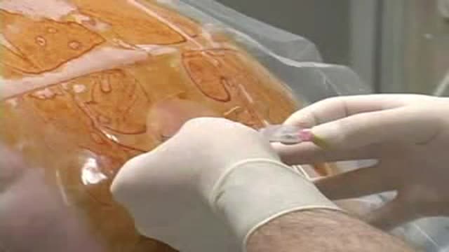

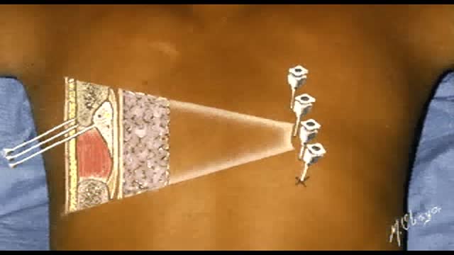
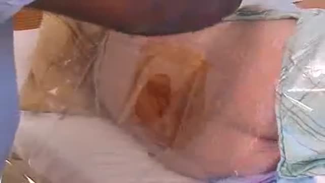
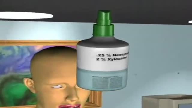
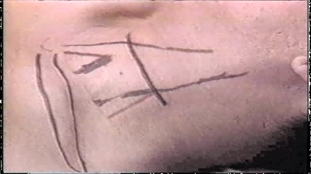
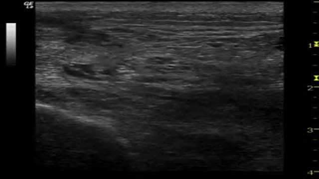
Internal jugular vein puncture

Educational only: videos on MedicalVideos.com are for general information and are not medical advice. Always consult your own doctor about your health.
Central venous access is essential in providing quality medical care to many patients for whom intensive therapy is required. In many situations, a semipermanent tunneled central line is preferred (see Indications). An anterior approach to the internal jugular vein (IJV) is the best option in this situation because it offers the easiest route with a low risk of complications. In this procedure, a tunneled catheter is surgically inserted into a vein in the neck or chest and passed under the skin. Only the end of the catheter is brought through the skin; medicines and intravenous (IV) fluid can be administered through this catheter; other tasks, such as blood sampling, can also be performed. The fact that the catheter is passed under the skin helps secure the catheter, reduces the rate of infection, and permits free movement of the catheter port. The placement of a tunneled catheter should be carried out by practitioners with specific experience in the procedure.
This video in 30 seconds
A Russian-language narration describes internal jugular vein catheterization, including turning the patient's head away, locating the sternocleidomastoid triangle, and displacing the carotid artery. It then describes needle advancement with aspiration, passing a guidewire, dilating the site, placing the catheter, checking its position and fixing it in place.
Key points
- Patient head positioning and anatomical landmarks are described
- The carotid artery is palpated and displaced medially
- Needle advancement with constant aspiration is described
- Guidewire passage, dilation and catheter placement are described
- Catheter position check, connection and fixation are described
Video Transcript
A Russian-language narration (the audio is in Russian) of internal jugular vein catheterization: turning the patient's head away, finding the triangle of the sternocleidomastoid, displacing the carotid artery, advancing the needle at 30 degrees toward the nipple with constant aspiration, then passing the guidewire, dilating, placing and fixing the catheter.
Read the full transcript
Для выполнения катетеризации еремной вены голова пациента должна быть повёрнута противоположную сторону, шея слегка разомнута. Находит точку, расположенную в верхушке треугольника, образованного двумя головками грудиноключично социевидной мышцы. Пальпаторна определяет сонную артерию и смещает ее медиально. Иглуб проводит луторальные артерии под углом 30 градусов к поверхности шеи в направлении момарного соска. При этом производится постоянная аспирация до идентификации вены. Шприц отсоединяют и через просвет иглы проводят гибкий проводник на 4-5 см за конец иглы. Иглу извлекают. Место пункции расширяется дилетатором. По проводнику в вену устанавливают катета. Проводник удаляют, повторно аспирирует кровь для контроля
положения катетер. Подсоединяют инфузионную систему или заполняют катетер гиперинизированным раствором. Фиксируют катетер кожей легатурами.
Transcript auto-generated from the video’s audio for accessibility and study. Educational content only — not medical advice.
Video Overview
From our Clinical Skills collection: Suturing, injections, catheterization, IV insertion and OSCE-style examination techniques demonstrated step by step. Browse all Clinical Skills videos →
Medical Videos
Free medical and surgery videos for patients, students and professionals — trusted since 2007.
Founded by Dr. Mohamed Ibrahim, MD, PhD — physician-scientist.
Follow us on Facebook · Instagram
SORT BY-
Top Comments
-
Latest comments