
- Physical Examination
- Surgical Examination
- Ophthalmology
- Clinical Skills
- Orthopedics
- Surgery Videos
- Laparoscopy
- Pediatrics
- Funny Videos
- Cardiothoracic Surgery
- Nursing Videos
- Plastic Surgery
- Otorhinolaryngology
- Histology and Histopathology
- Neurosurgery
- Dermatology
- Pediatric Surgery
- Urology
- Dentistry
- Oncology and Cancers
- Anatomy Videos
- Health and Fitness
- Radiology
- Anaesthesia
- Physical Therapy
- Pharmacology
- Interventional Radiology
- Cardiology
- Endocrinology
- Gynecology
- Emergency Medicine
- Psychiatry and Psychology
- Childbirth Videos
- General Medical Videos
- Nephrology
- Physiology
- Diet and Food Health
- Diabetes Mellitus
- Neurology
- Women Health
- Osteoporosis
- Gastroenterology
- Pulmonology
- Hematology
- Rheumatology
- Toxicology
- Nuclear Medicine
- Infectious Diseases
- Vascular Disease
- Reproductive Health
- Burns and Wound Healing
- Kidney Zone Podcast
- Other


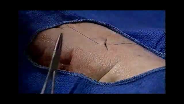


Abscess incision and drainage

Educational only: videos on MedicalVideos.com are for general information and are not medical advice. Always consult your own doctor about your health.
A video showing abscess incision and drainage
This video in 30 seconds
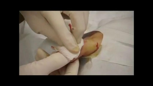
A clinical teaching video on incision and drainage of skin abscesses covers diagnosis by examination, needle aspiration or bedside ultrasound, and the equipment used. The narration then shows skin preparation and local anesthesia, the incision and breaking up of loculations, culturing, irrigation, packing, and follow-up wound care.
Key points
- Diagnosis by examination, aspiration or ultrasound is discussed
- Equipment, skin cleansing and local anesthesia are described
- The clinician makes an incision and breaks up loculations with a hemostat
- A sample is taken for bacterial culture and the cavity is irrigated
- The cavity is packed, dressed, and follow-up wound care is described
Chapters
- 0:03 — Introduction to incision and drainage
- 0:53 — Diagnosing a skin abscess
- 2:40 — Equipment and anesthesia preparation
- 4:01 — Preparation and local anesthesia
- 4:52 — Incision and breaking loculations
- 5:46 — Culture drainage and packing
- 8:14 — Dressing and follow-up care
Video Transcript
A New England Journal of Medicine clinical video on incision and drainage of skin abscesses. The narration covers diagnosis by examination, needle aspiration or bedside ultrasound, the equipment and local anesthetics used, the stab incision and breaking up of loculations, culturing the pus, packing the cavity, and the follow-up wound care over the following days.
Read the full transcript
This is a video in clinical medicine from the New England Journal of Medicine.
Incision and drainage is the primary therapy for cutaneous abscess management, as antibiotic treatment alone is inadequate for treating many of these lock-related collections of infectious material. Most localized skin abscesses without associated cellulitis can be managed with simple incision and drainage and do not require antibiotic treatment. Cutaneous abscesses have been described in all areas of the body, but are most commonly found in the axilla, buttocks, and extremities. This outpatient procedure is appropriate for many office settings, as well as for urgent care and emergency department practice environments. Diagnosis of a skin abscess is the first step in a successful procedure. This can be accomplished in three ways.
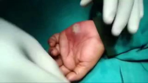
Physical examination of the affected area will often allow diagnosis of an underlying abscess based on swelling, pain, redness, and fluctuents. Spontaneously draining skin abscesses are also amenable to diagnosis by physical examination alone. Needle aspiration of a suspected skin abscess can assist the clinician in making the diagnosis of a localized abscess when results of physical examination are equivocal. Bedside ultrasound is a valuable adjunctive tool for identification of localized areas of fluid under the skin that may represent isolated areas of infection. An abscess that is diagnosed in one of these three ways may be appropriate for incision and drainage if it is larger than approximately 5 mm and found in an accessible location.
Extremely large or deep abscesses in areas difficult to anesthetize may be more appropriate to treat in a formal operating room setting. Abscesses of the palms, soles, or nasolabial folds can be associated with complications and may require consultation with an appropriate specialist. Incision and drainage is not indicated for cutaneous cellulitis without an underlying abscess. The need for preoperative antibiotics for conditions such as abnormal or artificial heart valves may require reconsideration of the timing for the procedure. Input from an appropriate specialist may be important for areas of the body with cosmetic concerns because of the expected scar formation after an abscess drainage.
Appropriate universal precautions for potential exposure to bodily fluids should always be used. Materials needed for the incision and drainage of an abscess are similar to those needed for a laceration repair. A preassembled laceration kit may contain many of the necessary items. For preparation in anesthesia, obtain a skin cleansing agent, sterile gauze, local anesthetic, and a 5-10 mL syringe with a 25-30 gauge needle. 1% lidocaine is an appropriate anesthetic for this procedure. Lidocaine with epinephrine offers advantages such as reduced bleeding and an extended duration of action, but is typically avoided in areas with a single blood supply.
Bupivocaine is another option that offers an increased duration of action for the anesthesia. Items important for the incision and drainage itself include a scalpel blade with handle, a small curved chemostat, normal saline with a sterile bowl, and a large syringe with a splash guard or a needle-less 18-gauge angiocatheter for irrigation of the wound. Swabs for bacterial culture, wound-packing material, scissors, gauze, and tape should all be available to complete the procedure and dress the wound. Obtain and form consent by discussing the risks and benefits of this procedure, including pain, bleeding, and scar formation.
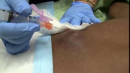
Wash your hands with antibacterial soap before beginning the procedure. Protect yourself from exposure to bodily fluids as many abscesses are under pressure. A face shield and gloves should be used. Place all equipment on a bedside table that is easy to reach. Position the patient so that the area for drainage is fully exposed. Apply a skin cleanser, such as chlorhexidine or povidone iodine, in a circular motion starting with the peak of the abscess. Cover a wide area outside of the wound to prevent contamination of other equipment. Unesthetize the top of the wound. This should be done by inserting a 25 or 30-gauge needle parallel to the skin and injecting into the intradermal tissues.
Once the entire open bore of the needle is under the skin, gentle pressure should be used to infiltrate with the anesthetic agent. You will note blanching of the tissue as the anesthetic spreads out. Continue with infiltration until you have covered an area over the top of the abscess large enough to anesthetize the area of incision. Some abscesses may require additional injections of anesthetic in a local field block pattern or moderate procedural sedation for additional patient comfort. Hold the scalpel between your thumb and forefinger. Make an incision directly over the center of the abscess that is oriented along the long axis of the fluid collection.
Resistance may be felt as the incision is initiated, and steady firm pressure will allow a controlled entry into the subcutaneous tissues. Purulent drainage will begin when the abscess cavity has been entered successfully. Cosmetic results can be optimized if the incision is made parallel to existing skin tension lines. The incision should be extended to create an opening large enough to ensure adequate drainage and prevent recurrent abscess formation. It may need to extend the length of the abscess borders. Care must be taken to control the scalpel during the stab incision to prevent puncturing through the back wall of the abscess.
The goal is to allow enough access for introduction of a hemostat to break up loculations and place internal packing. Use a swab to obtain a sample from the interior of the cavity for bacterial culture. While most patients will not require antibiotics after an abscess drainage, culture information is useful if the patient's condition later worsens and antibiotic treatment becomes necessary. After allowing the wound to drain spontaneously, gently express any further contents. Additional injections of local anesthetic may be helpful during this portion of the procedure if there is significant patient discomfort.
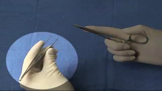
Use a curved hemostat for further blunt dissection to break loculations and allow the abscess cavity to be completely opened up. Insert the hemostat into the wound until you feel the resistance of normal tissue, then open the hemostat to perform blunt dissection of the internal portion of the abscess cavity. Continue this procedure of breaking up loculations in a circular motion until the entire abscess cavity has been explored and any deep tracks that extend into surrounding tissues have been identified. A gentle irrigation of the wound should be performed using sterile normal saline.
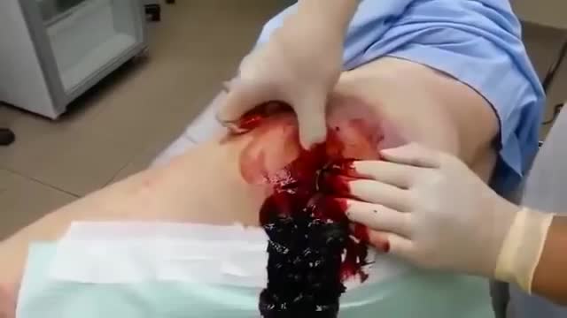
Appropriate wound incision size will enhance irrigation and prevent excess buildup of pressure within the abscess cavity. The irrigation should continue until the effluent is clear. Using wound packing material, such as quarter inch or half inch packing strips, gently pack the abscess by starting in one quadrant and gradually working around the entire cavity. Place sufficient packing to keep the walls of the abscess separated and to allow further drainage of infected debris. Overpacking may cause ischemia of the surrounding tissues or interfere with desired wound drainage and should be avoided.
Appropriate packing will allow healing by secondary intention and avoid premature closure of the wound which can lead to re-accumulation of bacteria and recurrent abscesses. For a simple abscess, the now openly draining wound allows the body's host defenses to clear the infection without the need to expose patients to the side effects or risks of antimicrobial therapy. After most incision and drainage procedures are performed in healthy patients, antibiotics are not required. Patients with extensive cellulitis beyond the abscess area or with significant comorbidities may require supplemental antibiotic treatment. Providers are encouraged to use their local bacterial culture susceptibility data to guide any such empiric therapy.
Community-acquired methicillin resistant staphylococcus aureus has garnered heightened attention because of increasing prevalence in skin infections. It is imperative to know and follow your regional management guidelines for this pathogen. Cover the abscess wound with a sterile non-adherent dressing. Topical antibiotics have a limited benefit and are not required. As with any wound, be certain that the patient's tetanus immunizations are up to date. All abscesses should have packing removed in a few days and most patients with wounds that have packing in place should be scheduled for a return visit for follow-up and packing removal two to three days after the procedure.
Patients should be given written instructions telling them to return earlier if there are any signs of worsening. On subsequent visits for wound care, packing should be removed to allow assessment of the ongoing healing by secondary intention. Use of fresh packing material may be necessary to continue the healing process if significant wound drainage is still ongoing. A follow-up visit should then be rescheduled for two to three days later. This is common for abscesses that require extensive drainage. In the absence of other complications, the need for repacking is not an indication for antibiotic treatment.
The acidic environment of infected tissue leads to difficulties with adequate anesthesia provided by local anesthetic agents. Using appropriate amounts of anesthetic, allowing sufficient time after injection or supplementing with oral and parenteral agents can increase patient comfort. Finally, additional complications to watch for include the progression to surrounding cellulitis, development of a fever, or other signs of clinical worsening. These may prompt consideration for repeat incision and drainage of an abscess or the need for antibiotic therapy. Most abscesses will respond well to simple incision and drainage and will not require treatment beyond packing changes and local wound care practices.
Transcript auto-generated from the video’s audio for accessibility and study. Educational content only — not medical advice.
Video Overview
From our Clinical Skills collection: Suturing, injections, catheterization, IV insertion and OSCE-style examination techniques demonstrated step by step. Browse all Clinical Skills videos →
Medical Videos
Free medical and surgery videos for patients, students and professionals — trusted since 2007.
Founded by Dr. Mohamed Ibrahim, MD, PhD — physician-scientist.
Follow us on Facebook · Instagram
SORT BY-
Top Comments
-
Latest comments