
- Physical Examination
- Surgical Examination
- Ophthalmology
- Clinical Skills
- Orthopedics
- Surgery Videos
- Laparoscopy
- Pediatrics
- Funny Videos
- Cardiothoracic Surgery
- Nursing Videos
- Plastic Surgery
- Otorhinolaryngology
- Histology and Histopathology
- Neurosurgery
- Dermatology
- Pediatric Surgery
- Urology
- Dentistry
- Oncology and Cancers
- Anatomy Videos
- Health and Fitness
- Radiology
- Anaesthesia
- Physical Therapy
- Pharmacology
- Interventional Radiology
- Cardiology
- Endocrinology
- Gynecology
- Emergency Medicine
- Psychiatry and Psychology
- Childbirth Videos
- General Medical Videos
- Nephrology
- Physiology
- Diet and Food Health
- Diabetes Mellitus
- Neurology
- Women Health
- Osteoporosis
- Gastroenterology
- Pulmonology
- Hematology
- Rheumatology
- Toxicology
- Nuclear Medicine
- Infectious Diseases
- Vascular Disease
- Reproductive Health
- Burns and Wound Healing
- Kidney Zone Podcast
- Other

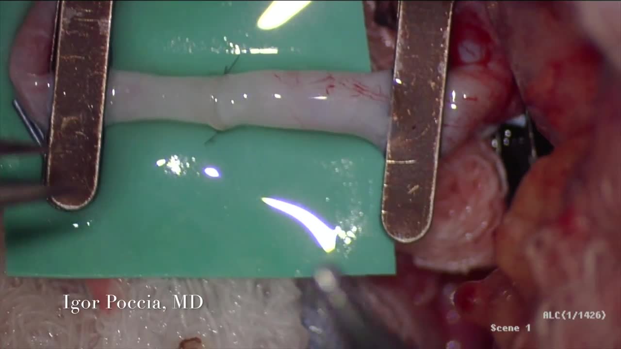
Microvascular Anastomosis

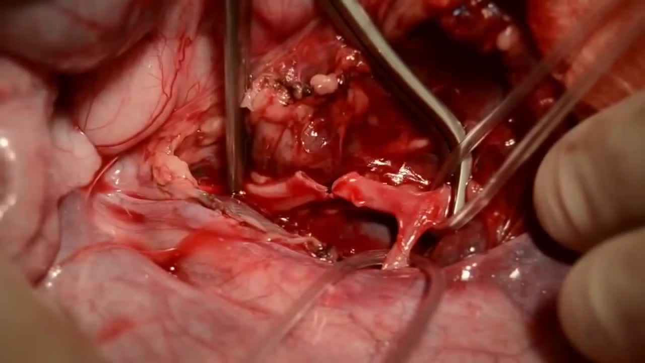
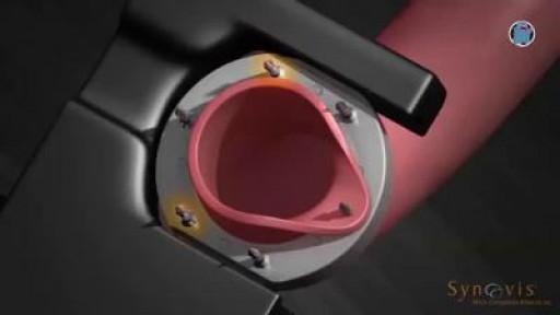
Simple microinstruments and a medical school laboratory microscope were used for anastomosis training. Chicken blood vessels were used as a material for this study. A long segment of blood vessel from the proximal brachial artery to the distal radial artery was used for training. End-to-side anastomosis was practiced first, and the training continued with end-to-end anastomosis of the appropriate segments.
This video in 30 seconds
This video shows microvascular anastomosis training using simple microinstruments and a medical school laboratory microscope. Chicken blood vessels were used as the training material, with a long segment from the proximal brachial artery to the distal radial artery used, end-to-side anastomosis practiced first and training continuing with end-to-end anastomosis.
Key points
- Microvascular anastomosis training is shown
- Simple microinstruments and a medical school laboratory microscope are used
- Chicken blood vessels are the training material
- End-to-side anastomosis is practiced first, followed by end-to-end anastomosis
Video Overview
This video presents “Microvascular Anastomosis” — Simple microinstruments and a medical school laboratory microscope were used for anastomosis training. Chicken blood vessels were used as a material for this study. A long segment of blood vessel from the proximal brachial artery to the. Whether you are preparing for a procedure, studying the technique, or learning how a condition is assessed, the recording shows the process as it is performed in practice.
From our Surgery Videos collection: Surgery videos: watch real operations step by step — general, laparoscopic and specialist surgery performed in the OR, explained for students and surgeons. Browse all Surgery Videos videos →
Educational content only — not a substitute for professional medical advice.
SORT BY-
Top Comments
-
Latest comments