- Physical Examination
- Surgical Examination
- Ophthalmology
- Clinical Skills
- Orthopedics
- Surgery Videos
- Laparoscopy
- Pediatrics
- Funny Videos
- Cardiothoracic Surgery
- Nursing Videos
- Plastic Surgery
- Otorhinolaryngology
- Histology and Histopathology
- Neurosurgery
- Dermatology
- Pediatric Surgery
- Urology
- Dentistry
- Oncology and Cancers
- Anatomy Videos
- Health and Fitness
- Radiology
- Anaesthesia
- Physical Therapy
- Pharmacology
- Interventional Radiology
- Cardiology
- Endocrinology
- Gynecology
- Emergency Medicine
- Psychiatry and Psychology
- Childbirth Videos
- General Medical Videos
- Nephrology
- Physiology
- Diet and Food Health
- Diabetes Mellitus
- Neurology
- Women Health
- Osteoporosis
- Gastroenterology
- Pulmonology
- Hematology
- Rheumatology
- Toxicology
- Nuclear Medicine
- Infectious Diseases
- Vascular Disease
- Reproductive Health
- Burns and Wound Healing
- Other
Histology and Histopathology

© 2023 Elsevier. All rights reserved. What are lymph nodes? Lymph nodes are small secondary lymphoid organs that are found along lymphatic vessels throughout the body.
Find our full video library only on Osmosis Prime: http://osms.it/more.
Join over 3 million current & future clinicians who learn by Osmosis, and over 130 universities around the world who partner with us to make medical and health education more engaging and efficient. We have unparalleled tools and materials to prepare you to succeed in school, on board exams, and as a future clinician. Sign up for a free trial at http://osms.it/more. If you're interested in exploring an institutional partnership, visit osmosis.org/educators to request a personalized demo.
Follow us on social:
Facebook: http://osms.it/facebook
Twitter: http://osms.it/twitter
Instagram for med: http://osms.it/instagram
Instagram for nursing: https://osms.it/ignursing
Linkedin: https://osms.it/linkedin
Our Vision: Everyone who cares for someone will learn by Osmosis.
Our Mission: To empower the world’s clinicians and caregivers with the best learning experience possible. Learn more here: http://osms.it/mission
Medical disclaimer: Knowledge Diffusion Inc (DBA Osmosis) does not provide medical advice. Osmosis and the content available on Osmosis's properties (Osmosis.org, YouTube, and other channels) do not provide a diagnosis or other recommendation for treatment and are not a substitute for the professional judgment of a healthcare professional in diagnosis and treatment of any person or animal. The determination of the need for medical services and the types of healthcare to be provided to a patient are decisions that should be made only by a physician or other licensed health care provider. Always seek the advice of a physician or other qualified healthcare provider with any questions you have regarding a medical condition. © 2023 Elsevier. All rights reserved.

A complete organized library of all my videos, digital slides, pics, & sample pathology reports is available here: https://kikoxp.com/posts/5084 (dermpath) & https://kikoxp.com/posts/5083 (bone/soft tissue sarcoma pathology)
Topics discussed:
Epidermis:
Layers of epidermis: 0:10
Melanocytes vs Keratinocytes: 5:16
Langerhans cells: 10:10 & 33:30 & 57:30
Dermis:
Papillary and reticular dermis: 11:50
Three types of white empty spaces on a slide: vessels, glands/ducts/cysts, or artifact: 15:25
Blood vessels & nerves: 18:24 & 48:50 & 58:59
Arrector pili & other dermal smooth muscle: 20:00
Adnexal:
Sebaceous gland: 21:10
Hair follicle 23:14
Eccrine sweat glands and ducts 24:45 & 50:00
Gland/duct vs blood vessel 27:20 & 48:50
Apocrine glands: this video https://kikoxp.com/posts/7837 (at 12:30)
Acrosyringium: this video https://kikoxp.com/posts/7837 (at 10:00)
Three types of pink bundles: smooth muscle, nerve, dense connective tissue: 27:50
Acral skin (palm sole) with contact dermatitis 29:37
Parakeratosis 30:00
Perivascular lymphocytes 30:40
Eosinophils vs neutrophils 31:20
Spongiosis with desmosome keratinocyte spines 32:10
Spongiotic vesicles with Langerhans cells 33:30
Normal acral skin (palm & sole) with stratum lucidum 34:20
Normal glomus body/apparatus (canal of Sucquet-Hoyer) 35:40
Nerve 36:46 & 51:50
Adipose tissue (white fat cells) in subcutis with Lochkern 37:55
Normal scalp skin with large anagen hair follicles: 39:30
Hair follicle anatomy (bulb/matrix, inner root sheath, outer root sheath, hair shaft, isthmus, infundibulum): 40:55 (labeled images):
https://kikoxp.com/posts/3661 & https://kikoxp.com/posts/7899
Pacinian corpuscle 50:40
Meissner corpuscle 1:02:28
Dense regular connective tissue (Fascia/Tendon/Ligament) vs Smooth Muscle 53:00
Basic Normal Skin Immunohistochemistry:
-cytokeratin in epidermis: 55:33
-S100 in melanocytes and Langerhans cells and adipocytes: 57:30
-Desmin in smooth muscle (arrector pili and blood vessels): 58:59
-CD31 in endothelial cells of blood vessels: 59:33
-SOX-10 in melanocytes: 1:00:40
Digit/Finger/Toe histology (amputation for subungual acral melanoma) 1:04:10 & 1:08:30
-bone 1:05:40
-glomus body 1:05:15
-tendon/ligament 1:06:10
-artery 1:06:58
-fingernail/toenail 1:08:54
-acrosyringium 1:10:45
Solar elastosis (what wrinkles look like microscopically!) 1:11:50
Other videos you might like:
Tendon vs Nerve Histology Made Simple with the Ramen Noodle Sign (of Fulton) video: https://kikoxp.com/posts/4466
Melanocytes vs Keratinocytes made easy video: https://kikoxp.com/posts/3802
Blood Vessel vs Gland vs Artifact Made Easy video: https://kikoxp.com/posts/4808
The basic normal structures of the skin discussed and described by a dermatopathologist. This material is intended for use by medical students, junior pathology or dermatology residents, or for anyone else studying normal human histology. Special thanks to two of my medical students at UAMS for helping make this video possible. Miki Lindsey convinced me that I really needed to sit down and record this video. Akash Patel took time to edit the video and make it ready for YouTube. My sincere thanks to both of them for helping me overcome procrastination.
Huge thanks to Abigail Cline, a medical student at Medical College of Georgia, for volunteering to type a transcript of this ENTIRE video (over 14,000 words!) so that I could provide closed caption subtitles for those with hearing impairments and for those who may need assistance in understanding spoken English (particularly given how quickly I speak!). You can access a text version of her transcript of my video here: https://kikoxp.com/posts/5390
Correction - I made a mistake in the video. I said that sebaceous gland secretions are turned into smelly substances by bacteria and that this makes body odor. That is incorrect. That is actually true of APOCRINE gland secretions not sebaceous secretions.
Also, in the past I used "keratinocyte" and "squamous cell" interchangeably (this is because in dermatopathology, we see and talk about squamous cell carcinomas all the time, and those tumors are composed of keratinocytes). But technically, in normal skin histology, "squamous cell" refers only to the flattened keratinocytes in the superficial epidermis. Thankfully, a histology PhD colleague pointed this out to me and corrected my lazy nomenclature!
Please check out my Soft Tissue Pathology & Dermatopathology survival guide textbooks: http://bit.ly/2Te2haB
This video is geared towards medical students, pathology or dermatology residents, or practicing pathologists or dermatologists. Of course, this video is for educational purposes only and is not formal medical advice or consultation.
Presented by Jerad M. Gardner, MD. Please subscribe to my channel to be notified of new pathology teaching videos.
Follow me on:
Snapchat: JMGardnerMD
Twitter: @JMGardnerMD
Instagram: @JMGardnerMD
Facebook: https://www.facebook.com/JMGardnerMD/

Access my FREE Online Membership today → https://www.thenotedanatomist.com
___
Unlock my Premium Tutoring Memberships → https://www.thenotedanatomist.com/premium-memberships
Lifetime Access to Online Anatomy Course
Foundational Q&A Cards Per Video
Notes and Key Takeaways
Downloadable Documents
Flashcards for Each Course
Weekly Group Tutoring Sessions
Direct Tutoring Sessions
___
Discover A Simplified Approach to Master the Complexity of Anatomy with me, Dr. David Morton ... The Noted Anatomist!
This video tutorial discusses an Introduction to Histology (study of tissues):
0:00. Intro
0:35. Hierarchical organization of living matter
1:56. H&E stains
3:00. Epithelium overview (characteristics and classifying scheme)
- 9:12. Simple squamous epithelium
- 11:05. Simple cuboidal epithelium
- 12:20. Simple columnar epithelium
- 13:36. Stratified squamous epithelium
- 15:51. Urinary epithelium (transitional epithelium)
- 16:45. Pseudo-stratified ciliated columnar epithelium (respiratory epithelium)
18:55. Connective tissue overview (characteristics and classifying scheme)
- 21.14. Connective tissue proper (loose CT, dense irregular CT, dense regular CT, adipose tissue)
- 24:50. Cartilage (hyaline cartilage, elastic cartilage, fibrocartilage)
- 26:04. Bone (osteoblasts, osteocytes, osteoclasts, calcium ...)
- 27:34. Blood (RBC, WBC, platelet, plasma)
28:54. Muscle tissue (skeletal muscle, cardiac muscle, smooth muscle)
32:54. Nervous tissue (neurons and glial cells)
36:58. In-a-Nutshell
37:07. Acknowledgements
For a more detailed study of histology go to The Histology Wizard: https://www.youtube.com/channe....l/UCAeLLruy9RkUWaW_r
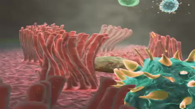
Cells may have slender extensions of the cell membrane to form cilia or the smaller extensions called microvilli. The microscopic microvilli effectively increase the surface area of the cell and are useful for absorption and secretion functions. A dramatic example is the human small intestine. The tissue has small fingerlike extensions called villi which are collections of cells, and those cells have many microvilli to even further increase the available surface area for the digestion process. According to Audesirk & Audesirk, this can give an effective surface area of about 250 square meters for absorption.
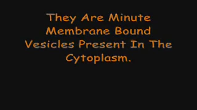
Cell Organelles in 3D
http://milagroparaelacne.plus101.com
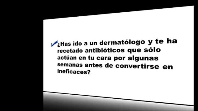
---Como Quitar Acne Cara. Existe una CAUSA PRINCIPAL DEL ACNÉ. Y no siempre un producto para el acné ataca esta causa. Esto significa que, si bien podrías obtener un beneficio de lociones, u otros productos o terapias, nunca curarás tu acné con ellos.
Es un poco como tener un techo con goteras y "solucionarlo" poniendo recipientes para recoger el agua de lluvia que cae del techo. Para solucionar adecuadamente el techo con goteras, se debe corregir la causa de raíz, que es el agujero en el techo.
Entonces... ¿Cuál Es La Principal
Causa Del ACNE?
La causa del acné es un DESEQUILIBRIO HORMONAL. Esto es algo que las empresas del acné nunca te dirán...
... Sin embargo, saber la causa del acné es una cosa. Descubrir la forma de eliminar eficazmente esta causa es otra cosa...
Como, Quitar, Acne, Cara, manchas de acne, quitar el acne, cicatrices de acne, como quitar granos, como quitar espinillas, cicatrices de acne, eliminar el acne, como eliminar barros, eliminar los barros,
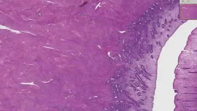
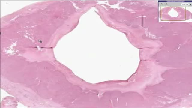
Histology of Uterus
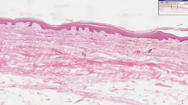
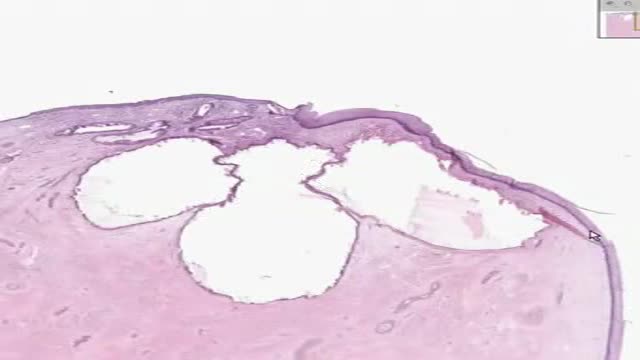
Histology of Thin Skin
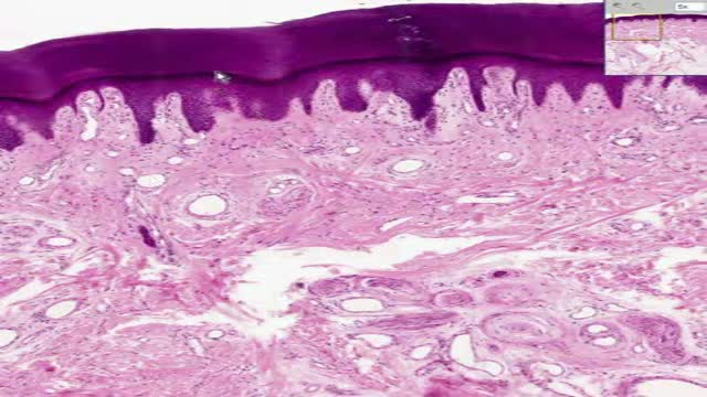
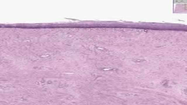
Histology of Thick Skin
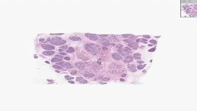
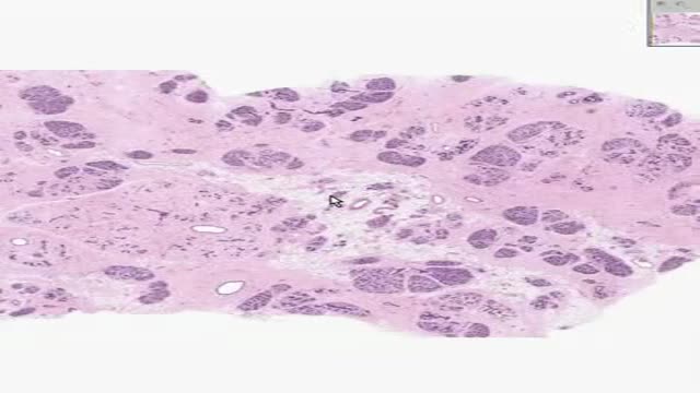
Histology of Active Breast
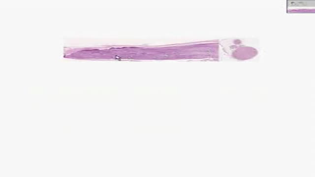
Histology of Vas Deferens
Histology of vagina
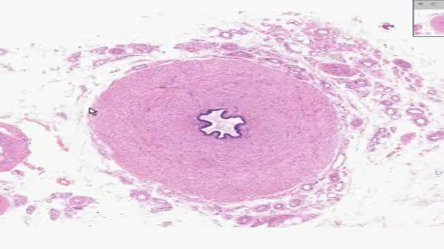
Histology of Corpus Luteum 2
Histology of Inactive Breast

Histology of Skin Appendages
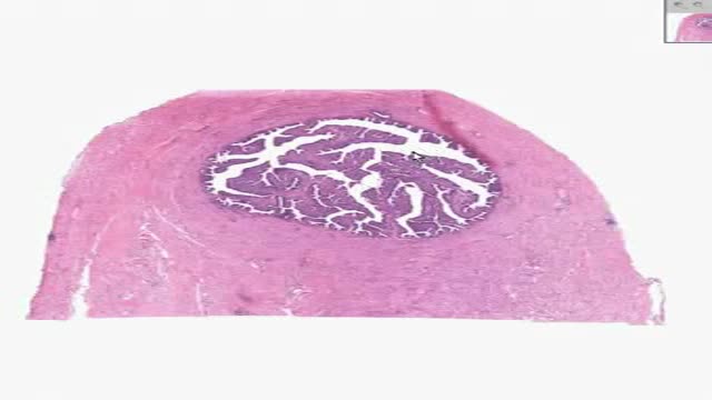
Histology of Fallopian Tube
Histology of Peripheral Nerve
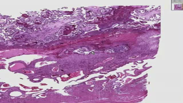
Histology of Placenta
Histology of Cervix
