- Physical Examination
- Surgical Examination
- Ophthalmology
- Clinical Skills
- Orthopedics
- Surgery Videos
- Laparoscopy
- Pediatrics
- Funny Videos
- Cardiothoracic Surgery
- Nursing Videos
- Plastic Surgery
- Otorhinolaryngology
- Histology and Histopathology
- Neurosurgery
- Dermatology
- Pediatric Surgery
- Urology
- Dentistry
- Oncology and Cancers
- Anatomy Videos
- Health and Fitness
- Radiology
- Anaesthesia
- Physical Therapy
- Pharmacology
- Interventional Radiology
- Cardiology
- Endocrinology
- Gynecology
- Emergency Medicine
- Psychiatry and Psychology
- Childbirth Videos
- General Medical Videos
- Nephrology
- Physiology
- Diet and Food Health
- Diabetes Mellitus
- Neurology
- Women Health
- Osteoporosis
- Gastroenterology
- Pulmonology
- Hematology
- Rheumatology
- Toxicology
- Nuclear Medicine
- Infectious Diseases
- Vascular Disease
- Reproductive Health
- Burns and Wound Healing
- Other
Other
A subdural hematoma is most often the result of a severe head injury. This type of subdural hematoma is among the deadliest of all head injuries. The bleeding fills the brain area very rapidly, compressing brain tissue. This often results in brain injury and may lead to death. Subdural hematomas can also occur after a minor head injury. The amount of bleeding is smaller and occurs more slowly. This type of subdural hematoma is often seen in older adults. These may go unnoticed for many days to weeks, and are called chronic subdural hematomas. With any subdural hematoma, tiny veins between the surface of the brain and its outer covering (the dura) stretch and tear, allowing blood to collect. In older adults, the veins are often already stretched because of brain shrinkage (atrophy) and are more easily injured.
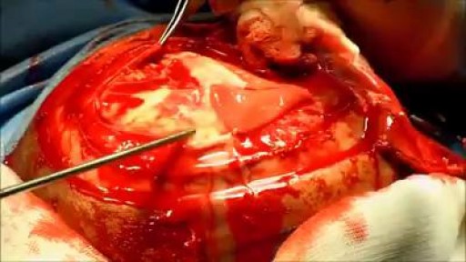
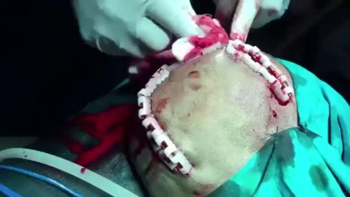
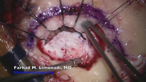
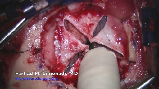
A craniotomy involves making an incision in the scalp and creating a hole known as a bone flap in the skull. The hole and incision are made near the area of the brain being treated. During open brain surgery, your surgeon may opt to: remove tumors. clip off an aneurysm

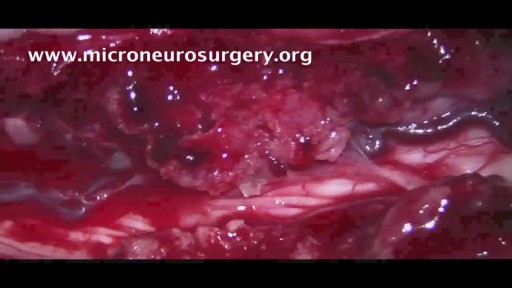
Retro-sigmoid craniotomy (often called "keyhole" craniotomy) is a minimally-invasive surgical procedure performed to remove brain tumors. This procedure allows for the removal of skull base tumors through a small incision behind the ear, providing access to the cerebellum and brainstem. Neurosurgeons may use this approach to reach certain tumors, such as meningiomas and acoustic neuromas (vestibular schwannomas).

Wet dreams occur when you ejaculate during your sleep. The medical term for a wet dream is “nocturnal emission.“ Most wet dreams are reported in teenage boys and young men, and sometimes they occur well into adulthood.

Callus Peel is a luxury, spa foot treatment that removes hard, callused skin leaving your feet feeling soft and revitalised. The treatment is a 15 minute...

Big wart blister after freezing with liquid nitrogen.
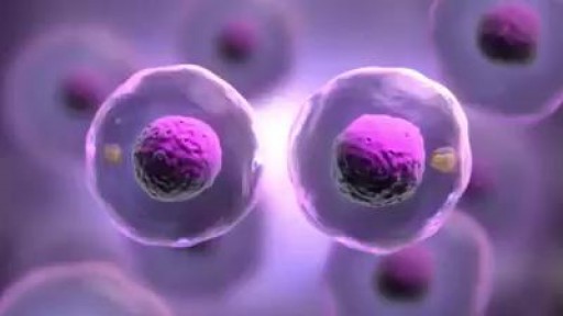
Mitosis, a process of cell duplication, or reproduction, during which one cell gives rise to two genetically identical daughter cells. Strictly applied, the term mitosis is used to describe the duplication and distribution of chromosomes, the structures that carry the genetic information.

Epidermoid cysts, also called sebaceous, keratin, or epithelial cysts, are small, hard lumps that develop under the skin. These cysts are common. They grow slowly. They do not cause other symptoms and are nearly never cancerous. Epidermoid cysts are often found on the face, head, neck, back, or genitals
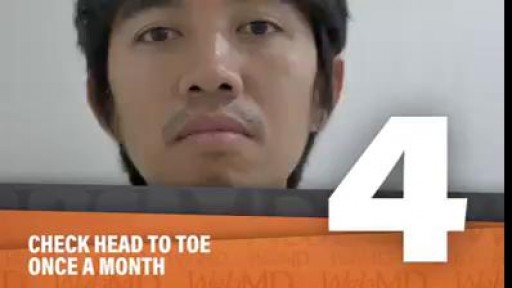
Skin isn't just your body's biggest organ-- it's also the fastest growing.
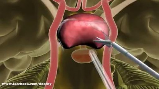
Minimally Invasive Brain Surgery To Remove Brain Tumors.
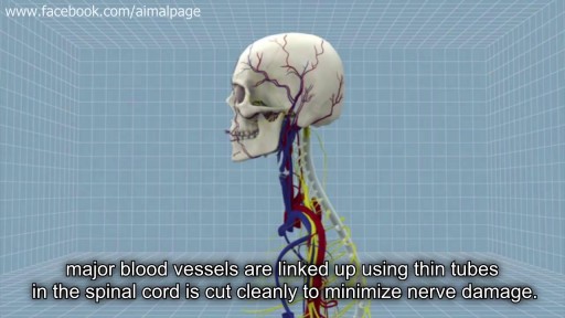
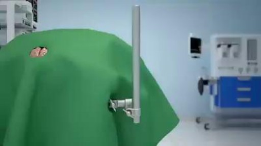
First Head Transplant Surgery
Epidural hematoma (EDH) is a traumatic accumulation of blood between the inner table of the skull and the stripped-off dural membrane. EDH results from traumatic head injury, usually with an associated skull fracture and arterial laceration.The inciting event often is a focused blow to the head, such as that produced by a hammer or baseball bat. In 85-95% of patients, this type of trauma results in an overlying fracture of the skull. Blood vessels in close proximity to the fracture are the sources of the hemorrhage in the formation of an epidural hematoma. Because the underlying brain has usually been minimally injured, prognosis is excellent if treated aggressively. Outcome from surgical decompression and repair is related directly to patient's preoperative neurologic condition. [1]
TUMOR FRONTO ORBITARIO NEUROCIRUGÍA
Removal of spinal cord tumor (meningioma): Spine Tumor Surgery
Brain Surgery: Microvascular Decompression of facial nerve for hemifacial spasm
Glioblastoma is a type of astrocytoma, a cancer that forms from star-shaped cells in the brain called astrocytes. In adults, this cancer usually starts in the cerebrum, the largest part of your brain
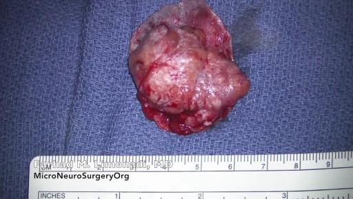
A meningioma is a tumor that arises from the meninges — the membranes that surround your brain and spinal cord. Most meningiomas are noncancerous (benign), though rarely a meningioma may be cancerous (malignant). Some meningiomas are classified as atypical, meaning they're neither benign nor malignant but, rather, something in between.
There is nothing that compares to the fresh-faced feeling you have when you leave the aesthetician after a facial. There is just something so freeing about knowing that nasty little buggers like blackheads, whiteheads and all other kinds of heads have been given the heave-ho. That could be why this Facebook video of a blackhead being removed has gone viral. With more than 24 million views, the popular video is weirdly difficult to stop watching.

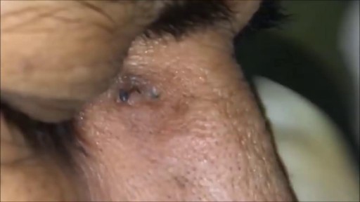
Squamous cell carcinomas typically appear as persistent, thick, rough, scaly patches that can bleed if bumped, scratched or scraped. They often look like warts and sometimes appear as open sores with a raised border and a crusted surface. In addition to the signs of SCC shown here, any change in a preexisting skin growth, such as an open sore that fails to heal, or the development of a new growth, should prompt an immediate visit to a physician.
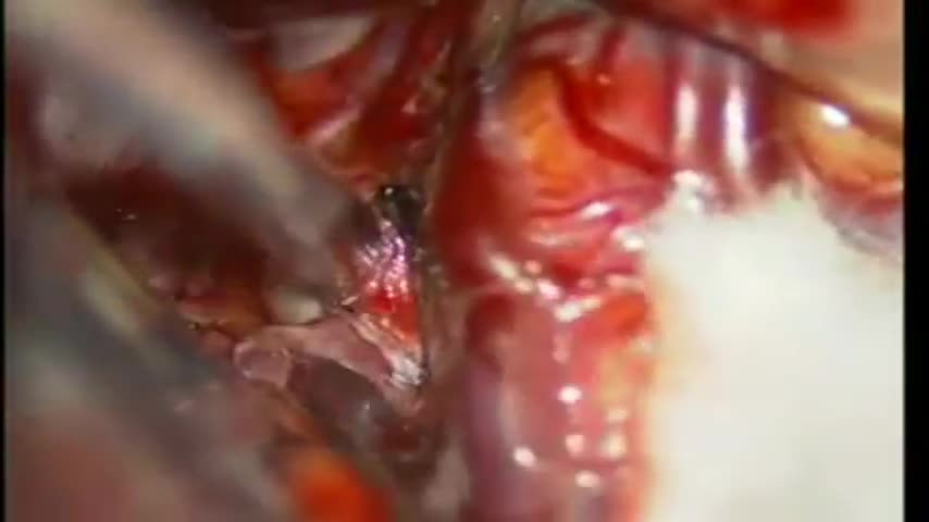
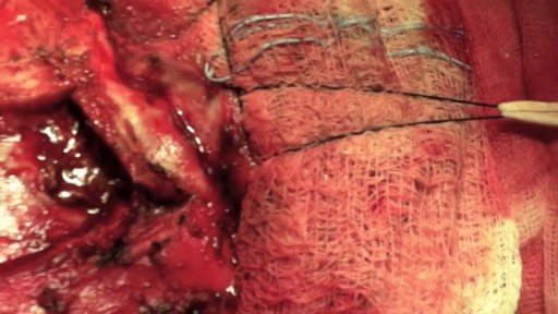
in this patient the aneurysm wasarising from middle cerebral artery M1 segment dividng into three branches,it is mandatory topreserve all three divisions,as was done in this case,this pt 25 yrs young man presented with sub arachnoid haemorrhage
