- Physical Examination
- Surgical Examination
- Ophthalmology
- Clinical Skills
- Orthopedics
- Surgery Videos
- Laparoscopy
- Pediatrics
- Funny Videos
- Cardiothoracic Surgery
- Nursing Videos
- Plastic Surgery
- Otorhinolaryngology
- Histology and Histopathology
- Neurosurgery
- Dermatology
- Pediatric Surgery
- Urology
- Dentistry
- Oncology and Cancers
- Anatomy Videos
- Health and Fitness
- Radiology
- Anaesthesia
- Physical Therapy
- Pharmacology
- Interventional Radiology
- Cardiology
- Endocrinology
- Gynecology
- Emergency Medicine
- Psychiatry and Psychology
- Childbirth Videos
- General Medical Videos
- Nephrology
- Physiology
- Diet and Food Health
- Diabetes Mellitus
- Neurology
- Women Health
- Osteoporosis
- Gastroenterology
- Pulmonology
- Hematology
- Rheumatology
- Toxicology
- Nuclear Medicine
- Infectious Diseases
- Vascular Disease
- Reproductive Health
- Burns and Wound Healing
- Other
Top videos

Sodium levels are tightly controlled in a healthy individual by regulation of urine concentration and an intact thirst mechanism. Hypernatremia (defined as a serum sodium level >145 mEq/L) is rare in patients with preserved thirst mechanism. When hypernatremia does occur, it is associated with a high mortality rate (>50% in most studies). Given this high mortality rate, the emergency physician must be able to recognize and treat this condition. This article discusses the patients in whom hypernatremia should be suspected and how to initiate workup and administer appropriate treatment. In general, hypernatremia can be caused by derangement of the thirst response or altered behavioral response thereto (primarily psychiatric patients, and elderly patients who are institutionalized), impaired renal concentrating mechanism (diabetes insipidus [DI]) secondary to kidney pathology (nephrogenic DI) or difficulty with the neurohormonal control of this concentrating mechanism (central DI), or by losses of free water from other sources.
Hyponatremia is a condition that occurs when the level of sodium in your blood is abnormally low. Sodium is an electrolyte, and it helps regulate the amount of water that's in and around your cells. In hyponatremia, one or more factors — ranging from an underlying medical condition to drinking too much water during endurance sports — causes the sodium in your body to become diluted. When this happens, your body's water levels rise, and your cells begin to swell. This swelling can cause many health problems, from mild to life-threatening. Hyponatremia treatment is aimed at resolving the underlying condition. Depending on the cause of hyponatremia, you may simply need to cut back on how much you drink. In other cases of hyponatremia, you may need intravenous fluids and medications
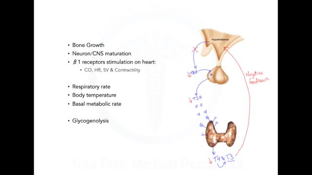
Graves disease is an autoimmune disorder that leads to an overactive thyroid gland (hyperthyroidism). An autoimmune disorder is a condition that occurs when the immune system mistakenly attacks healthy tissue. Causes The thyroid gland is an important organ of the endocrine system. The gland is located at the front of the neck above where the collarbones meet. This gland releases the hormones thyroxine (T4) and triiodothyronine (T3), which control body metabolism. Controlling metabolism is important for regulating mood, weight, and mental and physical energy levels. When the body makes too much thyroid hormone, the condition is called hyperthyroidism. (An underactive thyroid leads to hypothyroidism.) Graves disease is the most common cause of hyperthyroidism. It is due to an abnormal immune system response that causes the thyroid gland to produce too much thyroid hormone. Graves disease is most common in women over age 20. But the disorder can occur at any age and can affect men as well. Symptoms Younger people may have these symptoms: Anxiety or nervousness, as well as problems sleeping Breast enlargement in men (possible) Problems concentrating Fatigue Frequent bowel movements Hair loss Heat intolerance and increased sweating Increased appetite, despite having weight loss Irregular menstrual periods in women Muscle weakness of the hips and shoulders Moodiness, including irritability and anger Rapid or irregular heartbeat Shortness of breath with activity Tremor Many people with Graves disease have problems with their eyes: The eyeballs may seem to be bulging out and may be painful. Eyes can feel irritated and be tearing. Double vision may be present. Older people may have these symptoms: Rapid or irregular heartbeat Chest pain Memory loss Weakness and fatigue

Dehydration is a condition that occurs when the loss of body fluids, mostly water, exceeds the amount that is taken in. With dehydration, more water is moving out of our cells and bodies than what we take in through drinking. We lose water every day in the form of water vapor in the breath we exhale and in our excreted sweat, urine, and stool. Along with the water, small amounts of salts are also lost.
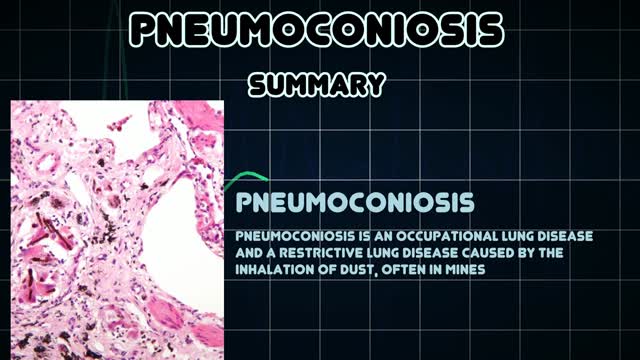
Pneumoconiosis is a general term given to any lung disease caused by dusts that are breathed in and then deposited deep in the lungs causing damage. Pneumoconiosis is usually considered an occupational lung disease, and includes asbestosis, silicosis and coal workers' pneumoconiosis (CWP), also known as "Black Lung Disease."
Every 10 minutes, someone is added to the national transplant waiting list, and every day, 22 people on average die waiting for a match, according to the United Network for Organ Sharing. But, thanks to innovations in bioengineering, all of that could change. Conceived nearly 60 years ago, the total artificial heart (TAH) has helped sustain the sickest biventricular failure patients waiting for a transplant. While the design of the primary TAH used today has mostly remained stagnant since the ’80s, when it was first implanted in a patient, new models and clinical trials may lead to a better device and, one day, a permanent solution. “We are still many years away from that,” Dr. Nader Moazami, director of the Cardiac Transplantation and Ventricular Assist Device Therapy Program at the Cleveland Clinic, told FoxNews.com of a permanent artificial heart. “Although tremendous strides have been made, biocompatibility will always remain a challenge.”
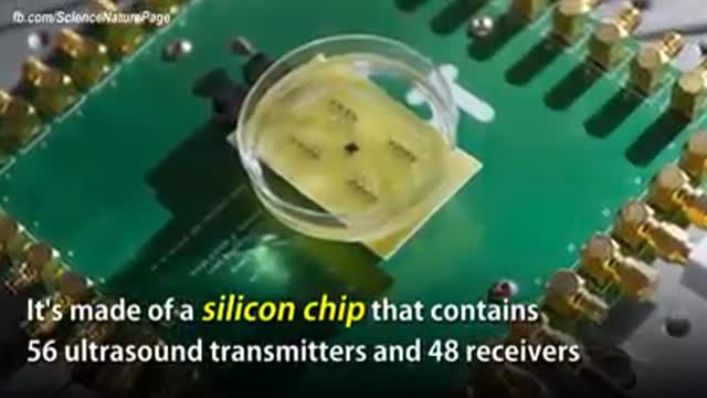
The term "miniaturization" is widely accepted in our vernacular as a positive step in product development. Reducing components to create less space, product footprint and more affordable medical devices are ongoing objectives for manufacturers today. Jabil strives to integrate new innovative technologies into product design and manufacturing as continual miniaturization of medical devices is a focus of the healthcare thought process. Miniaturization is a constantly moving target. Once a novel, new technology sets a higher bar for miniaturization standards, the next ambitious goal is to achieve an even thinner and smaller device. Industry trends, including minimally invasive surgical devices and home health care delivery, demand more sophisticated medical portable devices and easy-to-use electronics which may not be a core competency of medical device manufacturers.
Cholesterol is a fat-like, waxy substance that can be found in all parts of your body. It helps your body make cell membranes, many hormones, and vitamin D. The cholesterol in your blood comes from two sources: the foods you eat and your liver. But your liver makes all the cholesterol your body needs.
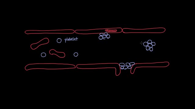
How do we make blood clots?
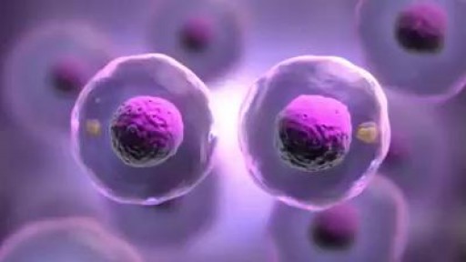
Mitosis, a process of cell duplication, or reproduction, during which one cell gives rise to two genetically identical daughter cells. Strictly applied, the term mitosis is used to describe the duplication and distribution of chromosomes, the structures that carry the genetic information.
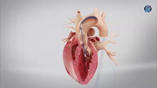
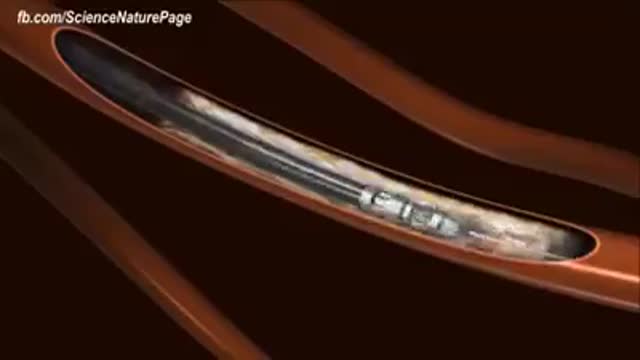
This minimally invasive surgical procedure repairs the valve without removing the old, damaged valve. Instead, it wedges a replacement valve into the aortic valve’s place. The surgery may be called a transcatheter aortic valve replacement (TAVR) or transcatheter aortic valve implantation (TAVI). Valve-within-valve — How does it work? Somewhat similar to a stent placed in an artery, the TAVR approach delivers a fully collapsible replacement valve to the valve site through a catheter. Once the new valve is expanded, it pushes the old valve leaflets out of the way and the tissue in the replacement valve takes over the job of regulating blood flow.

http://healthy-weight-loss.good-info.co ------- How To Lose Belly Fat, Fat Burning Workouts, How To Lose Belly Fat Fast For Men, Women. What kind of belly fat do you have? John has got something that just blew me away. It’s all about belly fat. You’ve probably heard and seen every ad talking about “one weird food” that causes belly fat. But this is totally different. And that’s probably why it’s so effective in making belly fat disappear. I’m talking MASSIVE fat loss. And in the most difficult area of our body. There are actually different KINDS of belly fat. 6 kinds, to be exact. And it’s different for men than women. Why? Because men and women use hormones differently. And hormones are one of the MAIN causes of belly fat. In SOME cases, what we think is fat. actually ISN’T. Take this strange but TRUE, “Finger Test” Click Here for the FREE Belly TEST. http://healthy-weight-loss.good-info.co
http://tinnitus-solution.info-pro.co --- Ear Infection, Loud Ringing In Ears, Tinnitus Suicide, Ear Is Ringing, Tinnitus One Ear, Tinnitus. Do you suddenly get up in the middle of the night hearing strange noises? Yes it can definitely be frightening, more so when you cannot find the source. Now consider for a moment that these sounds are coming from within you. Most people would be stunned to know that. Many of us do not even know that our internal organs can make sounds. Let us try to see whether you actually heard these noises or not, and if you did, where did they come from. Now before anything, let us get this straight - yes, you actually heard those noises. No, they are not a result of a creative mind that imagined things in slumber. But having said that, it is also true that there is indeed no source of the sounds you heard. So what is it? Confused? This is a classic case of tinnitus. What is tinnitus? What you experienced last night (or did you just get up from sleep and switch on the computer) is a classic case of tinnitus. This is a medical condition wherein a person hears all kinds of strange clicking, ringing, buzzing, whistling or hissing sounds within the ear. What's so worrisome about this condition for a lot of people is that, there's actually no physical source of these sounds. What makes it even worse is that, no one else seems to hear them. Frankly, these people cannot be really blamed. Naturally, if you cannot see where the sound is coming from, and if you keep hearing it, you are bound to get worried. In tinnitus, the sounds a person hears are actually perceptions. Since there's no actual source, they are often referred to as "phantom noises". Will it help you if you knew that about 8% of all people in the US suffer from tinnitus? Perhaps not, but at least now you know that you are not alone who hears these strange noises. Cure tinnitus. this simple, but effective system to erasing Tinnitus out of your life for good has now helped cure over 105,302 people of all the frustrating ringing, hissing, buzzing. Even if you’ve tried every single tinnitus treatment or remedy under the sun. 100% natural tinnitus cure click here: http://tinnitus-solution.info-pro.co

Women Heart Attack Symptoms, Healthy Living Tips, Heart Attack Symptoms Arm, Nutritional Tips, Heart.---- http://grow-younger-blood-good-info-co --- Why doctors let your family die One thing that keeps getting missed in that debate is how doctors, are so controlled by insurance companies, that they hold back from running life-saving blood tests. If you ever get a chance to ask a doctor, how he feels about HMO's, you'll get a glimpse into the sinister reality, of how corrupt and flat-out homicidal our healthcare system really is. This is why it's so important, to watch the new John video to get the REAL scoop on your health. It turns out that there are some life saving (or at least life-lengthening), tests you NEED to take but that doctors are not allowed to tell you about. And if you are under a big corporate health plan, you're even more likely to have been. cruelly lied to by the health industry. Watch this video. It's time to take your life and your family's health into your own hands. Once you have this information, no one not even the President himself can stop you Click Here. http://grow-younger-blood-good-info-co
Acufene Dopo Discoteca, Cosa Fare Quando Ti Fischiano Le Orecchie, Pressione Bassa E Acufeni---- http://acufeni-cura.plus101.com/ --- Ipnosi per l'acufene, L'ipnosi funziona veramente? Molte persone se lo chiederanno perchè è un tema avvolto da grande mistero. La medicina convenzionale spesso disdegna la pratica dell'ipnosi ma ci sono anche voci positive. Per cui a chi bisogna credere? Sapevi che la scienza medica convenzionale oggi guarda all'ipnosi come risposta a varie malattie? Si, è vero. Psichiatri e psicologi stanno sempre più passando all'ipnosi per raggiungere la mente del paziente. Incominciano a credere che, dal momento che la mente può controllare il corpo, può essere utilizzata per curare una malattia. Se non altro la mente può essere esercitata a rimanere positiva e un atteggiamento positivo spesso aiuta le cure. Per l'acufene - può l'ipnosi essere di aiuto? L'ipnosi può di fatto funzionare bene nei casi di acufene. Ciò perchè i rumori che una persona sente di fatto non sono veri rumori, ma solo suoni fantasma. E'in realtà la percezione del sentirli che infastidisce la persona - non c'è assolutamente causa fisica per tali rumori. Per cui l'ipnosi può funzionare in modo che la mente della persona non li senta più. I risultati possono essere stupefacenti. Si dice che dal 50% al 76% degli affetti da acufene può ottenere sollievo dagli acufeni solo con poche sedute di ipnosi. Per alcuni i rumori svaniscono del tutto, per altri si abbassano considerevolmente. Il problema dell'ipnosi è il mito che la circonda In realtà la maggioranza della gente non capisce l'ipnosi e come funziona. E' spesso fuorviata dalle sedute di ipnosi viste alla tv. Qui il terapista mette a dormire una persona e gli ordina di fare ogni tipo di stranezza. Naturalmente nessuno vuole essere completamente vulnerabile alla volontà del terapista, per questo l'ipnosi non è mai diventata una cura tra quelle principali. L'unico modo per sbarazzarsi per sempre degli acufeni è seguire un approccio di cura olistico. Utilizzando un trattamento multidimensionale per l'acufene, affrontiamo tutti i fattori che lo causano,eliminando alla radice questi elementi scatenanti. Questa è l'unica strada per ottenere una libertà definitiva dall'acufene. inserisci ora: http://acufeni-cura.plus101.com/

Pigmentflecken, Vitiligo Komplett Geheilt, Pigmentflecken Oberlippe, Vitiligo Heilung, Vitiligo--- http://vitiligo-heilung.info-pro.co/ --- Was ist Vitiligo? Vitiligo ist ein medizinischer Zustand, der die Haut befällt. Die Haut entwickelt dabei an unterschiedlichen Körperstellen nach und nach weiße Flecken, weshalb die Hautstörung im Deutschen auch als "Weißfleckenkrankheit" bekannt ist. Vitiligo macht keine Unterschiede bei Geschlecht oder Rasse und kann jeden befallen. Es wird geschätzt, dass gegenwärtig mehr als 100 Millionen Menschen weltweit in mehr oder minderem starken Maße an Vitiligo leiden. In den Vereinigten Staaten ist die Prävalenzrate etwa 1 % der Bevölkerung; Europa weist ähnliche Raten auf. Was verursacht den Zustand? Vitiligo tritt auf, wenn die Melanozyten in der Haut aus irgendeinem Grund zerstört werden oder ihre Funktion einstellen. Als Melanozyten bezeichnet man jene Hautzellen, die für die Hautfarbe verantwortlich sind. Werden sie zerstört oder anderweitig kompromittiert, stellen produzieren sie kein Melanin mehr, das Hautpigment. Das führt dann zu weißen Hautflecken. Es gibt verschiedene Faktoren, welche die Melanozyten zerstören oder beeinträchtigen können. Dennoch bleibt die Ursache bei den meisten Fällen von Vitiligo ungeklärt. Es wird außerdem angenommen, dass es sich bei Vitiligo um eine Autoimmunerkrankung handelt, welche das Immunsystem die Melanozyten angreifen lässt. Vitiligo kann zudem das Resultat einer Melanozytenstörung sein, welche diese Zellen praktisch veranlasst, "Sebstmord" zu begehen. Laut mancher Forscher kann Vitiligo aber auch durch chronischen Stress und Sonnenbrand hervorgerufen werden. Was sind die Symptome? Das offensichtlichste Symptom von Vitiligo sind die weißen Hautflecken, die sich im Laufe der Zeit vergrößern und auch weiter ausbreiten können. Die Rate, mit der die Hautstörung voranschreitet, unterscheidet sich dabei von Patient zu Patient. "Gratis-Präsentation enthüllt einen ziemlich ungewöhnlichen Tipp zur Beseitigung von Vitiligo für alle Zeiten und in nur 45-60 Tagen - Garantiert!" http://vitiligo-heilung.info-pro.co

Flecken Auf Der Haut, Braune Flecken Auf Der Haut Pilz, Homöopathie Bei Pigmentflecken--- http://vitiligo-heilung.info-pro.co --- Vitiligo Heilung für weiß gefleckte Haut, Zuerst treten die weißen Flecken punktuell auf, schließlich verbreiten sie sich über den ganzen Körper: Die Weißfleckenkrankheit ist belastend – und jetzt heilbar. Das Hautleiden ist weder gefährlich noch ansteckend – aber psychisch sehr belastend. Viele Betroffene trauen sich nicht mehr, in öffentliche Bäder zu gehen oder kurze Kleidung zu tragen. Schätzungen zufolge leiden bis zu zwei Prozent der Weltbevölkerung unter Vitiligo („Scheckhaut“). Statistisch gesehen sind die Hautpartien an Unterarmen, Handgelenken, Händen, Fingern, Ellbogen, Füßen und Genitalien am häufigsten betroffen. Die genauen Ursachen sind nicht bekannt. Wissenschaftler vermuten, dass Stress die Pigmentstörung auslösen kann. Klar ist aber, dass die Hautzellen einen zu hohen Anteil an Wasserstoffperoxid aufweisen. Es verhindert die Bildung des Hautfarbstoffs Melanin. Zudem ist bei den Betroffenen das Enzym Katalase beschädigt, das normalerweise den Abbau von Wasserstoffperoxid steuert. Die bessere Behandlungswahl bei Vitiligo Ein weitaus effektiverer und sicherer Weg zur Behandlung von Vitiligo ist die Nutzung holistischer Methoden. Dies umfasst die Verwendung von Kräuterextrakten - welche die weitere Ausbreitung der weißen Flecken behindern - zusammen mit der Einnahme bestimmter Vitaminergänzungen. Die Methode stimuliert den Hautpigmentierungsprozess. Damit diese natürliche Therapie aber in vollstem Umfange wirken kann, muss der Patient gewillt sein, Änderungen in seinem Lebensstil und seinen Essgewohnheiten einzuführen und konsequent zu befolgen, um so den Heilungsprozess zu beschleunigen. Erfahren Sie mehr darüber, indem Sie diese Webseite besuchen: http://vitiligo-heilung.info-pro.co
Watch that video of Horrifying Things Found Living Inside Human Body

Homemade Body Wraps For Weight Loss, Cheap Body Wraps That Work, Body Wraps To Lose Weight ---- http://do-body-wraps-work.plus101.com -- Cellulite and loose and flabby skin can often drain your confidence. You have maybe considered plastic surgery, but the risks and costs are just not reasonable. Fortunately, there are body wraps, which can offer alternative to plastic surgery. Getting a body wrap at a salon can be very expensive - multiple sessions can cost hundreds of dollars. The WrapYourselfSlim ebook shows readers how to get rid of their flabby stomaches and cellulite through the recipes made in the home kitchen. Which recipe is the best when wanting to seek drastic results? What Is The Best Body Wrap Recipe? One of the best body wrap recipes contains the ingredients of olive oil, green clay, water, herbs sea salt, and essential oils. You can select the oils and herbs such as lemongrass, sage, rosemary, rose petal powder, neem powder, lavender, ground basil, grapefruit, ginger root powder, alfalfa leaf powder, and rosehip powder. Measure out two tablespoons of olive oil, 1 cup of green clay, 2 cups of water, and a 1/2 cup of sea salt. Boil the water, then thoroughly stir inside of the sea salt until it has dissolved. Add your selected herbs, olive oil, and green clay, then stir til a paste has formed for you to set this mixture aside for cooling. Once the paste has cooled enough for your skin to handle, rub it on to your body, then cover it with sheets or towels cut into strips. Keep it on for one full hour before removing. A great tip that you should keep in mind when wanting to see the best results is to detoxify your body a couple days before applying the wrap on to yourself. Drink plenty of water before you apply the wrap on to yourself so you can see the highest results possible. You will definitely see significant results right after taking the wrap off, as the skin will instantly absorb the nutrients of the ingredients. This is better than the typical weight loss cream that you find today. Use this easy-to-prepare affordable formula to help contour your abs, thighs, hips buttocks, etc ... in less than two weeks! Stop procrastinating! Click Here for instant access to my recipes & start losing inches, detoxifying your body, and improving your skin right away! http://do-body-wraps-work.plus101.com

goldensleather video
