
- Physical Examination
- Surgical Examination
- Ophthalmology
- Clinical Skills
- Orthopedics
- Surgery Videos
- Laparoscopy
- Pediatrics
- Funny Videos
- Cardiothoracic Surgery
- Nursing Videos
- Plastic Surgery
- Otorhinolaryngology
- Histology and Histopathology
- Neurosurgery
- Dermatology
- Pediatric Surgery
- Urology
- Dentistry
- Oncology and Cancers
- Anatomy Videos
- Health and Fitness
- Radiology
- Anaesthesia
- Physical Therapy
- Pharmacology
- Interventional Radiology
- Cardiology
- Endocrinology
- Gynecology
- Emergency Medicine
- Psychiatry and Psychology
- Childbirth Videos
- General Medical Videos
- Nephrology
- Physiology
- Diet and Food Health
- Diabetes Mellitus
- Neurology
- Women Health
- Osteoporosis
- Gastroenterology
- Pulmonology
- Hematology
- Rheumatology
- Toxicology
- Nuclear Medicine
- Infectious Diseases
- Vascular Disease
- Reproductive Health
- Burns and Wound Healing
- Kidney Zone Podcast
- Other










Examination of the heart

Educational only: videos on MedicalVideos.com are for general information and are not medical advice. Always consult your own doctor about your health.
Examination of the heart
This video in 30 seconds
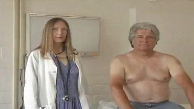
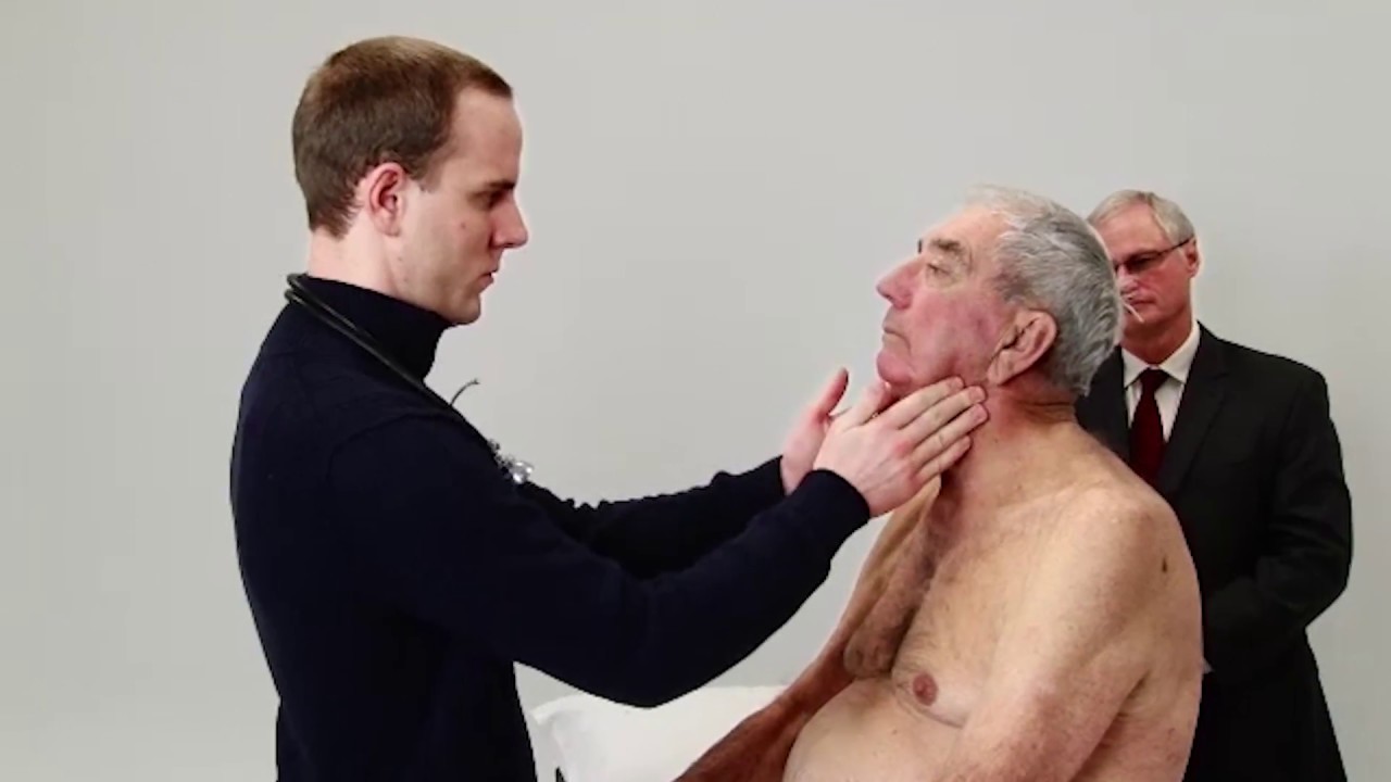
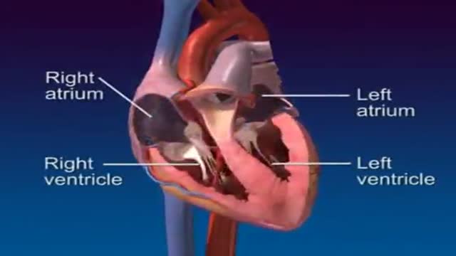
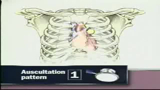
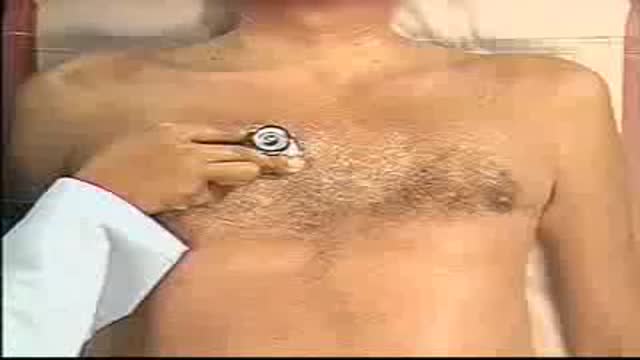
A Bates-style demonstration of the cardiac examination. The narration covers inspecting the precordium, palpating the interspaces and the apical impulse for its location, diameter, amplitude and duration, rolling the patient partly onto the left side to feel for it, and percussing the left border of cardiac dullness to estimate heart size.
Key points
- The examiner inspects the precordium for pulsations, heaves and retractions
- Fingertips palpate the interspaces for pulsations and the right ventricular impulse
- The apical impulse is assessed for location, diameter, amplitude and duration
- The patient rolls partly onto the left side to make the impulse easier to feel
- Percussion of the left border of cardiac dullness is used to estimate heart size
Chapters
- 0:00 — Positioning and inspecting precordium
- 0:27 — Palpating interspaces and impulses
- 1:21 — Assessing the apical impulse
- 2:43 — Percussing cardiac dullness border
Video Transcript
A Bates-style demonstration of the cardiac examination. The narration covers inspecting the precordium, palpating in the interspaces and for the apical impulse — its location, diameter, amplitude and duration — rolling the patient partly onto the left side to feel for it, and percussing the left border of cardiac dullness to estimate the size of the heart.
Read the full transcript
To examine the heart, stand at the patient's right side. Have the patient remain supine with the upper body raised to about 30 degrees. Inspect the precordium, noting any pulsations, heaves or retractions. There are none here. Look for the apical impulse, which when visible, is normally seen in the left-fifth interspace at or medial to the mid-clavicular line. Using your fingertips, palpate for pulsations or heart sounds in the right-second interspace, left-second interspace, and left-third interspace. Next, palpate for the systolic impulse of the right ventricle. While keeping one finger in the third interspace, place additional fingertips in the fourth and fifth interspaces.
If an impulse is palpable, note its location, duration and amplitude. If the patient's chest has an increased antroposterior diameter, palpate for the right ventricular impulse high in the epigastric area where it may be easier to feel. Finally, palpate the apical impulse. If it is not visible, feel for its location with your palm and fingers. When you find it, assess it with your fingertips. Observe its location, diameter, amplitude and duration. Identify its location by the interspaces in which you feel the apical impulse and by its distance in centimeters from the mid-sternal or mid-clavicular line.
Measure the diameter of the impulse in centimeters. Feel for the amplitude of the apical impulse. It is usually small and feels like a gentle tap. To estimate the duration of the apical impulse, feel it as you listen to the heart sounds with a stethoscope. The normal impulse may last through the first two-thirds of systole, but not longer. Please turn over to your left side. If you can't feel the apical impulse, ask the patient to roll partly onto his left side and try again. Now I'd like to tap on your chest to see the size of your heart.
If you still can't feel the apical impulse, estimate heart size by percussion. Starting on the far left of the chest, percuss toward the left border of cardiac dullness in the third, fourth, fifth and possibly sixth interspaces. Normally, percussion reveals pulmonary resonance laterally and cardiac dullness medially.
Transcript auto-generated from the video’s audio for accessibility and study. Educational content only — not medical advice.
Video Overview
From our Cardiology collection: ECG interpretation, echocardiograms, angioplasty, stents and heart conditions explained by cardiologists. Browse all Cardiology videos →
SORT BY-
Top Comments
-
Latest comments