
- Physical Examination
- Surgical Examination
- Ophthalmology
- Clinical Skills
- Orthopedics
- Surgery Videos
- Laparoscopy
- Pediatrics
- Funny Videos
- Cardiothoracic Surgery
- Nursing Videos
- Plastic Surgery
- Otorhinolaryngology
- Histology and Histopathology
- Neurosurgery
- Dermatology
- Pediatric Surgery
- Urology
- Dentistry
- Oncology and Cancers
- Anatomy Videos
- Health and Fitness
- Radiology
- Anaesthesia
- Physical Therapy
- Pharmacology
- Interventional Radiology
- Cardiology
- Endocrinology
- Gynecology
- Emergency Medicine
- Psychiatry and Psychology
- Childbirth Videos
- General Medical Videos
- Nephrology
- Physiology
- Diet and Food Health
- Diabetes Mellitus
- Neurology
- Women Health
- Osteoporosis
- Gastroenterology
- Pulmonology
- Hematology
- Rheumatology
- Toxicology
- Nuclear Medicine
- Infectious Diseases
- Vascular Disease
- Reproductive Health
- Burns and Wound Healing
- Kidney Zone Podcast
- Other







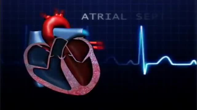
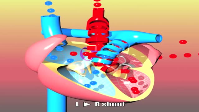




Pediatric Heart Examination

Educational only: videos on MedicalVideos.com are for general information and are not medical advice. Always consult your own doctor about your health.
This cardiology video demonstrates the heart examination in a child, covering the key steps of pediatric cardiac assessment. The examination includes inspection and palpation of the chest and listening to the heart sounds with a stethoscope. Heart examination is a routine part of child health assessments.
This video in 30 seconds
This narrated teaching video demonstrates the paediatric cardiac examination. It covers inspection and palpation of the chest, the order and locations for auscultation, where murmurs from each valve are best heard, and how normal heart rate varies by age. It closes with how murmurs are described and graded for loudness.
Key points
- Inspection notes chest asymmetry and any bulge over the heart
- Palpation locates the point of maximum impulse and checks for thrills
- Auscultation follows a set order of chest locations
- The narration links each valve's murmur to where it is best heard
- Heart rate by age and murmur grading are described
Chapters
- 0:06 — Inspection of the thorax
- 0:27 — Palpation and maximum impulse
- 1:28 — Order of auscultation
- 2:06 — Where each murmur is heard
- 2:56 — Heart rate by age
- 3:57 — Describing and grading murmurs
Video Transcript
A narrated teaching video on the pediatric cardiac examination. It covers inspection and palpation of the chest, the order and locations for auscultation, how murmurs from each valve are best heard, normal heart rates by age, and how murmurs are graded for loudness and described.
Read the full transcript
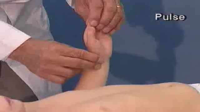
Every examination of the heart begins by inspecting the thorax. Thoracic asymmetries must be noted. The chest may sometimes show a bulge over the heart which indicates cardiomegaly. During palpation the first thing to locate is the point of maximum impulse which is normally found in the left medoclavicular line. In babies and young children the point of maximum impulse is in the fourth intercostal space. In older children it is between the fourth and fifth intercostal space. During palpation assess if the maximum impulse is raised or widened or too far to the left.
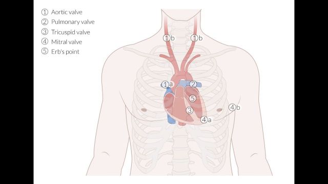
These findings would indicate left ventricular hypertrophy. A raise on the lower left sternal border may indicate right ventricular hyperactivity and hypertrophy. Sometimes a palpable cardiac thrill indicates circulatory pathology. The thrill should also be sought in the suprasternal notch. With young children cardiac percussion is hardly ever performed. The heart is auscultated in the following order. Second right and left intercostal space. Third left intercostal space. Herbs point. Fourth left intercostal space. Lower left sternum border. As well as over the heart apex and in special cases the fourth right
intercostal space. This illustration shows the relationship between the points of auscultation, the skeleton, and the heart valves. Murmers which originate in the pulmonary valve can best be heard in the second left intercostal space. Murmers radiate from the aortic valve via the aortic arch to the second right intercostal space. Murmers of mitral origin can be heard at herbs point and at the heart apex. A ventricular septal defect can be heard best at the lower left sternum border and at herbs point. Murmers which originate at the tricuspid valve radiate
peristernally to the lower right sternal border. During auscultation note the following points. Heart rate, rhythm, that is the regularity of the heartbeat, quality of the heart sounds, and the presence of additional heart sounds, murmurs or clicks. The heart rate changes with age. In the first year of life the heart rate is about 140 beats per minute. In the second year 120. In the fourth 100, in the eighth 90. Towards the end of puberty the heart rate approaches that of an adult. When listening to heart sounds we note their intensity.
Possible splitting. Whether there are additional sounds in the form of a third or fourth heart sound and clicks. Murmers can be diastolic or systolic. Can you hear this holocystolic murmur? In addition to the duration of a murmur we also judge its loudness on a scale of one to six. We further describe the quality of the murmur, that is whether it is soft or rough, the point of maximum intensity, as well as possible radiation to other areas, especially the back and the carotid arteries.
Transcript auto-generated from the video’s audio for accessibility and study. Educational content only — not medical advice.
Video Overview
From our Cardiology collection: ECG interpretation, echocardiograms, angioplasty, stents and heart conditions explained by cardiologists. Browse all Cardiology videos →
Medical Videos
Free medical and surgery videos for patients, students and professionals — trusted since 2007.
Founded by Dr. Mohamed Ibrahim, MD, PhD — physician-scientist.
Follow us on Facebook · Instagram
SORT BY-
Top Comments
-
Latest comments