
- Physical Examination
- Surgical Examination
- Ophthalmology
- Clinical Skills
- Orthopedics
- Surgery Videos
- Laparoscopy
- Pediatrics
- Funny Videos
- Cardiothoracic Surgery
- Nursing Videos
- Plastic Surgery
- Otorhinolaryngology
- Histology and Histopathology
- Neurosurgery
- Dermatology
- Pediatric Surgery
- Urology
- Dentistry
- Oncology and Cancers
- Anatomy Videos
- Health and Fitness
- Radiology
- Anaesthesia
- Physical Therapy
- Pharmacology
- Interventional Radiology
- Cardiology
- Endocrinology
- Gynecology
- Emergency Medicine
- Psychiatry and Psychology
- Childbirth Videos
- General Medical Videos
- Nephrology
- Physiology
- Diet and Food Health
- Diabetes Mellitus
- Neurology
- Women Health
- Osteoporosis
- Gastroenterology
- Pulmonology
- Hematology
- Rheumatology
- Toxicology
- Nuclear Medicine
- Infectious Diseases
- Vascular Disease
- Reproductive Health
- Burns and Wound Healing
- Kidney Zone Podcast
- Other

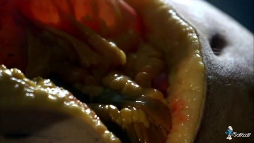
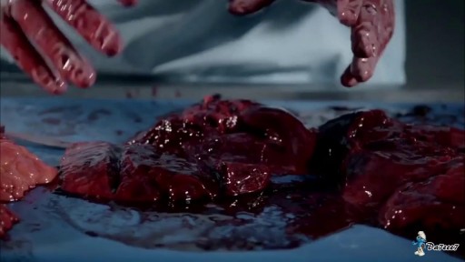
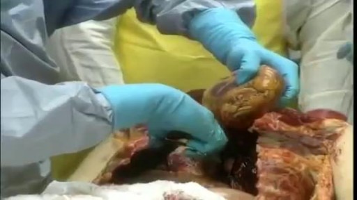
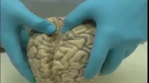
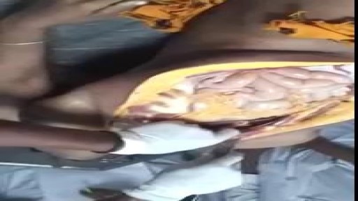
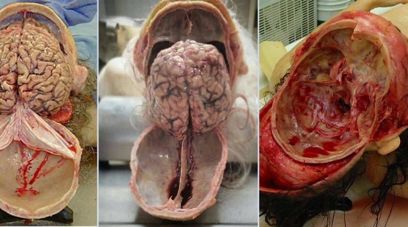



Full Female Body Medical Anatomy Autopsy

Watch that Full Female Body Medical Anatomy Autopsy
Video Transcript
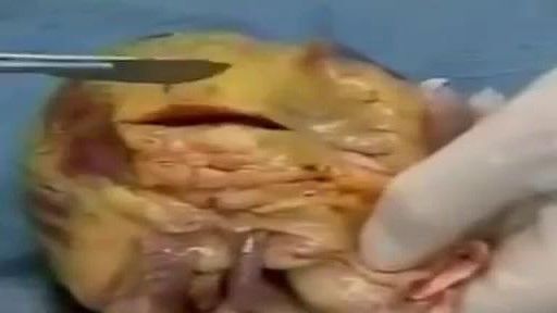
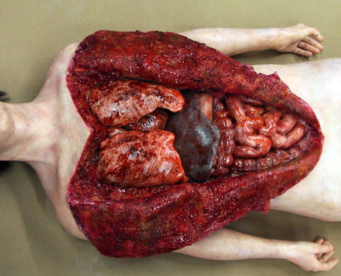
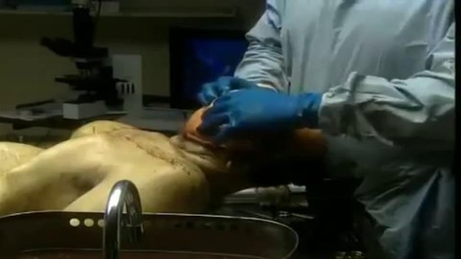
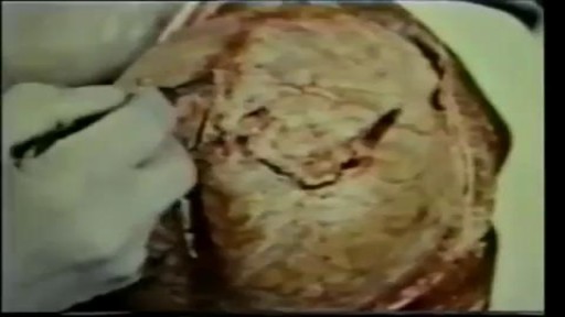
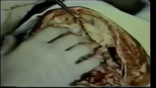
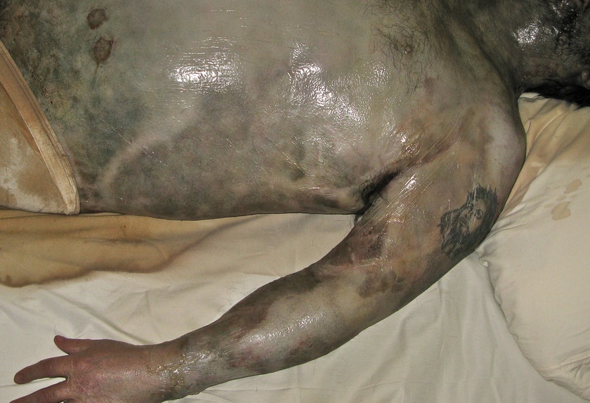
A narrated autopsy demonstration following the examination of a female body. The prosector talks through the abdominal incision through the peritoneum, removal of the intestines, cutting through the ribs and removing the chest plate, and then removal of the chest organs — dividing structures such as the inferior vena cava and pulmonary veins as the heart and lungs are taken out.
Read the full transcript
down to the pubic bone. And then the incision is continued through what we call the peritoneum, which is a lining of the abdomen or belly. And that is open and that exposes everything within the abdomen. And then the intestines are removed and that's one of the messier parts of the autopsies you might imagine. Then we actually cut through the ribs and remove a portion of the bone plate, the front plate. And then the incision is here. That's the big heart. Once that is completed we will proceed to remove the organs from the chest.
So we're cutting the inferior vena cava and the pulmonary veins. The last thing we cut is the superior vena cava. We'll take out the heart and we'll take out the lungs. We'll take out the different organs of the abdomen. And as these are removed they're handed to the resident or pathologist who then examines each organ individually. So you can see the medulla is a little dark. Was she in shock? Throughout this we are always constantly looking for any abnormalities, any anomalies that may be present in the remains and make any notation.
It looks like a benign cyst of the ovary here. Here's the uterus. Here's the other ovary. Looks normal. We will carefully dissect the organs in much more detail and look for specific lesions if you will or problems with the organs which may have contributed to the person's death. The liver amazingly doesn't look that congested. Autopsies of dissection will be the end of autopsy, not for the pathologist because additional follow-up such as microscopic examination to make sure that a heart attack is not the type of a heart attack you would expect from an infection.
Really normal including the deep nuclei. It's a spinal cord that looks normal. So again in this case there aren't specific questions about the neurologic status. Well I'm a neuropathologist so my main interest is removing the brain and that involves reflecting the scalp over the top of the head, removing the top of the skull and then removing the brain from the contact it has with the spinal cord and with the base of the skull. I believe the skull cap is put back. The scalp is pushed back and sewed back together so you really can't even tell
that the brain has been removed when you look at the person's face. Finally when this is all completed we will sew it up, repair all of the incisions and clean the cadaver, place it back in. There's a lot of benefits. This may sound corny but the autopsy is your friend. It can only help you so don't be afraid to consent to one. I think one of the big benefits to the autopsy for the general public is that sometimes we can give people answers, particularly in cases where you've got a person
who is healthy according to the family and then all of a sudden they're dead. And this family is torn up. They're saying why did he die? And they don't know. And if you can tell them you can give them an answer. Even if it's an answer that's not very pleasant to hear, at least it's something. It's closure and it'll give them some peace of mind. It's important for the public to understand the value of the autopsy for a couple of reasons. Prior to the 1960s we thought Alzheimer's disease was a very rare curious phenomenon
that afflicted very few people. From the 1970s on we've understood that it's a very common disorder, in fact it's an epidemic. And that would not have occurred unless the brain tissue had been very carefully examined at autopsy. Another tremendous value of the autopsy has been in the AIDS epidemic. We now know that there are certain strains of HIV that have a real tendency to infect the brain. And it was really the result of autopsy studies that defined an understanding of viruses that do infect the brain preferentially.
Well, even when we think we know the cause of death and with all our modern imaging techniques of CT scans and MRI, every study ever done on this subject has shown that in 20 to 30 percent of cases the clinical diagnosis is incorrect. That is, we find something at autopsy that was not expected clinically. This is a post-mortem change. Another aspect of doing an autopsy is that it can give information about genetic diseases, runaline diseases, which might help other family members in terms of diagnosing. Do they have that disease? Are they a carrier of that disease?
Will their children or other family members have that disease? And if they do, it might dictate how they should be treated or how they might be treated. In the coroner's office, we perform some sort of a watchdog function for product malfunction. For example, occasionally you get a tragic case of a baby who slips down through a hole in a baby's seat or a swing or something like that and then strangles. Then you have to evaluate that product. Is that product inherently dangerous or was it misused? So sometimes we can be sentinels for that kind of thing.
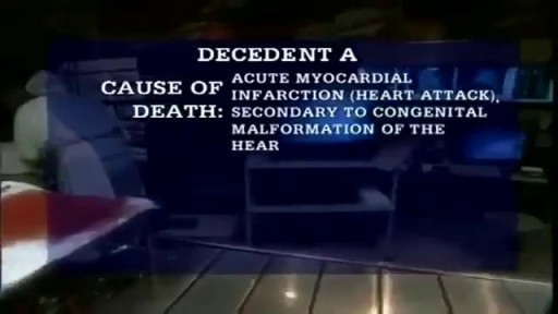
This could be 15 years old, but this is not. This is more recent. See, here we have this subindocardial infarction in the septum. We learn a lot from every autopsy. The autopsy, I think, still benefits the family. It benefits doctors and it benefits society as a whole. The autopsy is the last word and it's always going to be. We're in the midst now of what I would call a crisis in medical education. In 1950, about half of all people in this country that died were autopsy. Now the autopsy rate nationwide is between 10 and 15 percent.
Transcript auto-generated from the video’s audio for accessibility and study. Educational content only — not medical advice.
SORT BY-
Top Comments
-
Latest comments