
- Physical Examination
- Surgical Examination
- Ophthalmology
- Clinical Skills
- Orthopedics
- Surgery Videos
- Laparoscopy
- Pediatrics
- Funny Videos
- Cardiothoracic Surgery
- Nursing Videos
- Plastic Surgery
- Otorhinolaryngology
- Histology and Histopathology
- Neurosurgery
- Dermatology
- Pediatric Surgery
- Urology
- Dentistry
- Oncology and Cancers
- Anatomy Videos
- Health and Fitness
- Radiology
- Anaesthesia
- Physical Therapy
- Pharmacology
- Interventional Radiology
- Cardiology
- Endocrinology
- Gynecology
- Emergency Medicine
- Psychiatry and Psychology
- Childbirth Videos
- General Medical Videos
- Nephrology
- Physiology
- Diet and Food Health
- Diabetes Mellitus
- Neurology
- Women Health
- Osteoporosis
- Gastroenterology
- Pulmonology
- Hematology
- Rheumatology
- Toxicology
- Nuclear Medicine
- Infectious Diseases
- Vascular Disease
- Reproductive Health
- Burns and Wound Healing
- Kidney Zone Podcast
- Other
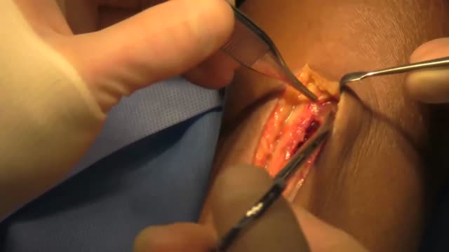
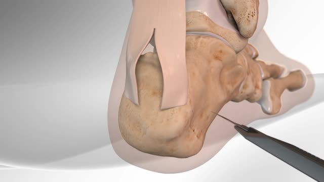
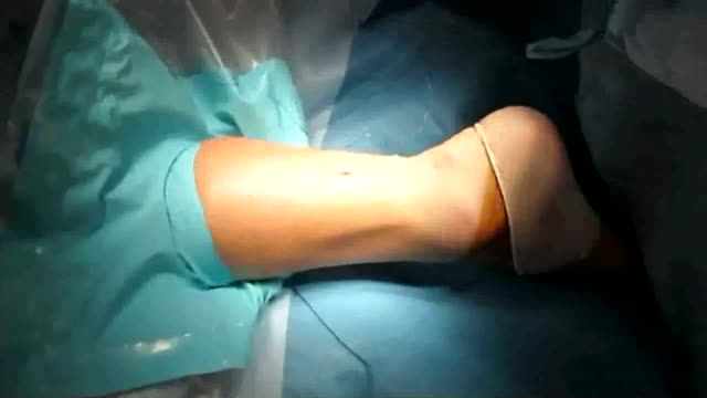
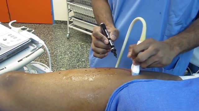
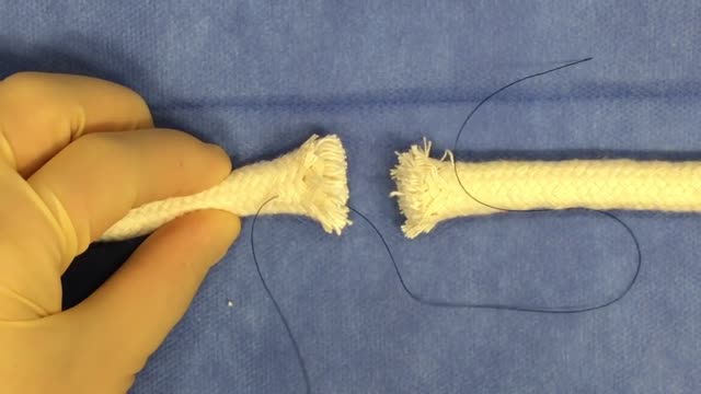
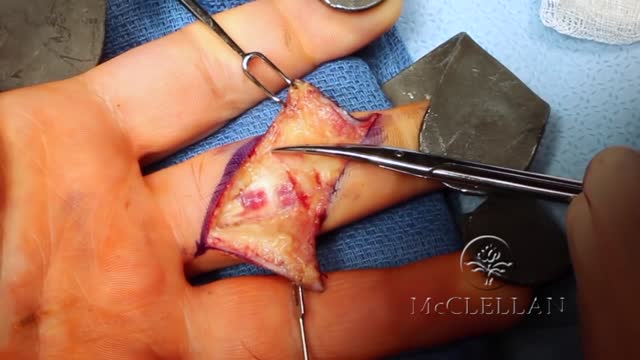
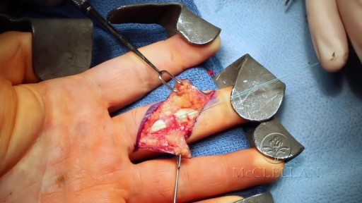

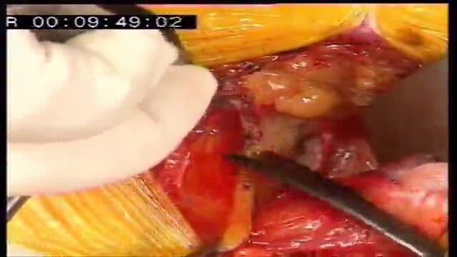


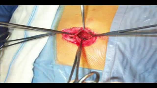
Mini-invasive surgical repair of a ruptured Achilles tendon

Mini-invasive surgical repair of a ruptured Achilles tendon
This video in 30 seconds
This narrated video shows minimally invasive repair of a ruptured Achilles tendon using the Dresden instrument technique. Sutures are passed percutaneously through the tendon ends, the ends are drawn together, and the plantar-flexion angle is compared with the uninjured foot. The fascia and skin are then closed.
Key points
- The rupture site is marked and a short incision is made medial to the tendon
- A Dresden instrument is introduced between the fascia and tendon sheath
- Needles pass sutures percutaneously through the tendon ends
- Tightening the sutures brings the tendon ends together
- Fascia and skin closure follows a check of plantar-flexion angle and tendon integrity
Chapters
- 0:01 — Marking rupture and incision
- 1:15 — Introducing the Dresden instrument
- 1:59 — Second instrument and suture passage
- 2:32 — Sutures drawn out and anchored
- 4:30 — Approximating the tendon ends
- 5:02 — Closure and angle check
Video Transcript
A narrated step-by-step of minimally invasive repair of a ruptured Achilles tendon using the Dresden instrument technique: passing sutures percutaneously through the tendon ends, drawing the ends together, comparing the plantar-flexion angle with the uninjured foot, and closing the fascia and skin.
Read the full transcript
The localization of the rupture is identified and demarcated with hatched lines. A short longitudinal incision is made medial to the tendon, about 3 cm proximal to the rupture. The skin is sharply divided. Subcutaneous bleeding is stopped by cauterization. The incision is carried sharply through the fascia and the layer between the fascia and tendon sheath is carefully tunneled. Without injuring the tendon sheath, the dresden instrument is then introduced into the layer between the fascia and tendon sheath, ensuring that the tip of the instrument is correctly positioned, just proximal to the calcaneal insertion.
A needle is pierced through the skin and then through the aperture of the instrument. Correct position of the needle is confirmed by gently pulling the instrument proximally. The second dresden instrument is then introduced into the subfascial layer. The needle is pierced through the tendon and the aperture of the second instrument. And finally through the skin on the opposite side. Correct position of the needle is yet again confirmed by carefully pulling the instruments proximally. A second needle is then introduced using the same procedure. When the dresden instruments are pulled out, the sutures are pulled out together with the instruments.
The instrument to the right has already been removed, and the instrument to the left is just being pulled out. The sutures are anchored by simple interrupted sutures. The tendon is approximated by tightening the knots, and an assistant holds the ankle implant half flexion comparing tension to the non-injured foot. The knots are buried by tying up both sutures and inserting them next to the knots. Finally, the fascia and skin are closed. The plantar flexion angle is now satisfactory, and tendon integrity has been re-established.
Transcript auto-generated from the video’s audio for accessibility and study. Educational content only — not medical advice.
Video Overview
From our Surgery Videos collection: Surgery videos: watch real operations step by step — general, laparoscopic and specialist surgery performed in the OR, explained for students and surgeons. Browse all Surgery Videos videos →
SORT BY-
Top Comments
-
Latest comments