
- Physical Examination
- Surgical Examination
- Ophthalmology
- Clinical Skills
- Orthopedics
- Surgery Videos
- Laparoscopy
- Pediatrics
- Funny Videos
- Cardiothoracic Surgery
- Nursing Videos
- Plastic Surgery
- Otorhinolaryngology
- Histology and Histopathology
- Neurosurgery
- Dermatology
- Pediatric Surgery
- Urology
- Dentistry
- Oncology and Cancers
- Anatomy Videos
- Health and Fitness
- Radiology
- Anaesthesia
- Physical Therapy
- Pharmacology
- Interventional Radiology
- Cardiology
- Endocrinology
- Gynecology
- Emergency Medicine
- Psychiatry and Psychology
- Childbirth Videos
- General Medical Videos
- Nephrology
- Physiology
- Diet and Food Health
- Diabetes Mellitus
- Neurology
- Women Health
- Osteoporosis
- Gastroenterology
- Pulmonology
- Hematology
- Rheumatology
- Toxicology
- Nuclear Medicine
- Infectious Diseases
- Vascular Disease
- Reproductive Health
- Burns and Wound Healing
- Kidney Zone Podcast
- Other


Vaginal Tape Vault Surgery: Pelvic Repair

Laparoscopic-assister percutaneous vaginal tape vault suspension, a minimally invasive prolapse repair with post-hysterectomy and uterine-sparing options
This video in 30 seconds
A narrated surgical lecture describes vaginal tape vault suspension, a laparoscopic-assisted repair for vaginal vault prolapse. It contrasts the technique with traditional sacrocolpopexy, describes passing mesh strips along the vaginal walls and anchoring them at the sacrum, and covers a uterine-sparing version and reported outcomes.
Key points
- Traditional sacrocolpopexy and its apical-only support limits are discussed
- Two mesh strips are passed from the perineal body along the vagina to the apex
- Laparoscopic ports, bowel retraction and exposure of the sacral promontory are described
- Mesh is sutured to the vaginal wall and anchored to the sacrum
- A uterine-sparing approach and follow-up outcome figures are discussed
Chapters
- 0:09 — Traditional sacrocolpopexy limits discussed
- 0:46 — Vaginal tape technique overview
- 1:32 — Mesh strips and port placement
- 2:19 — Laparoscopic exposure of sacrum
- 3:13 — Passing mesh and reducing prolapse
- 4:04 — Anchoring mesh to sacrum
- 4:43 — Uterine sparing approach described
Video Transcript
A narrated surgical lecture on vaginal tape vault suspension — a laparoscopic-assisted, minimally invasive repair for vaginal vault prolapse. It contrasts the approach with traditional sacrocolpopexy and its Y-shaped mesh, discusses the limits of apical-only support and the added procedures often needed for cystoceles and rectoceles, and covers post-hysterectomy and uterine-sparing options and their risks.
Read the full transcript
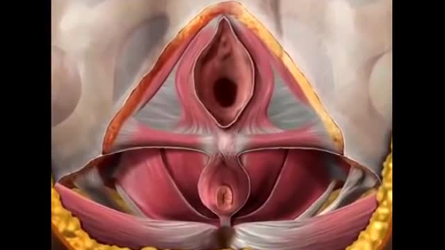
Traditional sacrocopopexy has involved placement of a Y-shaped mesh along the anterior and posterior vaginal walls. This tends to provide support only at the very apex of the vagina, and concomitant vaginal procedures are often required to correct cysticeals and recticeals and perineal descent. Utterine preserving modifications require placement of mesh material anterior, or sutures into the cervix, creating increased urinary tract injury risk and a risk of cervical stenosis. Furthermore, support of the anterior vaginal wall distal to the uterus can be difficult to address. The laparoscopic assisted vaginal tape, vault, or uterine suspension,
involves passage of two polypropylene mesh strips from the perineal body along the lateral walls of the vagina to the vaginal apex in order to provide full-length support along the entire vaginal axis. This unique technique recreates the natural lateral support of the vagina provided by the arches tendinia fasci pelvis that has been lost in severe prolapse cases. Here we demonstrate our technique with this procedure, which we have previously demonstrated to be equally efficacious with a lower risk of bladder injury when compared to traditional laparoscopic or open forms of sacrocopopexy.
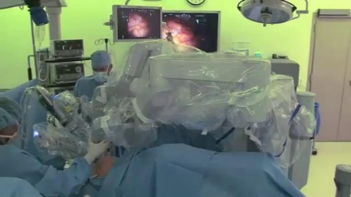
The procedure is demonstrated here in a woman with stage 3 vault prolapse who has status post hysterectomy. To construct a percutaneous vaginal tape or PVT, a standard sheet of polypropylene mesh is cut into strips 11 millimeters wide and 20 centimeters long. A nylon suture is passed through one end of the mesh strip and tied and left long. Four ports are placed in a fan array. An umbilical 10 millimeter port is used for the zero-degree camera. Two 12 millimeter ports are placed at the lateral rectus borders on each side,
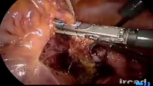
through which the primary surgeon dissects and sutures. A right lower quadrant 5 millimeter port is used to allow retraction and suction by the surgical assistant. After port insertion and placing the patient in steep Trendelenburg position, the bowel contents are retracted out of the pelvis. A looped suture on a CTX needle is passed into the left lower quadrant and used to grasp an epiploic appendage, retracting the sigmoid colon anterolaterally. This exposes the sacral promontory, iliac vessels, and right ureter without the need for a third assistant. The posterior peritoneum is ensized with hook electrocotery
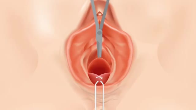
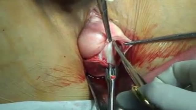
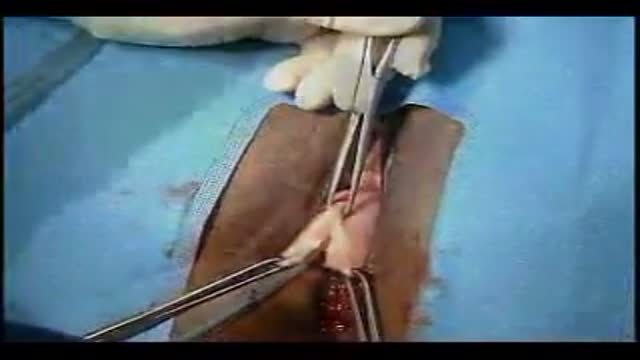
and a trough is created by opening the posterior peritoneum from the sacral promontory down to the vaginal apex, here reduced using an endoanal sizer. Next, small stab incisions are made at the 5 and 7 o'clock positions in the labial crease. A speculum can then be used to reduce the prolapse while allowing transvaginal visualization of the needle's subvaginal wall passage. An endoanal sizer can provide additional prolapse reduction. The mesh strip is loaded onto the stamey needle and passed using direct visual and laparoscopic guidance. The mesh strips are sutured to the vaginal wall with permanent suture just lateral to the apex on either side.
The knots are tied with a closed knot pusher or a lap clip may be applied, and a third optional stitch is placed anchoring both meshes to each other and the vaginal apex. The mesh is then pulled to the level of the sacral promontory. Two sutures of zero braided nylon are passed through the sacral anterior longitudinal ligament to anchor the mesh to the sacrum or tacking screws may be used. An excess mesh is trimmed. After cinching these knots down, the peritoneum is closed over the mesh to extra peritonealize the graft and to prevent direct contact with bowel.
This simple maneuver also prevents bowel herniation around the graft material and prevents formation of an enteroseal. The procedure can also be performed with a uterine sparing approach. Here we have a middle-aged patient with symptomatic stage 2 uterine descent who desired a uterine sparing approach. The steps of the procedure are identical with the stamey needles passed beneath the vaginal wall while the uterus is reduced. The mesh is then sewn to the vagina lateral to the cervix on either side, pulled to the sacrum and attached to the sacrum in the same manner.
The prolapse is markedly reduced with recreation of a normal and troitus provided by the support of the entire vaginal organ and levators. The mesh strips are cut at the skin level and the skin is closed with simple, varied sutures. We have performed this procedure on 40 patients to date. Follow-up over one year of the first 32 showed a re-operation rate of 6.25% for two anterior cystoceles, a mean total operating room time of 234 minutes that includes ancillary procedures for slings and perineoplasties, and a mean post-operative stay of 1.8 days at a tertiary referral center.
These compare favorably with traditional open and laparoscopic sacrocopypexes with a simplicity of performance, reduction in surgical team requirements, and potential for better outcomes in the long term regarding prolapse recurrence, operative morbidity, and sexual dysfunction due to reduced graft use. Laparoscopic percutaneous vaginal tape vault, or uterine suspension, is a safe, effective, and simple procedure that represents a major paradigm shift in how we approach pelvic organ prolapse repairs. The technique can be accomplished with minimal laparoscopic skills compared to traditional or even robotic laparoscopic surgery. Furthermore, surgeons wishing to use an open approach for sacrocopypexy may wish to consider the use of the percutaneous vaginal tape graft placement technique
in lieu of traditional graft placement in the anterior and posterior positions.
Transcript auto-generated from the video’s audio for accessibility and study. Educational content only — not medical advice.
Video Overview
From our Gynecology collection: Pelvic exams, Pap smears, IUD insertion, hysteroscopy and women's health procedures explained step by step. Browse all Gynecology videos →
SORT BY-
Top Comments
-
Latest comments