
- Physical Examination
- Surgical Examination
- Ophthalmology
- Clinical Skills
- Orthopedics
- Surgery Videos
- Laparoscopy
- Pediatrics
- Funny Videos
- Cardiothoracic Surgery
- Nursing Videos
- Plastic Surgery
- Otorhinolaryngology
- Histology and Histopathology
- Neurosurgery
- Dermatology
- Pediatric Surgery
- Urology
- Dentistry
- Oncology and Cancers
- Anatomy Videos
- Health and Fitness
- Radiology
- Anaesthesia
- Physical Therapy
- Pharmacology
- Interventional Radiology
- Cardiology
- Endocrinology
- Gynecology
- Emergency Medicine
- Psychiatry and Psychology
- Childbirth Videos
- General Medical Videos
- Nephrology
- Physiology
- Diet and Food Health
- Diabetes Mellitus
- Neurology
- Women Health
- Osteoporosis
- Gastroenterology
- Pulmonology
- Hematology
- Rheumatology
- Toxicology
- Nuclear Medicine
- Infectious Diseases
- Vascular Disease
- Reproductive Health
- Burns and Wound Healing
- Kidney Zone Podcast
- Other
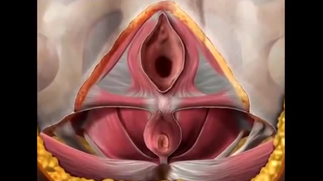
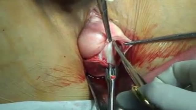






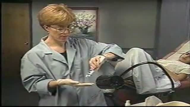
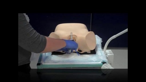
Anterior and Posterior Vaginal Repair Plus IVS Tunner

Educational only: videos on MedicalVideos.com are for general information and are not medical advice. Always consult your own doctor about your health.
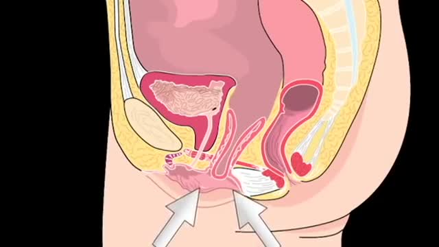
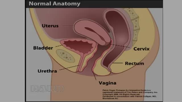
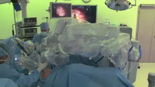
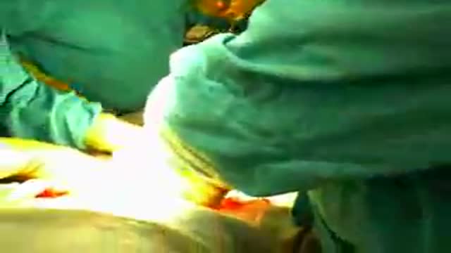
Surgical repair of pelvic organ prolapse is demonstrated in this video, combining anterior and posterior vaginal wall repair with a tension-free sling procedure. These operations restore support to the bladder and rectum in women with prolapse. It is an operative teaching resource for gynecology trainees. Educational only — not medical advice.
This video in 30 seconds
A narrated pelvic-floor operation combines anterior and posterior vaginal repair with a mesh procedure using an IVS tunneller. The surgeon describes dissecting the anterior wall, plicating the pubocervical fascia, anchoring a matrix with sutures, then the posterior dissection toward the ischial spines and the tunneller incisions placed near the anal verge.
Key points
- Anterior vaginal wall dissection exposes the cystocele in the midline
- Plication of the pubocervical fascia and reduction of the cystocele are shown
- A matrix is shaped and secured with four anchoring sutures
- The anterior site is trimmed and closed after the repair is checked
- Posterior dissection proceeds toward the ischial spines before the tunneller is used
Chapters
- 0:00 — Anterior vaginal wall dissection
- 1:04 — Plication of pubocervical fascia
- 1:33 — Anchoring sutures for matrix placement
- 2:44 — Securing matrix and checking reduction
- 3:37 — Trimming and closing anterior site
- 4:18 — Posterior vaginal repair dissection
- 5:37 — Using the IVS tunneller posteriorly
Video Transcript
A narrated pelvic-floor operation combining anterior and posterior vaginal repair with a mesh procedure using an IVS tunneller. The surgeon describes dissecting the anterior wall, plicating the pubocervical fascia, anchoring a matrix with four sutures, then the posterior dissection to the ischial spines and the tunneller incisions placed lateral and below the anal verge.
Read the full transcript
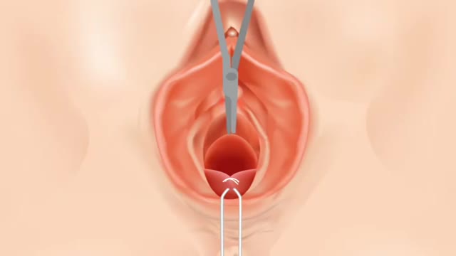
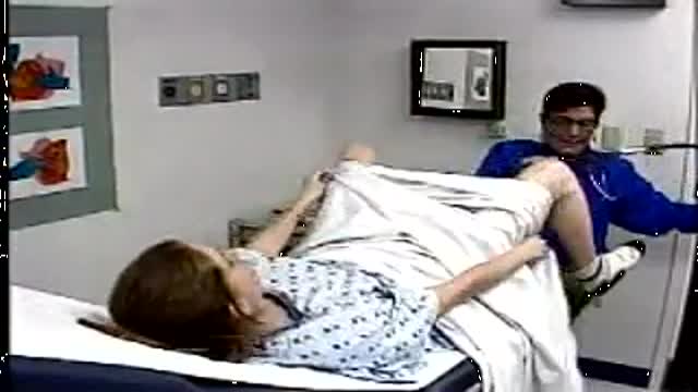
With the patient in the standard lathotomy position, the vaginal cuff is grasped bilaterally and injected with a diluted patrescent solution of lidocaine and epinephrine.
Next, the full thickness of the anterior vaginal wall is dissected vertically.
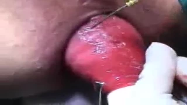
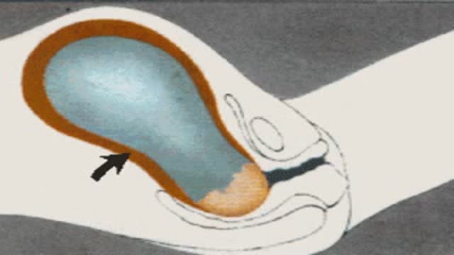
Continuing in the midline, dissection is carried out to expose the cystocial. Using sharp and blunt dissection, the cystocial is removed from the vaginal wall, exposing a nice plane of tissue. I like to take my dissection to the inferior portion of the pubic bone, pubic ramus, and to the ischial spine to expose the arches to the anus fascia pelvis.
Following completion on the right side, the same technique is employed on the left. Again, I'm taking this down bluntly with my finger to feel the ischial spine and the archis tendinus on our left side and up to the connective tissue of the pubic bone. With the bladder mobilized, plication of any separated pubocervical fascia is performed.
This offers slightly more protection between the symphysis graft and the bladder. A few simple interrupted sutures in this area are used to reduce the cystocial and replicate the fascia. Without going too far laterally and closing off the space, a good reduction of the cystocial is achieved. Next, connective tissues are located for placement of the anchoring sutures for the symphysis matrix. Absorbable sutures are placed with a needle driver on both sides. Four sutures will be used to secure the symphysis in place. Placement is similar to the area sutured for a para vaginal repair.
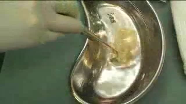
During preparation of the site and prior to use, the symphysis matrix must be rehydrated. What I like to do is cut this in a triangular pyramid shape. These two areas corresponding to the top two sutures, these going to the arches.
There's really very little I'll cut off here. There's various ways to fashion this. Aphillus works the best and mimics what the pubocervical fascia did. The beauty of this way also is you can leave this portion a little bit longer than you may need and then trim it at the appropriate time. It's good. Starting on the patient's upper left side, the free needles of the sutures are used to ligate the corresponding corners of the symphysis matrix. As the sutures are tied, the mesh is straightened to conform to the patient's anatomy.
It's helpful to put a throw in one side, go the other to secure that down and then come back and tie the remaining throws.
With all four corners secure, the site is inspected to confirm reduction of the cysticeal.
Excellent reduction of the cysticeal. No tension on the repair. Let's push well back. At this point, if this patient had urinary incontinence, can easily do a mid urethral sling through this incision, or some will close this incision and make a separate incision for the mid urethral sling. At this point, a minimal amount of excess vagina is trimmed and any small bleeders are cauterized. 2-0 vicaral is used to close the site. In this case, a running stitch will be used. However interrupted may also be used according to surgeon preference.
We have a nice full thickness cut here. It's easy just to dissect and take epithelium only, but you need some of this under tissue. I think it's very important if you're going to do mesh repairs.
And again, we're able to demonstrate a good suspension of the anterior segment. You can feel the mesh. There's no tension on the mesh and a good closure. Proceeding posteriorly, the patient also suffers from a recticeal with laxity to the rectovaginal septum. The posterior vaginal mucosa is injecting through the midline with 1% lidocaine and epinephrine to help create the plane and maintain hemostasis. The same principle of a full thickness dissection along the midline is employed posteriorly as well. Dissection is continued to just below the incision anteriorly of the vaginal cuff.
Sharp and blunt dissection is used on the rectovaginal septum to expose the recticeal. The target is the ischial spine bilaterally.
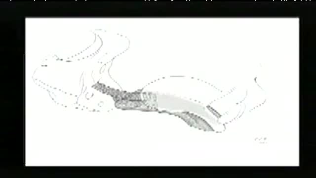
The venerosil sac is apparent here. That's an excellent dissection on this side. We'll start on the other side. Do the same. Good dissection to the ischial spine. We can see our rectivaginal septum. Well dissected venerosil. Traditional venerosil repair at this point would involve reduction, ligation of the sac and closure over the top. We're going to do something a little bit different using mesh. We're going to go ahead and use the IVS tunneller. The drape is expanded to permit use of the IVS tunneller. The first incision is made approximately two finger breaths lateral and two below the anal verge.
With a corresponding incision on the other side. Taking care to avoid the hemorrhoidal vaginal.
Transcript auto-generated from the video’s audio for accessibility and study. Educational content only — not medical advice.
Video Overview
From our Reproductive Health collection: Fertility, contraception, IVF and sexual health explained clearly by specialists. Browse all Reproductive Health videos →
Medical Videos
Free medical and surgery videos for patients, students and professionals — trusted since 2007.
Founded by Dr. Mohamed Ibrahim, MD, PhD — physician-scientist.
Follow us on Facebook · Instagram
SORT BY-
Top Comments
-
Latest comments